Home / Infrared Spectroscopy: A Quick Primer On Interpreting Spectra

Spectroscopy
By James Ashenhurst
- Infrared Spectroscopy: A Quick Primer On Interpreting Spectra
Last updated: October 31st, 2022 |
How To Interpret IR Spectra In 1 Minute Or Less: The 2 Most Important Things To Look For [Tongue and Sword]
Last post , we briefly introduced the concept of bond vibrations, and we saw that we can think of covalent bonds as a bit like balls and springs: the springs vibrate, and each one “sings” at a characteristic frequency, which depends on the strength of the bond and on the masses of the atoms. These vibrations have frequencies that are in the mid-infrared (IR) region of the electromagnetic spectrum.
We can observe and measure this “singing” of bonds by applying IR radiation to a sample and measuring the frequencies at which the radiation is absorbed. The result is a technique known as Infrared Spectroscopy , which is a useful and quick tool for identifying the bonds present in a given molecule.
We saw that the IR spectrum of water was pretty simple – but moving on to a relatively complex molecule like glucose (below) we were suddenly confronted with a forest of peaks!
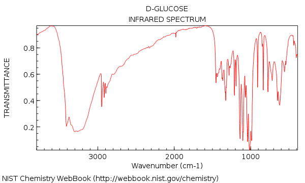
Your first impression of looking at that IR might be: agh! how am I supposed to make sense of that??
To which I want to say: don’t panic!
Table of Contents
- Let’s Correct Some Common Misconceptions About IR
- Starting With “Hunt And Peck” Is Not The Way To Go
- IR Spectroscopy: The Big Picture
- The Two Main Things To Look For In An IR Spectrum: “Tongues” and “Swords”.
- Alcohols and Carboxylic Acids: More Detail
- Specific Examples of IR Spectra of Carbonyl Functional Groups
- Less Crucial, But Still Useful: Two More Very Diagnostic Areas.
- Glucose, Revisited: The 1 Minute Analysis
1. Let’s Correct Some Common Misconceptions About IR
In this post, I want to show that a typical analysis of an IR spectrum is much simpler than you might think. In fact, once you learn what to look for, it can often be done in a minute or less. Why?
- IR is not generally used to determine the whole structure of an unknown molecule. For example, there isn’t a person alive who could look at the IR spectrum above and deduce the structure of glucose from it. IR is a tool with a very specific use. [Back in 1945 when IR was one of the few spectral techniques available, it was necessary to spend a lot more time trying to squeeze every last bit of information out of the spectrum. Today, with access to NMR and other techniques, we can do more cherry-picking]
- We don’t need to analyze every single peak ! (as we’ll see later, that’s what NMR is for : – ) ). Instead, IR is great for identifying certain specific functional groups , like alcohols and carbonyls. In this way it’s complimentary to other techniques (like NMR) which don’t yield this information as quickly.
With this in mind, we can simplify the analysis of an IR spectrum by cutting out everything except the lowest-lying fruit.
See that forest of peaks from 500-1400 cm -1 ? We’re basically going to ignore them all!
80% of the most useful information for our purposes can be obtained by looking at two specific areas of the spectrum : 3200-3400 cm -1 and 1650-1800 cm -1 . We’ll also see that there are at least two more regions of an IR spectrum worth glancing at, and thus conclude a “first-order” analysis of the IR spectrum of an unknown. [We might write a subsequent post which gets nittier and grittier about the finer points of analyzing an IR spectrum]
Bottom line: The purpose of this post is to show you how to prioritize your time in an analysis of an IR spectrum.
[BTW: all spectra are from the NIST database . Thank you, American taxpayers!]
2. Starting With “Hunt And Peck” Is Not The Way To Go
Confronted with an IR spectrum of an unknown (and a sense of rising panic), what does a typical new student do?
They often reach for the first tool they are given, which is a table of common ranges for IR peaks given to them by their instructor.
The next step in their analysis is to go through the spectrum from one side to the next, trying to match every single peak to one of the numbers in the table. I know this because this is exactly what I did when I first learned IR. I call it “hunting and pecking”.
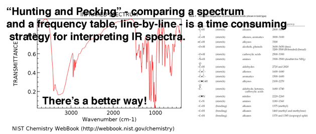
The only people who “hunt and peck” as their first step are people who have no plan (i.e. “newbies”).
So by reading the next few paragraphs you can save yourself a lot of time and confusion.
[Hunt and peck has its place, but only AFTER you’ve looked for “tongues” and “swords”, below. Hunting and pecking is great to make sure you didn’t miss anything big – but as a first step, it’s bloody awful!]
3. The Big Picture
In IR spectroscopy we measure where molecules absorb photons of IR radiation. The peaks represent areas of the spectrum where specific bond vibrations occur. [for more background, see the previous post, especially on the “ball and spring” model] . Just like springs of varying weights vibrate at characteristic frequencies depending on mass and tension, so do bonds.
Here’s an overview of the IR window from 4000 cm -1 to 500 cm -1 with various regions of interest highlighted.
An even more compressed overview looks like this: ( source )
Within these ranges, there are two high-priority areas to focus on , and two lesser-priority areas we’ll discuss further below.
4. The Two Main Things To Look For In An IR Spectrum: “Tongues” and “Swords”.
When confronted with a new IR spectrum, prioritize your time by asking two important questions:
- Is there a broad, rounded peak in the region around 3400-3200 cm -1 ? That’s where hydroxyl groups ( OH ) appear.
- Is there a sharp, strong peak in the region around 1850-1630 cm -1 ? That’s where carbonyl groups ( C=O ) show up.
First, let’s look at some examples of hydroxyl group peaks in the 3400 cm -1 to 3200 cm -1 region, which Jon describes vividly as “tongues”. The peaks below all belong to alcohols. Hydrogen bonding between hydroxyl groups leads to some variations in O-H bond strength, which results in a range of vibrational energies. The variation results in the broad peaks observed.
Hydroxyl groups that are a part of carboxylic acids have an even broader appearance that we’ll describe in a bit.
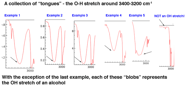
[Sometimes it helps to know what not to look for. On the far right hand side is included one example of a very weak peak on a baseline that you can safely ignore.]
The main point is that a hydroxyl group isn’t generally something you need to go looking for in the baseline noise.
Although hydroxyl groups are the most common type of broad peak in this region, N-H peaks can show up in this area as well (more on them in the Note 1 ). They tend to have a sharper appearance and may appear as one or two peaks depending on the number of N-H bonds.
Next, let’s look at some examples of C=O peaks, in the region around 1630-1800 cm -1. . These peaks are almost always the strongest peaks in the entire spectrum and are relatively narrow, giving them a somewhat “sword-like” appearance.
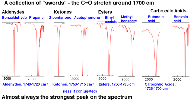
That sums up our 80/20 analysis: look for tongues and swords.
If you learn nothing else from this post, learn to recognize these two types of peaks!
Two other regions of the IR spectrum can quickly yield useful information if you train yourself to look for them.
3. The line at 3000 cm -1 is a useful “border” between alk ene C–H (above 3000 cm -1 ) and alk ane C–H (below 3000 cm -1 ) This can quickly help you determine if double bonds are present.
4. A peak in the region around 2200 cm -1 – 2050 cm -1 is a subtle indicator of the presence of a triple bond [C≡N or C≡C] . Nothing else shows up in this region.
A Common Sense Reminder
First, some obvious advice:
- if you’re given the molecular formula, that will determine what functional groups you should look for. It makes no sense to look for OH groups if you have no oxygens in your molecular formula, or likewise the presence of an amine if the formula lacks nitrogen.
- Less obviously, calculate the degrees of unsaturation if you are given the molecular formula, because it will provide important clues. Don’t look for C=O in a structure like C 4 H 10 O which doesn’t have any degrees of unsaturation.
5. Alcohols and Carboxylic Acids: More Detail
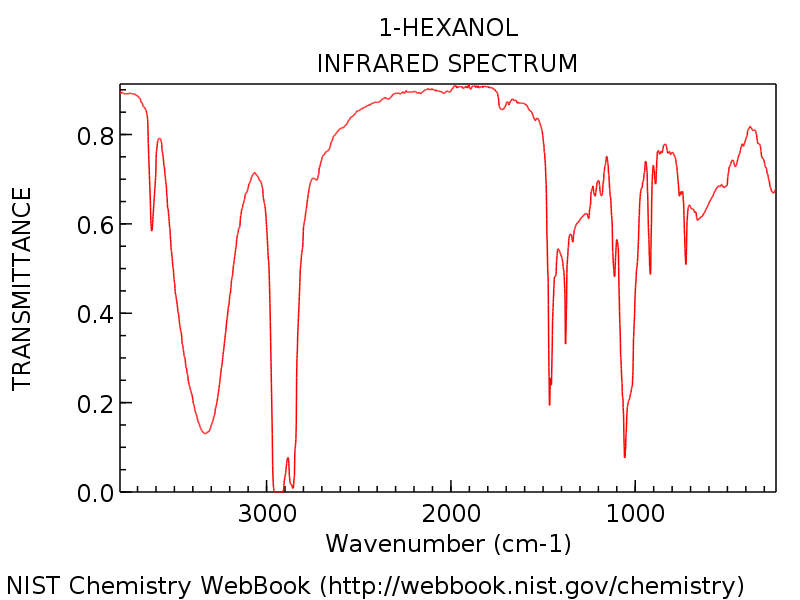
Let’s look at a specific example so we can see everything in perspective. The spectrum below is of 1-hexanol.
Note the hydroxyl group peak around 3300 cm -1 , typical of an alcohol (That sharp peak around 3600 cm -1 is a common companion to hydroxyl peaks: it represents non-hydrogen bonded O-H).
To gain some familiarity with variation, here’s some more examples of entire IR spectra of various alcohols.
- Cyclohexanol
Carboxylic Acids
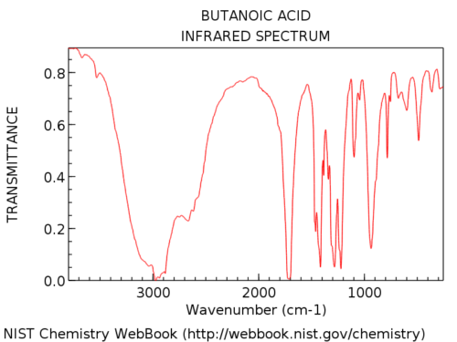
Hydroxyl groups in carboxylic acids are considerably broader than in alcohols. Jon calls it a “hairy beard”, which is a perfect description. Their appearance is also highly variable. The OH absorption in carboxylic acids can be so broad that it extends below 3000 cm -1 , pretty much “taking over” the left hand part of the spectrum.
Here’s an example: butanoic acid.
Here’s some more examples of full spectra so you can see the variation.
- Benzoic acid ,
- Pentanoic acid ,
- Acetic acid
The difference in appearance between the OH of an alcohol and that of a carboxylic acid is usually diagnostic. In the rare case where you aren’t sure whether the broad peak is due to the OH of an alcohol or a carboxylic acid, one suggestion is to check the region around 1700 cm for the C=O stretch. If it’s absent, you are likely looking at an alcohol.
[ Note 1 for more detail on the 3200-3500 cm -1 region : Amines, Amides, and Terminal Alkynes]
6. Specific Examples of IR Spectra of Carbonyl Functional Groups
The second important peak region is the carbonyl C=O stretch area at about 1630-1830 cm. Carbonyl stretches are sharp and strong.
Once you see a few of them they’re impossible to miss. Nothing else shows up in this region.
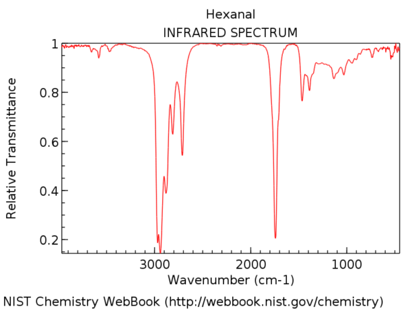
To put it in perspective, here’s the IR spectrum of hexanal. That peak a little after 1700 cm -1 is the C=O stretch. When it’s present, the C=O stretch is almost always the strongest peak in the IR spectrum and impossible to miss.
The position of the C=O stretch varies slightly by carbonyl functional group. Some ranges (in cm -1 ) are shown below:
- Aldehydes (1740-1690): benzaldehyde , propanal , pentanal
- Ketones (1750-1680): 2-pentanone , acetophenone
- Esters (1750-1735): ethyl acetate , methyl benzoate
- Carboxylic acids (1780-1710): benzoic acid , butanoic acid
- Amide (1690-1630): acetamide , benzamide , N,N -dimethyl formamide (DMF)
- Anhydrides (2 peaks; 1830-1800 and 1775-1740): acetic anhydride , benzoic anhydride
Conjugation will affect the position of the C=O stretch somewhat, moving it to lower wavenumber.
A decent rule of thumb is that you will never, ever see a C=O stretch below 1630. If you see a strong peak at 1500, for example, it is not C=O. It is something else.
7. Less Crucial, But Still Useful: Two More Very Diagnostic Areas.
- The C-H Stretch Boundary at 3000 cm -1
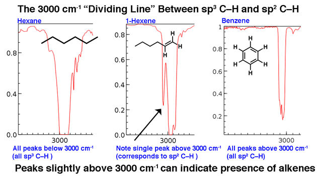
3000 cm -1 serves as a useful dividing line. Above this line is observed higher frequency C-H stretches we attribute to sp 2 hybridized C-H bonds. Two examples below: 1-hexene (note the peak that stands a little higher) and benzene.
For a molecule with only sp 3 -hybrized C-H bonds, the lines will appear below 3000 cm -1 as in hexane, below.
2. The Distinctive Triple Bond Region around 2200 cm -1
Molecules with triple bonds appear relatively infrequently in the grand scheme of things, but when they do, they do have a distinctive trace in the IR.
The region between 2000 cm -1 and 2400 cm -1 is a bit of a “ghost town” in IR spectra; there’s very little that appears in this region. If you do see peaks in this region, a likely candidate is a triple bonded carbon such as an alkyne or nitrile .
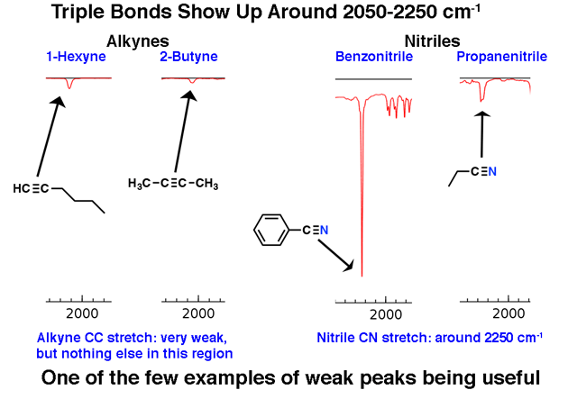
Note how weak the alkyne peaks are. This is one exception to the rule that one should ignore weak peaks. Still, caution is required: if you’re given the molecular formula, confirm that an alkyne is possible by calculating the degrees of unsaturation and ensuring that it is at least 2 or more.
Terminal alkynes (such as 1-hexyne) also have a strong C-H stretch around 3400 cm -1 that is more strongly diagnostic.
8. Glucose, Revisited: The 1 Minute Analysis
OK. We’ve gone over 4 regions that are useful for a quick analysis of an IR spectrum.
- (important!) O-H around 3200-3400 cm -1
- (important!) C=O around 1700 cm -1
- C-H dividing line at 3000 cm -1
- (rare) Triple bond region around 2050-2250 cm -1
Now let’s go back and look at the IR of glucose. What do we see?
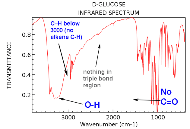
Here are the two big things to note:
- OH present around 3300 cm -1 . (in fact, this was included as one of the “swords” in section #3, above)
- No C=O stretch present. No strong peak around 1700 cm -1 . (The peak at 1450 cm -1 isn’t a C=O stretch).
Also, if we take a bit of extra time we can see:
- No alkene C-H (no peaks above 3000 cm -1 )
- Nothing in triple bonded region (rare, but still an easy thing to learn to check)
Now: If you were given this spectrum as an “unknown” along with its molecular formula, C 6 H 12 O 6 , what conclusions could you draw about its structure?
- The molecule has at least one OH group (and possibly more)
- The molecule doesn’t have any C=O groups
- The molecule *likely* doesn’t have any alkenes. If any alkenes are present, they don’t bear any C-H bonds, because we’d see their C-H stretch above 3000 cm -1 .
A molecule with one degree of hydrogen deficiency (C 6 H 12 O 6 ) but no C=O, and likely no C=C ?
A good guess would be that the molecule contains a ring . (We know this is the case, of course, but it’s nice to see the IR confirming what we already know).
This is what a 1-minute analysis of the IR of glucose can tell us. Not the whole structure, mind you, but certainly some important bits and pieces.
That’s enough for today. In the next post we’ll do some more 1-minute analyses and give more concrete examples of how to use the information in an IR spectrum to draw conclusions about molecular structure.
Related Articles
- IR Spectroscopy: 4 Practice Problems
- Bond Vibrations, Infrared Spectroscopy, and the “Ball and Spring” Model
- Introduction To UV-Vis Spectroscopy
- UV-Vis Spectroscopy: Practice Questions
- UV-Vis Spectroscopy: Absorbance of Carbonyls
- Degrees of Unsaturation (or IHD, Index of Hydrogen Deficiency)
More on the 3200 region: Amines, Amides, and Terminal Alkyne C-H
While we’re in the 3200 region…. Amines and Amides
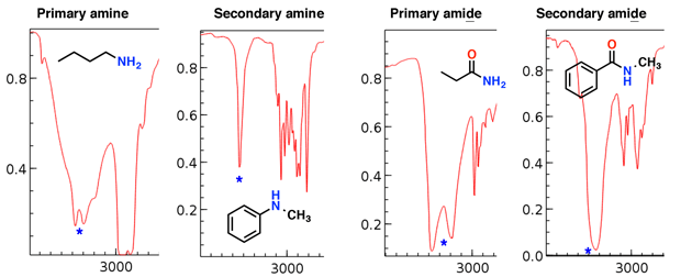
Amines and amides also have N-H stretches which show up in this region. [update: a comment from Paul Wenthold mentions some helpful advice about amides – they are rare – look for confirming evidence from the mass spectrum or other sources before assigning an amide based on a stretch in this region, as this region can also contain carbonyl “overtone” peaks]
Notice how the primary amine and primary amide have two “fangs”, while the secondary amine and secondary amide have a single peak.
The amine stretches tend to be sharper than the amide stretches; also the amides can be distinguished by a strong C=O stretch (see below).
Primary amines (click for spectra)
- Benzylamine
- Cyclohexylamine
Secondary amines:
- N-methylbenzylamine
- N,N-dibenzylamine
- N-methylaniline
Primary amides
- Propionamide
Secondary amides
- N-methyl benzamide
Terminal alkyne C-H
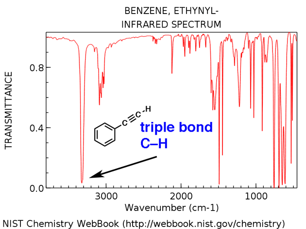
Terminal alkynes have a characteristic C-H stretch around 3300 cm -1 . Here it is for ethynylbenzene, below.
- Ethynylbenzene
00 General Chemistry Review
- Lewis Structures
- Ionic and Covalent Bonding
- Chemical Kinetics
- Chemical Equilibria
- Valence Electrons of the First Row Elements
- How Concepts Build Up In Org 1 ("The Pyramid")
01 Bonding, Structure, and Resonance
- How Do We Know Methane (CH4) Is Tetrahedral?
- Hybrid Orbitals and Hybridization
- How To Determine Hybridization: A Shortcut
- Orbital Hybridization And Bond Strengths
- Sigma bonds come in six varieties: Pi bonds come in one
- A Key Skill: How to Calculate Formal Charge
- The Four Intermolecular Forces and How They Affect Boiling Points
- 3 Trends That Affect Boiling Points
- How To Use Electronegativity To Determine Electron Density (and why NOT to trust formal charge)
- Introduction to Resonance
- How To Use Curved Arrows To Interchange Resonance Forms
- Evaluating Resonance Forms (1) - The Rule of Least Charges
- How To Find The Best Resonance Structure By Applying Electronegativity
- Evaluating Resonance Structures With Negative Charges
- Evaluating Resonance Structures With Positive Charge
- Exploring Resonance: Pi-Donation
- Exploring Resonance: Pi-acceptors
- In Summary: Evaluating Resonance Structures
- Drawing Resonance Structures: 3 Common Mistakes To Avoid
- How to apply electronegativity and resonance to understand reactivity
- Bond Hybridization Practice
- Structure and Bonding Practice Quizzes
- Resonance Structures Practice
02 Acid Base Reactions
- Introduction to Acid-Base Reactions
- Acid Base Reactions In Organic Chemistry
- The Stronger The Acid, The Weaker The Conjugate Base
- Walkthrough of Acid-Base Reactions (3) - Acidity Trends
- Five Key Factors That Influence Acidity
- Acid-Base Reactions: Introducing Ka and pKa
- How to Use a pKa Table
- The pKa Table Is Your Friend
- A Handy Rule of Thumb for Acid-Base Reactions
- Acid Base Reactions Are Fast
- pKa Values Span 60 Orders Of Magnitude
- How Protonation and Deprotonation Affect Reactivity
- Acid Base Practice Problems
03 Alkanes and Nomenclature
- Meet the (Most Important) Functional Groups
- Condensed Formulas: Deciphering What the Brackets Mean
- Hidden Hydrogens, Hidden Lone Pairs, Hidden Counterions
- Don't Be Futyl, Learn The Butyls
- Primary, Secondary, Tertiary, Quaternary In Organic Chemistry
- Branching, and Its Affect On Melting and Boiling Points
- The Many, Many Ways of Drawing Butane
- Wedge And Dash Convention For Tetrahedral Carbon
- Common Mistakes in Organic Chemistry: Pentavalent Carbon
- Table of Functional Group Priorities for Nomenclature
- Summary Sheet - Alkane Nomenclature
- Organic Chemistry IUPAC Nomenclature Demystified With A Simple Puzzle Piece Approach
- Boiling Point Quizzes
- Organic Chemistry Nomenclature Quizzes
04 Conformations and Cycloalkanes
- Staggered vs Eclipsed Conformations of Ethane
- Conformational Isomers of Propane
- Newman Projection of Butane (and Gauche Conformation)
- Introduction to Cycloalkanes (1)
- Geometric Isomers In Small Rings: Cis And Trans Cycloalkanes
- Calculation of Ring Strain In Cycloalkanes
- Cycloalkanes - Ring Strain In Cyclopropane And Cyclobutane
- Cyclohexane Conformations
- Cyclohexane Chair Conformation: An Aerial Tour
- How To Draw The Cyclohexane Chair Conformation
- The Cyclohexane Chair Flip
- The Cyclohexane Chair Flip - Energy Diagram
- Substituted Cyclohexanes - Axial vs Equatorial
- Ranking The Bulkiness Of Substituents On Cyclohexanes: "A-Values"
- Cyclohexane Chair Conformation Stability: Which One Is Lower Energy?
- Fused Rings - Cis-Decalin and Trans-Decalin
- Naming Bicyclic Compounds - Fused, Bridged, and Spiro
- Bredt's Rule (And Summary of Cycloalkanes)
- Newman Projection Practice
- Cycloalkanes Practice Problems
05 A Primer On Organic Reactions
- The Most Important Question To Ask When Learning a New Reaction
- Learning New Reactions: How Do The Electrons Move?
- The Third Most Important Question to Ask When Learning A New Reaction
- 7 Factors that stabilize negative charge in organic chemistry
- 7 Factors That Stabilize Positive Charge in Organic Chemistry
- Nucleophiles and Electrophiles
- Curved Arrows (for reactions)
- Curved Arrows (2): Initial Tails and Final Heads
- Nucleophilicity vs. Basicity
- The Three Classes of Nucleophiles
- What Makes A Good Nucleophile?
- What makes a good leaving group?
- 3 Factors That Stabilize Carbocations
- Equilibrium and Energy Relationships
- What's a Transition State?
- Hammond's Postulate
- Learning Organic Chemistry Reactions: A Checklist (PDF)
- Introduction to Free Radical Substitution Reactions
- Introduction to Oxidative Cleavage Reactions
06 Free Radical Reactions
- Bond Dissociation Energies = Homolytic Cleavage
- Free Radical Reactions
- 3 Factors That Stabilize Free Radicals
- What Factors Destabilize Free Radicals?
- Bond Strengths And Radical Stability
- Free Radical Initiation: Why Is "Light" Or "Heat" Required?
- Initiation, Propagation, Termination
- Monochlorination Products Of Propane, Pentane, And Other Alkanes
- Selectivity In Free Radical Reactions
- Selectivity in Free Radical Reactions: Bromination vs. Chlorination
- Halogenation At Tiffany's
- Allylic Bromination
- Bonus Topic: Allylic Rearrangements
- In Summary: Free Radicals
- Synthesis (2) - Reactions of Alkanes
- Free Radicals Practice Quizzes
07 Stereochemistry and Chirality
- Types of Isomers: Constitutional Isomers, Stereoisomers, Enantiomers, and Diastereomers
- How To Draw The Enantiomer Of A Chiral Molecule
- How To Draw A Bond Rotation
- Introduction to Assigning (R) and (S): The Cahn-Ingold-Prelog Rules
- Assigning Cahn-Ingold-Prelog (CIP) Priorities (2) - The Method of Dots
- Enantiomers vs Diastereomers vs The Same? Two Methods For Solving Problems
- Assigning R/S To Newman Projections (And Converting Newman To Line Diagrams)
- How To Determine R and S Configurations On A Fischer Projection
- The Meso Trap
- Optical Rotation, Optical Activity, and Specific Rotation
- Optical Purity and Enantiomeric Excess
- What's a Racemic Mixture?
- Chiral Allenes And Chiral Axes
- Stereochemistry Practice Problems and Quizzes
08 Substitution Reactions
- Introduction to Nucleophilic Substitution Reactions
- Walkthrough of Substitution Reactions (1) - Introduction
- Two Types of Nucleophilic Substitution Reactions
- The SN2 Mechanism
- Why the SN2 Reaction Is Powerful
- The SN1 Mechanism
- The Conjugate Acid Is A Better Leaving Group
- Comparing the SN1 and SN2 Reactions
- Polar Protic? Polar Aprotic? Nonpolar? All About Solvents
- Steric Hindrance is Like a Fat Goalie
- Common Blind Spot: Intramolecular Reactions
- The Conjugate Base is Always a Stronger Nucleophile
- Substitution Practice - SN1
- Substitution Practice - SN2
09 Elimination Reactions
- Elimination Reactions (1): Introduction And The Key Pattern
- Elimination Reactions (2): The Zaitsev Rule
- Elimination Reactions Are Favored By Heat
- Two Elimination Reaction Patterns
- The E1 Reaction
- The E2 Mechanism
- E1 vs E2: Comparing the E1 and E2 Reactions
- Antiperiplanar Relationships: The E2 Reaction and Cyclohexane Rings
- Bulky Bases in Elimination Reactions
- Comparing the E1 vs SN1 Reactions
- Elimination (E1) Reactions With Rearrangements
- E1cB - Elimination (Unimolecular) Conjugate Base
- Elimination (E1) Practice Problems And Solutions
- Elimination (E2) Practice Problems and Solutions
10 Rearrangements
- Introduction to Rearrangement Reactions
- Rearrangement Reactions (1) - Hydride Shifts
- Carbocation Rearrangement Reactions (2) - Alkyl Shifts
- Pinacol Rearrangement
- The SN1, E1, and Alkene Addition Reactions All Pass Through A Carbocation Intermediate
11 SN1/SN2/E1/E2 Decision
- Identifying Where Substitution and Elimination Reactions Happen
- Deciding SN1/SN2/E1/E2 (1) - The Substrate
- Deciding SN1/SN2/E1/E2 (2) - The Nucleophile/Base
- SN1 vs E1 and SN2 vs E2 : The Temperature
- Deciding SN1/SN2/E1/E2 - The Solvent
- Wrapup: The Key Factors For Determining SN1/SN2/E1/E2
- Alkyl Halide Reaction Map And Summary
- SN1 SN2 E1 E2 Practice Problems
12 Alkene Reactions
- E and Z Notation For Alkenes (+ Cis/Trans)
- Alkene Stability
- Alkene Addition Reactions: "Regioselectivity" and "Stereoselectivity" (Syn/Anti)
- Stereoselective and Stereospecific Reactions
- Hydrohalogenation of Alkenes and Markovnikov's Rule
- Hydration of Alkenes With Aqueous Acid
- Rearrangements in Alkene Addition Reactions
- Halogenation of Alkenes and Halohydrin Formation
- Oxymercuration Demercuration of Alkenes
- Hydroboration Oxidation of Alkenes
- m-CPBA (meta-chloroperoxybenzoic acid)
- OsO4 (Osmium Tetroxide) for Dihydroxylation of Alkenes
- Palladium on Carbon (Pd/C) for Catalytic Hydrogenation of Alkenes
- Cyclopropanation of Alkenes
- A Fourth Alkene Addition Pattern - Free Radical Addition
- Alkene Reactions: Ozonolysis
- Summary: Three Key Families Of Alkene Reaction Mechanisms
- Synthesis (4) - Alkene Reaction Map, Including Alkyl Halide Reactions
- Alkene Reactions Practice Problems
13 Alkyne Reactions
- Acetylides from Alkynes, And Substitution Reactions of Acetylides
- Partial Reduction of Alkynes With Lindlar's Catalyst
- Partial Reduction of Alkynes With Na/NH3 To Obtain Trans Alkenes
- Alkyne Hydroboration With "R2BH"
- Hydration and Oxymercuration of Alkynes
- Hydrohalogenation of Alkynes
- Alkyne Halogenation: Bromination, Chlorination, and Iodination of Alkynes
- Alkyne Reactions - The "Concerted" Pathway
- Alkenes To Alkynes Via Halogenation And Elimination Reactions
- Alkynes Are A Blank Canvas
- Synthesis (5) - Reactions of Alkynes
- Alkyne Reactions Practice Problems With Answers
14 Alcohols, Epoxides and Ethers
- Alcohols - Nomenclature and Properties
- Alcohols Can Act As Acids Or Bases (And Why It Matters)
- Alcohols - Acidity and Basicity
- The Williamson Ether Synthesis
- Ethers From Alkenes, Tertiary Alkyl Halides and Alkoxymercuration
- Alcohols To Ethers via Acid Catalysis
- Cleavage Of Ethers With Acid
- Epoxides - The Outlier Of The Ether Family
- Opening of Epoxides With Acid
- Epoxide Ring Opening With Base
- Making Alkyl Halides From Alcohols
- Tosylates And Mesylates
- PBr3 and SOCl2
- Elimination Reactions of Alcohols
- Elimination of Alcohols To Alkenes With POCl3
- Alcohol Oxidation: "Strong" and "Weak" Oxidants
- Demystifying The Mechanisms of Alcohol Oxidations
- Protecting Groups For Alcohols
- Thiols And Thioethers
- Calculating the oxidation state of a carbon
- Oxidation and Reduction in Organic Chemistry
- Oxidation Ladders
- SOCl2 Mechanism For Alcohols To Alkyl Halides: SN2 versus SNi
- Alcohol Reactions Roadmap (PDF)
- Alcohol Reaction Practice Problems
- Epoxide Reaction Quizzes
- Oxidation and Reduction Practice Quizzes
15 Organometallics
- What's An Organometallic?
- Formation of Grignard and Organolithium Reagents
- Organometallics Are Strong Bases
- Reactions of Grignard Reagents
- Protecting Groups In Grignard Reactions
- Synthesis Problems Involving Grignard Reagents
- Grignard Reactions And Synthesis (2)
- Organocuprates (Gilman Reagents): How They're Made
- Gilman Reagents (Organocuprates): What They're Used For
- The Heck, Suzuki, and Olefin Metathesis Reactions (And Why They Don't Belong In Most Introductory Organic Chemistry Courses)
- Reaction Map: Reactions of Organometallics
- Grignard Practice Problems
16 Spectroscopy
- Conjugation And Color (+ How Bleach Works)
- Bond Vibrations, Infrared Spectroscopy, and the "Ball and Spring" Model
- 1H NMR: How Many Signals?
- Homotopic, Enantiotopic, Diastereotopic
- Diastereotopic Protons in 1H NMR Spectroscopy: Examples
- C13 NMR - How Many Signals
- Liquid Gold: Pheromones In Doe Urine
- Natural Product Isolation (1) - Extraction
- Natural Product Isolation (2) - Purification Techniques, An Overview
- Structure Determination Case Study: Deer Tarsal Gland Pheromone
17 Dienes and MO Theory
- What To Expect In Organic Chemistry 2
- Are these molecules conjugated?
- Conjugation And Resonance In Organic Chemistry
- Bonding And Antibonding Pi Orbitals
- Molecular Orbitals of The Allyl Cation, Allyl Radical, and Allyl Anion
- Pi Molecular Orbitals of Butadiene
- Reactions of Dienes: 1,2 and 1,4 Addition
- Thermodynamic and Kinetic Products
- More On 1,2 and 1,4 Additions To Dienes
- s-cis and s-trans
- The Diels-Alder Reaction
- Cyclic Dienes and Dienophiles in the Diels-Alder Reaction
- Stereochemistry of the Diels-Alder Reaction
- Exo vs Endo Products In The Diels Alder: How To Tell Them Apart
- HOMO and LUMO In the Diels Alder Reaction
- Why Are Endo vs Exo Products Favored in the Diels-Alder Reaction?
- Diels-Alder Reaction: Kinetic and Thermodynamic Control
- The Retro Diels-Alder Reaction
- The Intramolecular Diels Alder Reaction
- Regiochemistry In The Diels-Alder Reaction
- The Cope and Claisen Rearrangements
- Electrocyclic Reactions
- Electrocyclic Ring Opening And Closure (2) - Six (or Eight) Pi Electrons
- Diels Alder Practice Problems
- Molecular Orbital Theory Practice
18 Aromaticity
- Introduction To Aromaticity
- Rules For Aromaticity
- Huckel's Rule: What Does 4n+2 Mean?
- Aromatic, Non-Aromatic, or Antiaromatic? Some Practice Problems
- Antiaromatic Compounds and Antiaromaticity
- The Pi Molecular Orbitals of Benzene
- The Pi Molecular Orbitals of Cyclobutadiene
- Frost Circles
- Aromaticity Practice Quizzes
19 Reactions of Aromatic Molecules
- Electrophilic Aromatic Substitution: Introduction
- Activating and Deactivating Groups In Electrophilic Aromatic Substitution
- Electrophilic Aromatic Substitution - The Mechanism
- Ortho-, Para- and Meta- Directors in Electrophilic Aromatic Substitution
- Understanding Ortho, Para, and Meta Directors
- Why are halogens ortho- para- directors?
- Disubstituted Benzenes: The Strongest Electron-Donor "Wins"
- Electrophilic Aromatic Substitutions (1) - Halogenation of Benzene
- Electrophilic Aromatic Substitutions (2) - Nitration and Sulfonation
- EAS Reactions (3) - Friedel-Crafts Acylation and Friedel-Crafts Alkylation
- Intramolecular Friedel-Crafts Reactions
- Nucleophilic Aromatic Substitution (NAS)
- Nucleophilic Aromatic Substitution (2) - The Benzyne Mechanism
- Reactions on the "Benzylic" Carbon: Bromination And Oxidation
- The Wolff-Kishner, Clemmensen, And Other Carbonyl Reductions
- More Reactions on the Aromatic Sidechain: Reduction of Nitro Groups and the Baeyer Villiger
- Aromatic Synthesis (1) - "Order Of Operations"
- Synthesis of Benzene Derivatives (2) - Polarity Reversal
- Aromatic Synthesis (3) - Sulfonyl Blocking Groups
- Birch Reduction
- Synthesis (7): Reaction Map of Benzene and Related Aromatic Compounds
- Aromatic Reactions and Synthesis Practice
- Electrophilic Aromatic Substitution Practice Problems
20 Aldehydes and Ketones
- What's The Alpha Carbon In Carbonyl Compounds?
- Nucleophilic Addition To Carbonyls
- Aldehydes and Ketones: 14 Reactions With The Same Mechanism
- Sodium Borohydride (NaBH4) Reduction of Aldehydes and Ketones
- Grignard Reagents For Addition To Aldehydes and Ketones
- Wittig Reaction
- Hydrates, Hemiacetals, and Acetals
- Imines - Properties, Formation, Reactions, and Mechanisms
- All About Enamines
- Breaking Down Carbonyl Reaction Mechanisms: Reactions of Anionic Nucleophiles (Part 2)
- Aldehydes Ketones Reaction Practice
21 Carboxylic Acid Derivatives
- Nucleophilic Acyl Substitution (With Negatively Charged Nucleophiles)
- Addition-Elimination Mechanisms With Neutral Nucleophiles (Including Acid Catalysis)
- Basic Hydrolysis of Esters - Saponification
- Transesterification
- Proton Transfer
- Fischer Esterification - Carboxylic Acid to Ester Under Acidic Conditions
- Lithium Aluminum Hydride (LiAlH4) For Reduction of Carboxylic Acid Derivatives
- LiAlH[Ot-Bu]3 For The Reduction of Acid Halides To Aldehydes
- Di-isobutyl Aluminum Hydride (DIBAL) For The Partial Reduction of Esters and Nitriles
- Amide Hydrolysis
- Thionyl Chloride (SOCl2)
- Diazomethane (CH2N2)
- Carbonyl Chemistry: Learn Six Mechanisms For the Price Of One
- Making Music With Mechanisms (PADPED)
- Carboxylic Acid Derivatives Practice Questions
22 Enols and Enolates
- Keto-Enol Tautomerism
- Enolates - Formation, Stability, and Simple Reactions
- Kinetic Versus Thermodynamic Enolates
- Aldol Addition and Condensation Reactions
- Reactions of Enols - Acid-Catalyzed Aldol, Halogenation, and Mannich Reactions
- Claisen Condensation and Dieckmann Condensation
- Decarboxylation
- The Malonic Ester and Acetoacetic Ester Synthesis
- The Michael Addition Reaction and Conjugate Addition
- The Robinson Annulation
- Haloform Reaction
- The Hell–Volhard–Zelinsky Reaction
- Enols and Enolates Practice Quizzes
- The Amide Functional Group: Properties, Synthesis, and Nomenclature
- Basicity of Amines And pKaH
- 5 Key Basicity Trends of Amines
- The Mesomeric Effect And Aromatic Amines
- Nucleophilicity of Amines
- Alkylation of Amines (Sucks!)
- Reductive Amination
- The Gabriel Synthesis
- Some Reactions of Azides
- The Hofmann Elimination
- The Hofmann and Curtius Rearrangements
- The Cope Elimination
- Protecting Groups for Amines - Carbamates
- The Strecker Synthesis of Amino Acids
- Introduction to Peptide Synthesis
- Reactions of Diazonium Salts: Sandmeyer and Related Reactions
- Amine Practice Questions
24 Carbohydrates
- D and L Notation For Sugars
- Pyranoses and Furanoses: Ring-Chain Tautomerism In Sugars
- What is Mutarotation?
- Reducing Sugars
- The Big Damn Post Of Carbohydrate-Related Chemistry Definitions
- The Haworth Projection
- Converting a Fischer Projection To A Haworth (And Vice Versa)
- Reactions of Sugars: Glycosylation and Protection
- The Ruff Degradation and Kiliani-Fischer Synthesis
- Isoelectric Points of Amino Acids (and How To Calculate Them)
- Carbohydrates Practice
- Amino Acid Quizzes
25 Fun and Miscellaneous
- A Gallery of Some Interesting Molecules From Nature
- Screw Organic Chemistry, I'm Just Going To Write About Cats
- On Cats, Part 1: Conformations and Configurations
- On Cats, Part 2: Cat Line Diagrams
- On Cats, Part 4: Enantiocats
- On Cats, Part 6: Stereocenters
- Organic Chemistry Is Shit
- The Organic Chemistry Behind "The Pill"
- Maybe they should call them, "Formal Wins" ?
- Why Do Organic Chemists Use Kilocalories?
- The Principle of Least Effort
- Organic Chemistry GIFS - Resonance Forms
- Reproducibility In Organic Chemistry
- What Holds The Nucleus Together?
- How Reactions Are Like Music
- Organic Chemistry and the New MCAT
26 Organic Chemistry Tips and Tricks
- Common Mistakes: Formal Charges Can Mislead
- Partial Charges Give Clues About Electron Flow
- Draw The Ugly Version First
- Organic Chemistry Study Tips: Learn the Trends
- The 8 Types of Arrows In Organic Chemistry, Explained
- Top 10 Skills To Master Before An Organic Chemistry 2 Final
- Common Mistakes with Carbonyls: Carboxylic Acids... Are Acids!
- Planning Organic Synthesis With "Reaction Maps"
- Alkene Addition Pattern #1: The "Carbocation Pathway"
- Alkene Addition Pattern #2: The "Three-Membered Ring" Pathway
- Alkene Addition Pattern #3: The "Concerted" Pathway
- Number Your Carbons!
- The 4 Major Classes of Reactions in Org 1
- How (and why) electrons flow
- Grossman's Rule
- Three Exam Tips
- A 3-Step Method For Thinking Through Synthesis Problems
- Putting It Together
- Putting Diels-Alder Products in Perspective
- The Ups and Downs of Cyclohexanes
- The Most Annoying Exceptions in Org 1 (Part 1)
- The Most Annoying Exceptions in Org 1 (Part 2)
- The Marriage May Be Bad, But the Divorce Still Costs Money
- 9 Nomenclature Conventions To Know
- Nucleophile attacks Electrophile
27 Case Studies of Successful O-Chem Students
- Success Stories: How Corina Got The The "Hard" Professor - And Got An A+ Anyway
- How Helena Aced Organic Chemistry
- From a "Drop" To B+ in Org 2 – How A Hard Working Student Turned It Around
- How Serge Aced Organic Chemistry
- Success Stories: How Zach Aced Organic Chemistry 1
- Success Stories: How Kari Went From C– to B+
- How Esther Bounced Back From a "C" To Get A's In Organic Chemistry 1 And 2
- How Tyrell Got The Highest Grade In Her Organic Chemistry Course
- This Is Why Students Use Flashcards
- Success Stories: How Stu Aced Organic Chemistry
- How John Pulled Up His Organic Chemistry Exam Grades
- Success Stories: How Nathan Aced Organic Chemistry (Without It Taking Over His Life)
- How Chris Aced Org 1 and Org 2
- Interview: How Jay Got an A+ In Organic Chemistry
- How to Do Well in Organic Chemistry: One Student's Advice
- "America's Top TA" Shares His Secrets For Teaching O-Chem
- "Organic Chemistry Is Like..." - A Few Metaphors
- How To Do Well In Organic Chemistry: Advice From A Tutor
- Guest post: "I went from being afraid of tests to actually looking forward to them".
Comment section
85 thoughts on “ infrared spectroscopy: a quick primer on interpreting spectra ”.
Ain’t this great! Man you’re an angel
this quick guide is awesome, I’ve learned so much reading it. To recall whatever you forgot over time, this is the best option. Thank you
Glad you found it useful for refreshing your memory!
This has really been helpful for my studies in chemistry
I am glad you find it helpful Cirona!
This is very helpful
Glad you find it helpful Anand
I love this analysis very much impressive thanks 👍
Glad you find it helpful!
A lifesaver if there was ever one. Infrared Spectroscopy was so confusing for me in undergrad,and post grad had me even more muddled.One look at this article on the morning of the test was enough to make me take my test confidently and do it well! The way you simplified it while highlighting important points is crazy. I was trying to remember all the values given from the typical IR frequency table which wasn’t working at all and was leaving me anxious. Tongues and Swords made it so simple and memorable. Thanks for all that you do and more! This is Monica Rao all the way from India!
Glad to hear you found it useful! I had a similar experience in undergraduate and glad that this simplified things for you!
i love you, you just saved my life
explanation is very easy to understand. thank you
Best Explanation so far. Really helpful
- Pingback: FTIR spectroscopy - Easy To Calculate
- Pingback: IR Spectroscopy and FTIR Spectroscopy: How an FTIR Spectrometer Works and FTIR Analysis - Technology Networks - Mubashir Khan.
- Pingback: IR spectroscopy - Easy To Calculate
This is so helpful thank you
Best Review on IR
Thank you Sagar.
Hi James, Thank you for your very clear tutorials on interpreting IR spectra. They have been really helpful to me.
I have a few questions regarding a compound with an unknown structure, which I am trying to decipher using FTIR. Would you be happy to have a look at this for me and confirm whether or not I have done it right, based on the information on your tutorials?
Thanks I am newbie and this finally made a pathway in my grey cells :)
Thanks a lot. This has really helped me I understood everything in it
Thank you so much for this great work. I have one problem: I used to work with polymers (in my particular case I am working with PVC films). Firstly, I do an FTIR spectrum of the “as received” PVC film. Next, I carry out a thermal treatment of the PVC film (below its Tg) and repeat the FTIR. The peaks have not change, however the intensity of them is different. I have tried to figure out an explanation for this phenomenon (searching in bibliography), but I didn´t found an answer. Do you have any idea of why this happen?
thank you very much.
You are absolutely amazing. I feel so happy and satisfied reading this. Your style of presenting the context is so good. Thank You for your hard work for us.
Thank you so much. Two month i have struggled about this topic. Full of detail in simple words with various example. Thank you again
I am really grateful this lesson is really awesome.
Thank you so much!! Your post really helped understangding IR :)
Thank you so much for this great information sir
Thank you!! This is so easy explained and helpful. I have one question: How much can I trust in my software suggestions? the software of my FTIR instrumen has some libraries included.
I’m not sure. There can be considerable variability between samples of the same molecule, depending on how the sample is prepared (thickness of film) and the amount of water present (which affects hydrogen bonding). The libraries are a good starting point but not a magic bullet, good when part of a more holistic approach to combine with other information (e.g. HRMS data)
Thanks Paul – I was unaware of the overtones in that region. Very helpful, thank you!
One thing you didn’t mention is the carbonyl overtone peaks, which result when the molecule absorbs two photons of IR light. These show up as weak peaks at 2 x the carbonyl frequency, so are in that 3300 – 3500 range.
It’s important know about this because beginning students very often assign those peaks to NH stretches. And it’s not crazy, because NH stretches in monosubstituted amides can be relatively weak, so it can be difficult to distinguish them.
This isn’t perfect, but, from an instructor perspective, my advice is to avoid the urge to assign them to amine or amide. If you see a carbonyl, expect to see that overtone and don’t call it an NH stretch. Now, this means you might miss an amide, but that alone is not sufficient to conclude it is amide. You would need to verify it by other means. As noted, amide C=O stretches tend to be lower energy than other functional groups, but even then I’d be careful about putting too fine a point on it (absorptions usually come in ranges, not in specific spots – the C=O is 1680: does that mean it’s amide? Could be, but it could also be a ketone at the edge of its range; it’s consistent with both). Now, if you have a mass spectrum that indicates the presence of a N (by having an odd molecular mass), so you know N is present, then sure, it could be NH stretching. But absent other information that indicates an amide, my advice is don’t go that direction.
This is the best review I have ever seen-splendid!
This is an excellent resource on IR for a newbie…love to give this to my students for reading. Looking for posts on mass spectrometry..
Thanks Anju – appreciate it. This is what I wish someone told me when I was learning how to interpret IR spectra.
Very clear, lots of examples and well thought out instructions. I feel so much more confident! Thank you soo much!!!
Great! So glad you feel more confident!
Symply excellent. Please, we need MOC Text book.
Not happening! But thank you
Seriously it is the best of all explanation I have seen ,it really helpful 💖
Very helpful. I can understand the materials much better
Thanks for the wonderful lecture, my question is how can one identify aromatic or the benzene ring absorption. Please I also need your email address
Look for the C-H bond stretch below 3000 cm-1. It is not specific for the aromatic ring but at least points to an sp2 hybridized carbon bonded to H.
saved my life honestly.
Honestly? Awesome!
Best explanation ever ! The only one I understood .. Thank you a lot!
Thanks Olivia! Glad you found it helpful!
I was completely lost at lecture on IR but after reading this, i realized its simple things made difficult. You saved me a failure.
So glad to hear it Josan.
Thanks! This article saved me. Recommended this to all my friends.
Thanks for letting me know Harshit!
Wow! Thanks – you will never know how much time this saved me.
So glad to hear it Freeman.
Excellent explanation! Thank you for all the hard work.
Thanks Zeke!
Thank you! you just saved my life
If it made IR less painful, that’s awesome Alejandra!
I see nothing about <500 cm-1 which is what I need to know
Really? I wish I had a better answer for you. The region below 500 cm-1 is an “enduring mystery” for many of us. https://amphoteros.com/2019/01/18/an-enduring-mystery/
Loved this!!!!!
THANK YOU!!!! THIS SAVED MY LIFE!!!!!!
I know your faculty plans did not work out, but you are so better than many professors! Thank you! Never stop chasing your dreams!
Beautifully explained Sir !!
Best explanation of IR spectra I’ve came across. Waiting for your next post :)
I completely agree with the above posts. You should Youtube as well my friend. Great job!
I do have a Youtube channel but it has been quite neglected!
I COMPLETELY AGREE 100% with the previous praises and comments – you have been a SAVING grace in my organic chemistry understanding and I appreciate your approach in simplifying the most complex things. I have honestly spent 4+hrs in attempting 2 problems in figuring out the structures and feel so much better moving forward. THANK YOU! Keep up the phenomenal job!
Thank you Maribel!
Thanks for such a great focused article. It’s really very helpful.
Tried to make it useful. If it succeeded, great!
This is very clear and understandable even to a layman. Thanks a lot
You’re welcome!
why alkenes group (3000 -3100) & alkyl halides (500 -539) are added to NORYL (PPE + PS) plastic? which properties are affected?
How do you know the peak in the 3000-3100 isn’t from the styrene?
Thank you so much for this guide! Very thorough approach and great explanation.
Meg – so glad you’ve found it helpful. Put a lot of work into it!
Great work! best I could find in all these years in fact.
Never using another website or youtube vid (unless its yours) for help again. You’re amazing
Beautifully explained!
This is the best review for IR Spectroscopy out there!
THIS IS SO HELPFUL!! so many different examples were used and I understand everything now! Will there be a quick tutorial for carbon and proton NMR as well?
Leave a Reply
Your email address will not be published. Required fields are marked *
Save my name, email, and website in this browser for the next time I comment.
Notify me via e-mail if anyone answers my comment.
This site uses Akismet to reduce spam. Learn how your comment data is processed .
last updated Mon, Apr 1, 2002
Infrared Spectroscopy Dr.A.Bacher
V ibrational modes within a molecule can be described using the anharmonic oscillator model. This model assumes that the two masses (with known weight) are connected with a spring (with known strength). With the help of quantum mechanical calculations (Schroedinger equation) you can find the frequencies of basic stretching and bending modes:
From this equation, one can deduce some basic trends can be deducted: a. If the force constant F (= bond strength) increases, the stretching frequency will increase as well (in cm -1 )
b. If the masses of the involved atoms increase, the peak will shift to lower wavenumbers e.g. H/D-exchange in labeling experiments although the bond strength remains the same.
Based on the equation, one would always expect a sharp lines at a well defined wavenumber. Unfortunately, the change in vibration modes is always accompanied with change in rotational mode (Stokes and Anti-Stokes). The required energy for this process is much smaller (1-5 cm -1 ) and causes together with some other effects the broading of the 'lines'. The number of basic stretching and bending modes expected for a molecule increases with the number of atoms in the molecule. For non-linear molecules 3N-6 (2N-5 bending, N-1 stretching) vibrations are observed (e.g. CH 2 Cl 2 ). For linear molecules e.g. CO 2 one expect to find 3N-5 (3*3-5=4) modes. If there is no symmetry in the molecule most of them will be observed the IR spectrum; the remaining modes will be observed in a Raman spectrum. The more complicated the molecule is (the more atoms it possesses and the lower the symmetry), the more peaks can be observed in the IR spectrum (see example 3 and 4 ). A signal is only observed in the IR spectrum, if the dipole momentum of the molecule changes during the interaction with the electromagnetic radiation. This is very likely for groups which already possess a significant dipole momentum to start with e.g. C-O, C-Cl, O-H, etc. (strong peaks). Groups with a small difference in electronegativity e.g. C-H, C-C, C=C, etc., will usually show weak or medium sized peaks in the IR spectrum. An interpretation of an IR spectrum should include a detailed assignment of the peaks: exact wavenumber from the spectrum (integer), the intensity (w/m/s/br) and which functional group it represents, and maybe in addition the corresponding literature value. However, it is not necessary to interpret every little peak in the IR spectrum. Also, be cautious when you compare spectra which were obtain with different techniques (solution, Nujol mull, KBr pellet). The actual number of your vibration changes quite at bit, especially for highly polar compounds (Why ?). 2. Interpretation of the spectrum The IR-spectrum can be divided into five ranges major ranges of interest for an organic chemist: a. From 2700-4000 cm -1 (E-H-stretching: E=B, C, N, O) In this range typically E-H-stretching modes are observed. The C-H-stretching modes can be found between 2850 and 3300 cm -1 , depending on the hydrization . The range from 2850-3000 cm -1 belongs to saturated systems (alkanes, sp 3 , example 1 ), while the peaks from 3000-3100 cm -1 indicate an unsaturated system (alkenes, sp 2 , example 2; aromatic ring, example 3,4 ). Latter ones are usually weak or medium in intensity. The CH-function on a C-C-triple bond (alkynes) will appear as a sharp, strong peak around 3300 cm -1 . The differences in wavenumbers are mainly due to different hybridization (=bond strength, the s-character in the bond, the stronger the bond is) and number of ligands on the carbon atom. The O-H-stretching modes from alcohols and phenols ( example 5 ) are mostly broad and very strong (3200-3650 cm -1 ) The O-H-peaks due to carboxylic acids ( example 6 ) show a very broad and less intense peak between 2500 and 3500 cm -1 . The change in peak shape is a result of the different degree of hydrogen bonds in alcohol and carboxylic acids. These peaks change significantly with the polarity of the solvent. They usually become sharper if the polarity of the solvent increases e.g. camphor in CCl 4 and CH 2 Cl 2 (see reader). P eaks that are due to N-H-stretching modes are sharper than O-H-peaks (3300-3500 cm -1 ). Primary amines ( example 7 ) have two peaks (sym./asym. vibration) in this range, while secondary amines ( example 8 ) have only one peak. There are no peaks in this area for tertiary amines. Why?
Two small or medium peaks at ~2750 and ~2850 cm -1 are a result of an aldehyde ( example 15 ). b. From 2000 - 2700 cm -1 (E-X-triple bonds: E=X=C, N, O) This range covers mainly the triple bond stretching modes. The C-C-triple bond of alkynes (2130-2150 cm -1 ) is usually fairly weak, if observed at all. The C-N-triple bond of nitriles ( example 10 ) (2100-2160 cm -1 ). In most cases a peak (with varying intensity) around 2349 cm -1 (together with 667 cm -1 ). This is due to the CO 2 in the beam (poor background correction). c. From 1500 - 2000 cm -1 (E-X-double bonds: E=X=C, N, O) This is the most important range in the entire IR spectrum for organic chemists. If there is a very strong peak between 1640 and 1850 cm -1 , there is most likely a carbonyl function in the molecule. Analysis of the exact peak position will reveal further what type of carbonyl function is present. The general rule is the more reactive the carbonyl compound is, the further to the right (=higher wavenumber) the C=O stretching frequency will be. The following sequence is observed:
acid chlorides > anhydrides > ester > aldehydes > ketones > carboxylic acids > amides
In order to identify a specific group additional information is needed from the other ranges of the IR spectrum:
Ring strain usually increases the C=O stretching frequency. Conjugation of teh carbonyl group with other double bonds (aromatics, alkenes) or the formation of hydrogen bonds decreases the bond strength (shift: 15-60 cm -1 to lower wavenumbers) as the following examples of cyclic ketones demonstrate.
For all 'anhydride type systems' (O=C-X-C=O, X=O, NR, S, CH 2 ) two peaks are observed in this region due to a symmetric and an asymmetric stretching mode.
In addition to the dominant C=O-mode, the C-C-double bond ( example 2 -4 )is also located in this area (1600-1660 cm -1 ). However, if there is a carbonyl group present, it might be difficult to locate a weak or medium sized peak right next to it. Sometimes it is only observed as a shoulder. If there is a coupling between a C=C-group and other double bonded systems e.g. C=O or aromatic systems, the intensity will increase due to the increase in dipole momentum in the double bond. A medium or strong peak in this area corresponds to aromatic ring. A nitro group shows two very intense peaks in the range between 1300-1400 cm -1 (sym.) and 1500-1600 cm -1 (asym.) ( example 17 ).In a good spectrum, it might be possible to deduct the substitution pattern on an aromatic system from the overtone combination vibrations in the range from 1660-2000 cm -1 e.g. four peaks with increasing intensity are characteristic for a monosubstitution on your ring ( example 4 and table for Arenes below). d. From 1000-1500 cm -1 (E-X-single bonds, deformation, rocking modes) A strong peak around 1450 cm -1 indicates the presence of methylene groups (CH 2 ), while an additional strong peak about 1375 cm -1 is caused by a methyl group (CH 3 ) ( examples 1, 8-10 ). A symmetric 'doublet' with medium intensity around 1370 cm -1 is characteristic for an isopropyl group, while an asymmetric 'doublet' between 1365 and 1390 cm -1 is often due to a t-Bu-group. The C-O-C-functions of ethers and esters are typically found as strong peaks in the range between 1000 and 1300 cm -1 ( example 13 ). Generally, assignments in this area have to be done with extreme care, because there are a lot of ring absorbances in this ‘ fingerprint area ’. e. Below 1000 cm -1 (out of plane modes, C-X: X=Cl, Br, I, heavier atoms) This range belongs to the ’fingerprint area’, where assignments are a little bit uncertain. In some cases you can find information about the substitution pattern of alkenes or aromatic ring systems, if the range between 700-900 cm -1 is analyzed correctly (oop bending).
4. Examples Example 1 : Alkane - Undecane
- Search Search Please fill out this field.
What Are Investor Relations (IR)?
- Understanding IR
- IR and Legislation
- IR Functions
The Bottom Line
- Investing Basics
Investor Relations (IR): Definition, Career Path, and Example
:max_bytes(150000):strip_icc():format(webp)/picture-53886-1440626964-5bfc2a89c9e77c005876da24.jpg)
Gordon Scott has been an active investor and technical analyst or 20+ years. He is a Chartered Market Technician (CMT).
:max_bytes(150000):strip_icc():format(webp)/gordonscottphoto-5bfc26c446e0fb00265b0ed4.jpg)
Investopedia / Hilary Allison
The investor relations (IR) department is a division of a business, usually a public company , whose job it is to provide investors with an accurate account of company affairs. This helps private and institutional investors make informed decisions on whether to invest in the company.
Key Takeaways
- The investor relations (IR) department is a division of a business whose job it is to provide investors with an accurate account of company affairs.
- IR departments are required to be tightly integrated with a company's accounting department, legal department, and executive management team.
- IR departments have to be aware of changing regulatory requirements and advise the company on what can and cannot be done from a PR perspective.
- Legislation, such as the Dodd-Frank Act, has strengthened investor relations by requiring more transparency in the financial marketplace.
Understanding Investor Relations (IR)
Investor relations ensures that a company's publicly traded stock is being fairly traded through the dissemination of key information that allows investors to determine whether a company is a good investment for their needs. IR departments are sub-departments of public relations (PR) departments and work to communicate with investors, shareholders, government organizations, and the overall financial community.
Companies normally start building their IR departments before going public. During this pre-initial public offering ( IPO ) phase, IR departments can help establish corporate governance, conduct internal financial audits , and start communicating with potential IPO investors.
For example, when a company goes on an IPO roadshow, it is common for some institutional investors to become interested in the company as an investment vehicle. Once interested, institutional investors require detailed information about the company, both qualitative and quantitative.
To obtain this information, the company's IR department is called upon to provide a description of its products and services, financial statements, financial statistics, and an overview of the company's organizational structure.
The IR department's largest role is its interactions with investment analysts who provide public opinion on the company as an investment opportunity.
Investor Relations and Legislation
The Sarbanes-Oxley Act , also known as the Public Company Accounting Reform and Investor Protection Act, was passed in 2002, increasing reporting requirements for publicly traded companies. This expanded the need for public companies to have internal departments dedicated to investor relations, reporting compliance, and the accurate dissemination of financial information.
In the aftermath of the financial crisis, the Obama Administration passed the Dodd-Frank Wall Street Reform and Consumer Protection Act of 2009 to prevent financial institutions from taking excessive risks and introduced new measures to prevent lenders from exploiting consumers. To achieve this, the legislation established an independent agency called the Consumer Financial Protection Burea (CFPB) tasked with setting and enforcing clear, standardized rules for companies providing financial services.
The legislation strengthened investor relations by requiring more transparency across the financial system. For example, the CFPB now requires that a mortgage disclosure comes in a single form that outlines associated risks and costs, allowing consumers to compare loans with other lenders. Similarly, the legislation strengthened the Credit Card Accountability, Responsibility, and Disclosure (CARD) Act of 2009 , requiring issuers to disclose rates and fees clearly to help customers make more informed financial decisions. Additionally, the reforms prohibit credit card companies from directly marketing promotions to young consumers.
Investor Relations Functions
IR teams are typically tasked with coordinating shareholder meetings and press conferences, releasing financial data, leading financial analyst briefings, publishing reports to the Securities and Exchange Commission (SEC), and handling the public side of any financial crisis.
In addition, IR departments have to be aware of changing regulatory requirements and advise the company on what can and cannot be done from a PR perspective. For example, IR departments have to lead companies in quiet periods , where it is illegal to discuss certain aspects of a company and its performance.
The IR department's largest role is its interactions with investment analysts who provide public opinion on the company as an investment opportunity. These opinions influence the overall investment community, and it is the IR department's job to manage analysts' expectations.
Goals of Investor Relations
IR is intended to increase and sustain investor and other stakeholder confidence by giving them accurate and timely information regarding the company's financial and operating performance. It's also used to communicate the strategic plans and objectives.
IR seeks to maximize shareholder value by giving investors a thorough picture of the business strategy and expansion objectives of the company. This may enhance interest from existing investors, boost demand for the company's shares, and ultimately raise the share price.
IR is also intended to enhance corporate governance by making sure that the business complies with pertinent laws, rules, and moral guidelines. This could increase the company's access to capital markets as well as its reputation and trust with investors. In this realm, IR is meant to invoke confidence that a company is adhering to all of the required laws set forth by governing bodies.
Communication is also made easier because to IR, which acts as a conduit between a firm's stakeholders and investors. This gives investors access to key decision-makers within the company. This can also support the company in addressing investor issues, giving management input, and fostering positive connections with its stakeholders.
Companies often have pages on their website dedicated to investor relations. This section will house financial statements, external disclosures, SEC filings, or annual reports.
Benefits of Investor Relations
By leveraging IR, companies can increase their access to capital markets which can enable them to obtain finance more effectively and at a reduced cost. This is done by developing relationships with investors and analysts.
IR contributes to greater transparency by delivering accurate and timely information to investors about a company's financial performance, strategic positioning, and other significant developments. Investor trust can be increased and the company's reputation can be enhanced as a result.
Effective IR may also assist businesses in growing their investor base and luring fresh capital. By attracting new investors and raising demand for the company's stock, effective IR can assist raise the liquidity of a company's shares. This could enhance the company's worth and make it easier for all investors to trade.
Companies can increase their access to capital markets, which can enable them to obtain finance more effectively and at a reduced cost, by developing relationships with investors and analysts. Effective IR may also assist businesses in growing their investor base and luring fresh capital.
Last, by ensuring that businesses adhere to pertinent laws, regulations, and moral standards, IR aids in enhancing corporate governance . This could increase the company's access to capital markets as well as its reputation and trust with investors.

Why Is It Important for a Company to Have an Investor Relations Division?
Companies require an investor relations division to provide current and prospective investors with relevant information so they can make informed investment decisions. Failure to disclose information that may have a material impact on a company’s share price could result in a fine or other disciplinary action from regulators.
What Are the Primary Functions of an Investor Relations Division?
The investor relations team oversees functions such as coordinating shareholder meetings and press conferences, releasing financial data, leading financial analyst briefings, publishing SEC filings, and handling the public relations of a company-specific financial crisis.
What Role Does an Investor Relations Division Play Before a Company Goes Public?
Before a company goes public, an investor relations division may assist with establishing corporate governance, conducting internal financial audits, and disseminating information to prospective IPO investors.
What Effect Does Government Legislation Have on Investor Relations?
Legislation such as the Sarbanes-Oxley Act and Dodd-Frank Act have strengthened investor relations by requiring financial institutions to provide greater transparency, particularly about fees and risk. Reforms have also increased reporting requirements for publicly traded companies.
Investor relations refers to a division within a company that provides investors with information about its corporate affairs, helping them make more informed investment decisions. The IR division typically works closely with accounting and legal departments along with executive management to ensure the dissemination of essential financial information.
Often companies establish an IR department before going public, with the division assisting in areas such as corporate governance, financial auditing, and communicating with prospective IPO investors. Since the 2008 financial crisis, legislation, such as the Sarbanes-Oxley Act and Dodd-Frank Act, has strengthened Investor relations by requiring greater reporting and transparency across the financial system to help consumers make more informed decisions.
U.S Congress. " H.R.3763 - Sarbanes-Oxley Act of 2002 ."
The White House Archives. " Jobs and The Economy: Putting America Back to Work ."
FTC.gov. " The Credit Card Accountability, Responsibility, and Disclosure Act of 2009 ," Page 16.
:max_bytes(150000):strip_icc():format(webp)/GettyImages-513676272-efd95f82492447f581517b4690d31421.jpg)
- Terms of Service
- Editorial Policy
- Privacy Policy
- Your Privacy Choices
Photometry & Reflectometry

Photometry is the measurement of light absorbed in the ultraviolet (UV) to visible (VIS) to infra-red (IR) range. This measurement is used to determine the amount of an analyte in a solution or liquid. Photometers utilize a specific light source and detectors that convert light passed through a sample solution into a proportional electric signal. These detectors may be for example photodiodes, photoresistors, or photomultipliers. Photometry uses Beer–Lambert’s law to calculate coefficients obtained from the transmittance measurement. A correlation between absorbance and analyte concentration is then established by a test-specific calibration function to achieve highly accurate measurements.
Photometry is a widely used quantitative analysis in research laboratories to determine the amounts of inorganic and organic compounds in solutions and other liquids. Photometry also has broad industrial applications in the determination of contaminants in drinking and wastewater, analysis of nutrients in soil, food and beverage samples, building material composition, and many other areas.
Construction and Working of Photometer
The components of a typical photometer include a light source, monochromator, sample, and detector. Light sources can be tungsten-halogen lamps (generally the source of light used for analyses in the visible light range) or LEDs. For measurements in the UV–Visible range a xenon flash lamp may be used. The monochromator filters the light radiated by the light source to allow only a very narrow spectrum to pass. The light then passes through the cuvette or the sample holding cell. Based on the amount of analyte (or a dye derived from it) that is present in the sample solution, some part of the light is absorbed by the solution and the remaining is transmitted. The transmitted light is directed towards the detectors, which produce an electric current proportional to the light intensity.
Beer–Lambert Law
Beer–Lambert law, also known as Beer’s law, states that the quantity of light absorbed by a sample is directly proportional to the concentration of the analyte present and path length of the light through the sample. “Sample” in this context means either the analyte itself (direct measurement) or a dye derived from the analyte (when using reagents or kits). The relationship is described by the formula:
Reflectometry
Reflectometry (also known as remission photometry) is a non-destructive analytical technique that uses the reflection of light by surfaces and interfaces to measure characteristics such as color intensity, film thickness and refractive index.
As with other photometers, the main elements of reflectometers include a light source, usually long-life LEDs of specific wavelengths that are focused onto a sample surface via a lens system and the reflected light is measured by detectors.
Reflectometers are often designed to measure physical characteristics of surfaces like color changes on a test strip. In this approach, a sample can be placed on a test strip and its remission (REM) value can be compared against appropriate controls and standards. As in photometry, the difference in intensity between emitted and reflected light allows a quantitative determination of the concentration of specific analytes.
Reflectometry is commonly used in industrial applications as a rapid, sensitive method for quantitating a wide variety of organic and inorganic parameters in water, food, beverages, and environmental samples as well as other diverse uses such as surface analysis of building materials and skin color quantification.
Related Technical Articles
- Transmittance to Absorbance Table A transmittance to absorbance table enables fast conversion from transmittance values to absorbance in the lab or in the field.
- Measuring Chemical Oxygen Demand in Water Treatment Facilities We are a global leader in the life science industry and have produced test kits to measure numerous analytes. Water Online spoke with us about advances in measuring chemical oxygen demand.
- Ammonium in Wastewater Step-by-step accurate reflectometric determination of ammonium in wastewater with Nessler’s reagent or Indophenol blue using Reflectroquant® system and test strips.
- Analytical Method: Analytical Quality Assurance, Standard for COD/Chloride Preparation of a standard solution for COD/chloride
- K268 Olive Oil Commission Regulation (EEC) No 2568/91 Annex IX Learn about the UV-spectrometric determination of the K268 nm value of olive oil, assessing the UV absorbance at 268 nm, to ensure quality and compliance with regulatory standards.
- See All (24)
Related Protocols
- Analytical Method: Analytical Quality Assurance, Standard for Hydrogen peroxide Preparation of a standard solution for Hydrogen peroxide
- Analytical Method: Aluminium (total) in effluents Photometric determination with Chromazurole S subsequent to acid mineralisation; DIN decomposition
- Analytical Method: Aluminium (total) in effluents, high organic content Photometric determination with Chromazurole S subsequent to acid mineralisation
- Analytical Method: Aluminium (total) in lime stone Photometric determination with Chromazurole S subsequent to fusion melting
- Ammonium in Effluents Step-by-step method describing the photometric determination of ammonium ions in effluents using Spectroquant® test kits and spectrophotometer.
- See All (33)
Find More Articles and Protocols

Join our webinar on June 15th to find out how to quickly analyze chemical disinfectant parameters to ensure the safety of your production line after disinfection & the different methods available.
To continue reading please sign in or create an account.
Research. Development. Production.
We are a leading supplier to the global Life Science industry with solutions and services for research, biotechnology development and production, and pharmaceutical drug therapy development and production.
© 2024 Merck KGaA, Darmstadt, Germany and/or its affiliates. All Rights Reserved.
Reproduction of any materials from the site is strictly forbidden without permission.
- English - EN
- Español - ES

Want to create or adapt books like this? Learn more about how Pressbooks supports open publishing practices.
6.3 IR Spectrum and Characteristic Absorption Bands
With a basic understanding of IR theory, we will now take a look at the actual output from IR spectroscopy experiments and learn how to get structural information from the IR spectrum. Below is the IR spectrum for 2-hexanone.

Notes for interpreting IR spectra:
- The vertical axis is ‘% transmittance’, which indicates how strongly light was absorbed at each frequency. The solid line traces the values of % transmittance for every wavelength passed through the sample. At the high end of the axis, 100% transmittance means no absorption occurred at that frequency. Lower values of % transmittance mean that some of the energy is absorbed by the compound and gives downward spikes. The spikes are called absorption bands in the IR spectrum. A molecule has a variety of covalent bonds, and each bond has different vibration modes, so the IR spectrum of a compound usually shows multiple absorption bands.
Please note that the direction of the horizontal axis (wavenumber) in IR spectra decreases from left to right. The larger wavenumbers (shorter wavelengths) are associated with higher frequencies and higher energy.
Stretching Vibrations
Generally, stretching vibrations require more energy and show absorption bands in the higher wavenumber/frequency region. The characteristics of stretching vibration bands associated with the bonds in some common functional groups are summarized in Table 6.1 .
Table 6.1 Characteristic IR Frequencies of Stretching Vibrations
The information in Table 6.1 can be summarized in the diagram for easier identification (Figure 6.3b) , in which the IR spectrum is divided into several regions, with the characteristic band of certain groups labelled.
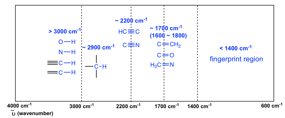
The absorption bands in IR spectra have different intensities that can usually be referred to as strong (s), medium (m), weak (w), broad and sharp. The intensity of an absorption band depends on the polarity of the bond, and a bond with higher polarity will show a more intense absorption band. The intensity also depends on the number of bonds responsible for the absorption, and an absorption band with more bonds involved has a higher intensity.
The polar O-H bond (in alcohol and carboxylic acid) usually shows strong and broad absorption bands that are easy to identify. The broad shape of the absorption band results from the hydrogen bonding of the OH groups between molecules. The OH bond of an alcohol group usually has absorption in the range of 3200–3600 cm -1 , while the OH bond of the carboxylic acid group occurs at about 2500–3300 cm -1 (Figure 6.4a and Figure 6.4c).
The polarity of the N-H bond (in amine and amide) is weaker than the OH bond, so the absorption band of N-H is not as intense or as broad as O-H, and the position is in the 3300–3500 cm -1 region.
The C-H bond stretching of all hydrocarbons occurs in the range of 2800–3300 cm -1 , and the exact location can be used to distinguish between alkane, alkene and alkyne. Specifically:
- ≡C-H (sp C-H) bond of terminal alkyne gives absorption at about 3300 cm -1
- =C-H (sp 2 C-H) bond of alkene gives absorption at about 3000-3100 cm -1
- -C-H (sp 3 C-H) bond of alkane gives absorption at about ~2900 cm -1 (see the example of the IR spectrum of 2-hexanone in Figure 6.3a; the C-H absorption band at about 2900 cm -1 )
A special note should be made for the C-H bond stretching of an aldehyde group that shows two absorption bands: one at ~2800 cm -1 and the other at ~ 2700 cm -1 . It is therefore relatively easy to identify the aldehyde group (together with the C=O stretching at about 1700 cm -1 ) since essentially no other absorptions occur at these wavenumbers (see the example of the IR spectrum of butanal in Figure 6.4d ).
The stretching vibration of triple bonds C≡C and C≡N have absorption bands of about 2100–2200 cm -1 . The band intensity is in a medium to weak level. The alkynes can generally be identified with the characteristic weak but sharp IR absorbance bands in the range of 2100–2250 cm -1 due to stretching of the C≡C triple bond, and terminal alkynes can be identified by their absorbance at about 3300 cm -1 due to stretching of sp C-H.
As mentioned earlier, the C=O stretching has a strong absorption band in the 1650–1750 cm -1 region. Other double bonds like C=C and C=N have absorptions in lower frequency regions of about 1550–1650 cm -1 . The C=C stretching of an alkene only shows one band at ~1600 cm -1 (Figure 6.4b) , while a benzene ring is indicated by two sharp absorption bands: one at ~1600 cm -1 and one at 1500–1430 cm -1 (see the example of the IR spectrum of ethyl benzene in Figure 6.4e ) .
You will notice in Figure 6.3a and 6.3b that a region with the lower frequency 400–1400 cm -1 in the IR spectrum is called the fi ngerprint region . Similar to a human fingerprint, the pattern of absorbance bands in the fingerprint region is characteristic of the compound as a whole. Even if two different molecules have the same functional groups, their IR spectra will not be identical, and such a difference will be reflected in the bands in the fingerprint region. Therefore, the IR from an unknown sample can be compared to a database of IR spectra of known standards in order to confirm the identification of the unknown sample.
Organic Chemistry I Copyright © 2021 by Xin Liu is licensed under a Creative Commons Attribution-NonCommercial-ShareAlike 4.0 International License , except where otherwise noted.
Share This Book
An official website of the United States Government
- Kreyòl ayisyen
- Search Toggle search Search Include Historical Content - Any - No Include Historical Content - Any - No Search
- Menu Toggle menu
- INFORMATION FOR…
- Individuals
- Business & Self Employed
- Charities and Nonprofits
- International Taxpayers
- Federal State and Local Governments
- Indian Tribal Governments
- Tax Exempt Bonds
- FILING FOR INDIVIDUALS
- How to File
- When to File
- Where to File
- Update Your Information
- Get Your Tax Record
- Apply for an Employer ID Number (EIN)
- Check Your Amended Return Status
- Get an Identity Protection PIN (IP PIN)
- File Your Taxes for Free
- Bank Account (Direct Pay)
- Payment Plan (Installment Agreement)
- Electronic Federal Tax Payment System (EFTPS)
- Your Online Account
Tax Withholding Estimator
- Estimated Taxes
- Where's My Refund
- What to Expect
- Direct Deposit
- Reduced Refunds
- Amend Return
Credits & Deductions
- INFORMATION FOR...
- Businesses & Self-Employed
- Earned Income Credit (EITC)
- Child Tax Credit
- Clean Energy and Vehicle Credits
- Standard Deduction
- Retirement Plans
Forms & Instructions
- POPULAR FORMS & INSTRUCTIONS
- Form 1040 Instructions
- Form 4506-T
- POPULAR FOR TAX PROS
- Form 1040-X
- Circular 230
How can we help you?
- Get your refund status
- Sign in to your account
- Get your tax record
- Make a payment
- File your taxes for free
- Find forms & instructions
- Get answers to your tax questions
- Check your amended return status
Tools & applications

Document Upload Tool
Upload documents in response to an IRS notice or letter.

Your Account
Access your individual, business or tax pro account.

IRS Free File
Prepare and file your federal income taxes online for free.

Where's My Refund?
Find the status of your last return and check on your refund.

Pay Directly From Your Bank Account
Use Direct Pay to securely pay your taxes from your checking or savings account.

Get Your Tax Records
Request your transcripts online or by mail.

Identity Protection PIN (IP PIN)
Protect yourself from tax-related identity theft.

Make sure you have the right amount of tax withheld from your paycheck.

Taxpayer Assistance Center Locator
Find your local office and see what services are available.
News & announcements

Free File will help you do your taxes online for free

Free tax preparation
Get free one-on-one tax preparation help nationwide if you qualify

Employee Retention Credit
You can withdraw incorrect ERC claims if you haven’t received the money

IRS offers face-to-face Saturday help
Many Taxpayer Assistance Centers will open one Saturday each month from February to May

Inflation Reduction Act Strategic Operating Plan
See how the IRS will deliver transformational change

Clean energy credits and deductions
Updates on credits and deductions under the Inflation Reduction Act

Tax Updates and News
Special updates and news for 2024

Penalty relief for many taxpayers
Prior year collection notices to resume in 2024

IRS operations status
Check our current processing times for returns, refunds and other services.
Preparing for disasters
Tips for safeguarding important documents in preparation for an emergency.

Follow @IRSnews on X for the latest news and announcements.
Jump to content
NIST Chemistry WebBook , SRD 69
- IUPAC identifier
- More options
- SRD Program
- Science Data Portal
- Office of Data and Informatics
- More documentation
Phosphine, triphenyl-
- Formula : C 18 H 15 P
- Molecular weight : 262.2855

- IUPAC Standard InChIKey: RIOQSEWOXXDEQQ-UHFFFAOYSA-N Copy
- CAS Registry Number: 603-35-0
- Other names: Triphenylphosphine; Triphenylphosphorus; Trifenylfosfin; Triphenylphosphide; Triphenylphosphane; Phosphorus triphenyl; NSC 10; NSC 215203; PP 360
- Permanent link for this species. Use this link for bookmarking this species for future reference.
Infrared Spectrum
- Condensed phase thermochemistry data
- Phase change data
- Gas phase ion energetics data
- IR Spectrum
- Mass spectrum (electron ionization)
- UV/Visible spectrum
- X-ray Photoelectron Spectroscopy Database, version 5.0
- Switch to calorie-based units
Data at NIST subscription sites:
- NIST / TRC Web Thermo Tables, professional edition (thermophysical and thermochemical data)
NIST subscription sites provide data under the NIST Standard Reference Data Program , but require an annual fee to access. The purpose of the fee is to recover costs associated with the development of data collections included in such sites. Your institution may already be a subscriber. Follow the links above to find out more about the data in these sites and their terms of usage.
Go To: Top , References , Notes
Data compilation copyright by the U.S. Secretary of Commerce on behalf of the U.S.A. All rights reserved.
Data compiled by: Coblentz Society, Inc.
Condensed Phase Spectrum
Notice: This spectrum may be better viewed with a Javascript and HTML 5 enabled browser.
- Help / Software credits
The interactive spectrum display requires a browser with JavaScript and HTML 5 canvas support.
Select a region with data to zoom. Select a region with no data or click the mouse on the plot to revert to the orginal display.
The following components were used in generating the plot:
- Resize (distributed with Flot)
- Selection (distributed with Flot)
- Axis labels
- Labels ( Modified by NIST for use in this application )
Additonal code used was developed at NIST: jcamp-dx.js and jcamp-plot.js .
Use or mention of technologies or programs in this web site is not intended to imply recommendation or endorsement by the National Institute of Standards and Technology, nor is it intended to imply that these items are necessarily the best available for the purpose.
Notice: Except where noted, spectra from this collection were measured on dispersive instruments, often in carefully selected solvents, and hence may differ in detail from measurements on FTIR instruments or in other chemical environments. More information on the manner in which spectra in this collection were collected can be found here.
Notice: Concentration information is not available for this spectrum and, therefore, molar absorptivity values cannot be derived.
Additional Data
View scan of original (hardcopy) spectrum .
View image of digitized spectrum (can be printed in landscape orientation).
View spectrum image in SVG format .
Download spectrum in JCAMP-DX format.
This IR spectrum is from the Coblentz Society's evaluated infrared reference spectra collection .
Go To: Top , Infrared Spectrum , Notes
No reference data available.
Go To: Top , Infrared Spectrum , References
- Data from NIST Standard Reference Database 69: NIST Chemistry WebBook
- The National Institute of Standards and Technology (NIST) uses its best efforts to deliver a high quality copy of the Database and to verify that the data contained therein have been selected on the basis of sound scientific judgment. However, NIST makes no warranties to that effect, and NIST shall not be liable for any damage that may result from errors or omissions in the Database.
- Customer support for NIST Standard Reference Data products.
- If you believe that this page may contain an error, please fill out the error report form for this page.

- school Campus Bookshelves
- menu_book Bookshelves
- perm_media Learning Objects
- login Login
- how_to_reg Request Instructor Account
- hub Instructor Commons
Margin Size
- Download Page (PDF)
- Download Full Book (PDF)
- Periodic Table
- Physics Constants
- Scientific Calculator
- Reference & Cite
- Tools expand_more
- Readability
selected template will load here
This action is not available.

5: Infrared Spectroscopy
- Last updated
- Save as PDF
- Page ID 332805

- Kate Graham
- College of Saint Benedict/Saint John's University
Infrared Spectroscopy
Infrared spectroscopy measures the absorption of energy that matches the vibrational frequency. The energies are affected by the strength of the bond and the masses of the atoms.
Definitions and Introduction to Hooke’s Law
Hooke's Law: \(\displaystyle \nu = \frac{1}{2\pi} \sqrt{\frac{k}{m}}\)
Where k = force constant
\(\nu\) = frequency
- What happens to \(\nu\) if k is increased?
- What happens to \(\nu\) if m is increased?
Hooke’s Law applied to IR Spectroscopy*
*Watch a video on IR and Hooke’s Law. [meta: LINK MISSING]
- The stronger the spring, the higher (faster) the frequency of bouncing.
- The weaker the spring, the higher (faster) the frequency of bouncing.
- The heavier the weight, the higher (faster) the frequency of bouncing.
- The lighter the weight, the higher (faster) the frequency of bouncing.
- Order the bonds shown below in order of strength:

- If we assume bonds are like springs, explain the frequencies in this table:
*IR absorbance is typically measured in wavenumbers (proportional to Hz); another system for measuring frequency.
- Explain the trends in this table. Hint: consider atomic weights of halides.
IR can therefore be a useful tool for determining what types of bonds (functional groups are present in a sample.
Infrared Spectroscopy: Functional Group Determination*
IR can be a useful tool for determining what types of bonds (functional groups are present in a sample.
* All spectra are either from SDBS (Japan National Institute of Advanced Industrial Science and Technology) or simulated.
Hydrocarbons
The spectrum of octane is shown below.

- List the different types of bonds present in this structure.
- How many peaks would you expect to see?

- Based on Hooke’s Law which peak corresponds to C-H stretches? Which peak corresponds to C-C stretches?
Hydrocarbons (cont.)
The spectrum of octene is shown below.
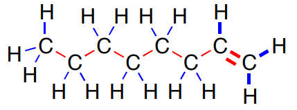
- There are two different types of C-H stretches: sp 3 C-H and sp 2 C-H. Based on the location of the sp 3 C-H in octane, which of these is the new sp 2 C-H?

- There are two different types of C-C stretches. C-C and C=C. Based on the location of the sp 3 C-H in octane, which of these is the new C=C?

Alkene Analysis (cont.)
The spectra of two different alkenes are shown below.

1, 11-dodecadiene

- How do these two spectra differ?
- Can you tell how many C-C bonds or C=C are in a structure from the IR spectrum?

- Find the sp 3 C-H and the C-C stretches in the spectrum below.
There are two new stretches: O-H and C-O.
Most OH stretches show up between 3200-3400 cm -1 .
- The shape of this OH peak is very distinctive. Describe this shape.

The C-O stretches show up between 1000-1300 cm -1 .
- Circle or highlight this peak.

Ethers are similar to alcohols. Here is the spectrum of butyl ether.

Ethers have sp 3 C-H stretches. C-C stretches and a C-O stretch.
- Find these key peaks in the spectrum below.
- They lack a __________ stretch that is present in an alcohol.

Amines are similar to alcohols, also. Let’s consider the spectrum of 1-hexylamine

Amines have sp 3 C-H stretches. C-C stretches and a C-N stretch.
- They also have an N-H stretch (or two) above 3200 cm -1 .

- The N-H stretch (or two) above 3200 cm -1 has a significantly different shape than the alcohol O-H stretch. Describe the difference.
Aldehydes, such as 1-octanal, have sp 3 C-H stretches and C-C stretches.

All carbonyl functional groups have a distinctive C=O stretch that absorbs strongly around 1700 cm -1 .
- Find the sp 3 C-H stretches and C-C stretches in the spectrum below.
- Circle or label the C=O peak.
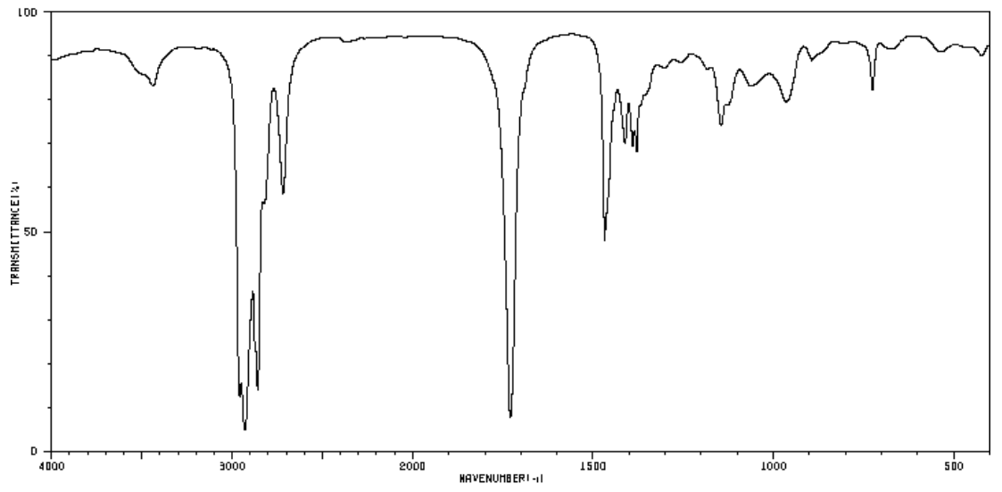
Aldehydes also have an sp 2 C-H stretch due to the aldehydic H. This peak usually has two bands around 2850 and 2750 cm −1 .

- How would the spectrum of 2-octanone differ from 1-octanal?
Similar Functional Groups
- Label 1-2 relevant peaks that will help you distinguish these two functional groups.
Esters vs Ketones

Alcohol vs Carboxylic Acids

Summary: IR Key Ranges

Summary: IR Key Ranges (cont)
Practice: IR Spectroscopy to Determine Functional Group
- Identify the most likely functional groups in these spectra.

IR and Functional Group Determination
Part 1. Match the following compounds with the corresponding spectra on the following pages.

Part 2. Label 2-3 relevant peaks for each IR spectrum with the correct bond type.

Infrared Spectroscopy: Analysis Beyond Stretching
A molecule can vibrate in many ways, and each way is called a vibrational mode. Simple diatomic molecules have only one bond and only one vibrational band. If the molecule is symmetrical, the band is not observed in the IR spectrum. More complex molecules have more possible vibrations many peaks in their IR spectra.
Some possible movements:

So far, we have looked the primary stretching frequencies for polar bonds.
There are some diagnostic bending and out-of-plane bending.
Aromatic Substitution Patterns
Here are typical out-of-plane (oop) bends for mono- and di-substituted aromatic compounds.

- Determine the substitution pattern of the following compound based on oop bends.
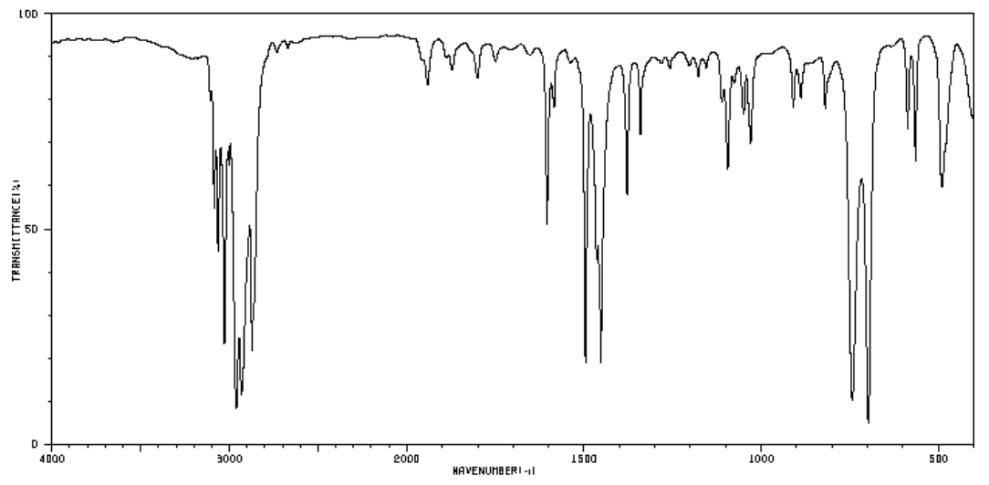
Aromatic Substitution Patterns (cont)
- These are the spectra of the three isomers of dimethylbenzene (xylenes). Identify the ortho, meta, and para isomers.
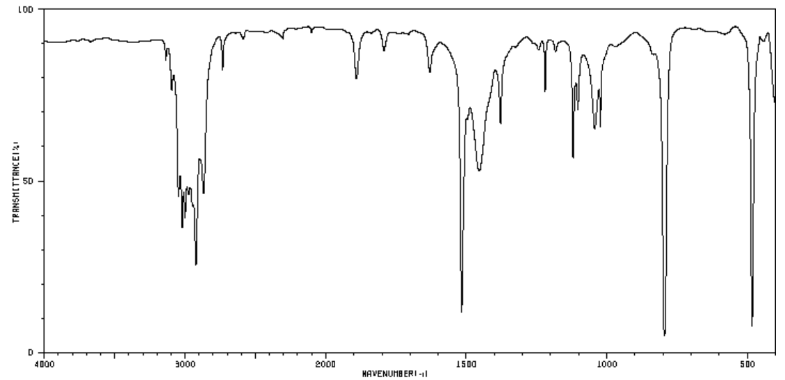
Alkene Substitution Patterns
- Alkene molecules have C-H stretches above ____________ cm -1 .
- Alkene molecules have C-C stretches around ____________ cm -1 .
These two stretches are very typical for ALL alkene structures and do not provide any information about the substitution patterns but the out-of-plane (oop) bends can be diagnostic.
Here are typical out-of-plane (oop) bends for cis, trans, and terminal alkenes.

Tetrasubstituted alkenes have no hydrogens attached to the C=C moiety, and thus have no alkene C-H stretching or wagging peaks. They do have a C=C stretching peak, which appears from 1680 to 1660.
- Is there a peak that would be diagnostic for a tetra-substituted alkene?
- Determine the substitution pattern of the following isomers of hexenes based on oop bends. (trans-3-hexene, cis-3-hexene, and 1-hexene)

N-H stretches fall in the same wavenumber range as O-H stretches, but that N-H stretches are easy to distinguish, because they will be less broad and less intense. This is due to N-H bonds being less polar than O-H bonds.
- O-H and N-H stretches show up around ____________ cm -1 .
An amine does contain a C-N bond and there will be a C-N stretching peak as a result. However, C-N stretches are not very polar, so the peak intensities are not strong.
- C-N stretches show up around ____________ cm -1 .
It is important to note that a primary amine has a scissors peak that appears from 1650 to 1580 cm -1 . Sometimes, people will confuse this peak with an alkene.
- N-H stretch
- C-N stretch
- NH 2 scissor

Summary of Infrared Spectroscopy:
- What information does IR provide?
- What are the limitations of IR?
- Briefly explain what information can be deduced from each of the IR regions below.

- For each set of compounds shown below, explain how IR Spectroscopy could help you tell them apart.

Another IR Matching Problem
Part 1. Identify the major functional group for the following compounds:

Part 3. Match all of the IR spectra with the correct compound (from Part 1).


IMAGES
VIDEO
COMMENTS
Generally, an AF Form 1288 Application for Reserve Assignment, signed by the IR's losing Commander, resume and last 3 OPRs/EPRs are needed to apply for an IR position. On-line applications via the RMVS system are not accepted. Once the Detachment has received all the applicable documents from the member, they will in-turn coordination with ...
The IR spectra for the major classes of organic molecules are shown and discussed. ... Assignment. Range (cm-1) Intensity. Assignment. Alkanes. 2850-3000: str: CH 3, CH 2 & CH 2 or 3 bands: 1350-1470 1370-1390 720-725: med med wk: CH 2 & CH 3 deformation CH 3 deformation CH 2 rocking: Alkenes. 3020-3100 1630-1680
\( \newcommand{\vecs}[1]{\overset { \scriptstyle \rightharpoonup} {\mathbf{#1}} } \) \( \newcommand{\vecd}[1]{\overset{-\!-\!\rightharpoonup}{\vphantom{a}\smash {#1 ...
This condition can be summarized in equation (2) form as follows: (2) Vibrations that satisfy this equation are said to be infrared active. The H-Cl stretch of hydrogen chloride and the asymmetric stretch of CO 2 are examples of infrared active vibrations. Infrared active vibrations cause the bands seen in an infrared spectrum.
Infrared spectroscopy ( IR spectroscopy or vibrational spectroscopy) is the measurement of the interaction of infrared radiation with matter by absorption, emission, or reflection. It is used to study and identify chemical substances or functional groups in solid, liquid, or gaseous forms. It can be used to characterize new materials or ...
Here's an overview of the IR window from 4000 cm -1 to 500 cm -1 with various regions of interest highlighted. An even more compressed overview looks like this: ( source) 3600 - 2700 cm -1. X-H (single bonds to hydrogen) 2700 - 1900 cm -1. X≡X (triple bonds) 1900 - 1500 cm -1. X=X (double bonds) 1500 - 500 cm -1.
An infrared spectroscopy correlation table (or table of infrared absorption frequencies) is a list of absorption peaks and frequencies, typically reported in wavenumber, for common types of molecular bonds and functional groups. [1] [2] In physical and analytical chemistry, infrared spectroscopy (IR spectroscopy) is a technique used to identify ...
An interpretation of an IR spectrum should include a detailed assignment of the peaks: exact wavenumber from the spectrum (integer), the intensity (w/m/s/br) and which functional group it represents, and maybe in addition the corresponding literature value. However, it is not necessary to interpret every little peak in the IR spectrum.
Find the structure from 1H spectrum. 1H exercise generator. Assign 1H NMR spectra to molecule. 13C NMR. 1H NMR spectra of small molecules. 1H NMR spectra of Boc amino acids. Number of different Hs. 1H NMR integrate and find the structure. 1H number of signals.
This organic chemistry video tutorial provides a basic introduction into IR spectroscopy. It explains how to identify and distinguish functional groups such...
Infrared Spectroscopy. 1. Introduction. As noted in a previous chapter, the light our eyes see is but a small part of a broad spectrum of electromagnetic radiation. On the immediate high energy side of the visible spectrum lies the ultraviolet, and on the low energy side is the infrared. The portion of the infrared region most useful for ...
IR spectroscopy is a great method for identification of compounds, especially for identification of functional groups. Therefore, we can use group frequencies for structural analysis. ... One of the most importance applications of IR spectroscopy is structural assignment of the molecule depending on the relationship between the molecule and ...
%PDF-1.6 %âãÏÓ 157 0 obj > endobj 176 0 obj >/Filter/FlateDecode/ID[29E5AF2A7F9D0F4196F4DB70B6DBCA78>89F80795035FDF4A95A9FB23652E2839>]/Index[157 33]/Info 156 0 R ...
Investor Relations - IR: Investor relations (IR) is a department, present in most medium-to-large public companies , that provides investors with an accurate account of company affairs. This helps ...
Photometry. Photometry is the measurement of light absorbed in the ultraviolet (UV) to visible (VIS) to infra-red (IR) range. This measurement is used to determine the amount of an analyte in a solution or liquid. Photometers utilize a specific light source and detectors that convert light passed through a sample solution into a proportional electric signal.
The wavenumber is defined as the reciprocal of wavelength (Formula 6.3), and the wavenumbers of IR radiation are normally in the range of 4000 cm-1 to 600 cm-1 (approximate corresponds the wavelength range of 2.5 μm to 17 μm of IR radiation). Please note that the direction of the horizontal axis (wavenumber) in IR spectra decreases from left ...
Notice: Except where noted, spectra from this collection were measured on dispersive instruments, often in carefully selected solvents, and hence may differ in detail from measurements on FTIR instruments or in other chemical environments. More information on the manner in which spectra in this collection were collected can be found here. Notice: Concentration information is not available for ...
When answering assignment questions, you may use this IR table to find the characteristic infrared absorptions of the various functional groups. However, you should be able to indicate in broad terms where certain characteristic absorptions occur. ... Alkynes have characteristic IR absorbance peaks in the range of 2100-2250 cm-1 due to ...
Notice: Except where noted, spectra from this collection were measured on dispersive instruments, often in carefully selected solvents, and hence may differ in detail from measurements on FTIR instruments or in other chemical environments. More information on the manner in which spectra in this collection were collected can be found here. Notice: Concentration information is not available for ...
Sign in to your account. Get your tax record. Make a payment. File your taxes for free. Find forms & instructions. Get answers to your tax questions. Apply for an Employer ID Number (EIN) Check your amended return status.
The wavenumber is defined as the reciprocal of wavelength ( Formula 6.3 ), and the wavenumbers of infrared radiation are normally in the range of 4000 cm -1 to 600 cm -1 (approximate corresponds the wavelength range of 2.5 μm to 17 μm of IR radiation). Formula 6.3 Wavenumber. Please note the direction of the horizontal axis (wavenumber) in IR ...
www.itochu.co.jp
Notice: Except where noted, spectra from this collection were measured on dispersive instruments, often in carefully selected solvents, and hence may differ in detail from measurements on FTIR instruments or in other chemical environments. More information on the manner in which spectra in this collection were collected can be found here. Notice: Concentration information is not available for ...
This page titled 5: Infrared Spectroscopy is shared under a not declared license and was authored, remixed, and/or curated by Kate Graham. Infrared spectroscopy measures the absorption of energy that matches the vibrational frequency. The energies are affected by the strength of the bond and the masses of the atoms.