If you're seeing this message, it means we're having trouble loading external resources on our website.
If you're behind a web filter, please make sure that the domains *.kastatic.org and *.kasandbox.org are unblocked.
To log in and use all the features of Khan Academy, please enable JavaScript in your browser.

High school biology
Course: high school biology > unit 8.
- Meet the heart!
- Circulatory system and the heart
- The circulatory system review
- Meet the lungs!
- The lungs and pulmonary system
The respiratory system review
- The circulatory and respiratory systems
The respiratory system
Common mistakes and misconceptions.
- Physiological respiration and cellular respiration are not the same. People sometimes use the word "respiration" to refer to the process of cellular respiration, which is a cellular process in which carbohydrates are converted into energy. The two are related processes, but they are not the same.
- We do not breathe in only oxygen or breathe out only carbon dioxide. Often the terms "oxygen" and "air" are used interchangeably. It is true that the air we breathe in has more oxygen than the air we breathe out, and the air we breathe out has more carbon dioxide than the air that we breathe in. However, oxygen is just one of the gases found in the air we breathe. (In fact, the air has more nitrogen than oxygen!)
- The respiratory system does not work alone in transporting oxygen through the body. The respiratory system works directly with the circulatory system to provide oxygen to the body. Oxygen taken in from the respiratory system moves into blood vessels that then circulate oxygen-rich blood to tissues and cells.
Want to join the conversation?
- Upvote Button navigates to signup page
- Downvote Button navigates to signup page
- Flag Button navigates to signup page


- school Campus Bookshelves
- menu_book Bookshelves
- perm_media Learning Objects
- login Login
- how_to_reg Request Instructor Account
- hub Instructor Commons
- Download Page (PDF)
- Download Full Book (PDF)
- Periodic Table
- Physics Constants
- Scientific Calculator
- Reference & Cite
- Tools expand_more
- Readability
selected template will load here
This action is not available.

16.2: Structure and Function of the Respiratory System
- Last updated
- Save as PDF
- Page ID 16817

- Suzanne Wakim & Mandeep Grewal
- Butte College
Seeing Your Breath
Why can you “see your breath” on a cold day? The air you exhale through your nose and mouth is warm, like the inside of your body. Exhaled air also contains a lot of water vapor because it passes over moist surfaces from the lungs to the nose or mouth. The water vapor in your breath cools suddenly when it reaches the much colder outside air. This causes the water vapor to condense into a fog of tiny droplets of liquid water. You release water vapor and other gases from your body through the process of respiration.
.jpg?revision=1&size=bestfit&width=399&height=241)
What is Respiration?
Respiration is the life-sustaining process in which gases are exchanged between the body and the outside atmosphere. Specifically, oxygen moves from the outside air into the body; and water vapor, carbon dioxide, and other waste gases move from inside the body into the outside air. Respiration is carried out mainly by the respiratory system. It is important to note that respiration by the respiratory system is not the same process as cellular respiration that occurs inside cells, although the two processes are closely connected. Cellular respiration is the metabolic process in which cells obtain energy, usually by “burning” glucose in the presence of oxygen. When cellular respiration is aerobic, it uses oxygen and releases carbon dioxide as a waste product. Respiration by the respiratory system supplies the oxygen needed by cells for aerobic cellular respiration and removes the carbon dioxide produced by cells during cellular respiration.
Respiration by the respiratory system actually involves two subsidiary processes. One process is ventilation or breathing. This is the physical process of conducting air to and from the lungs. The other process is gas exchange. This is the biochemical process in which oxygen diffuses out of the air and into the blood while carbon dioxide and other waste gases diffuse out of the blood and into the air. All of the organs of the respiratory system are involved in breathing, but only the lungs are involved in gas exchange.
Respiratory Organs
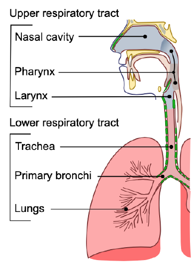
The organs of the respiratory system form a continuous system of passages called the respiratory tract, through which air flows into and out of the body. The respiratory tract has two major divisions: the upper respiratory tract and the lower respiratory tract. The organs in each division are shown in Figure \(\PageIndex{2}\). In addition to these organs, certain muscles of the thorax (the body cavity that fills the chest) are also involved in respiration by enabling breathing. Most important is a large muscle called the diaphragm, which lies below the lungs and separates the thorax from the abdomen. Smaller muscles between the ribs also play a role in breathing. You can learn more about breathing muscles in the concept of Breathing .
Upper Respiratory Tract
All of the organs and other structures of the upper respiratory tract are involved in the conduction or the movement of air into and out of the body. Upper respiratory tract organs provide a route for air to move between the outside atmosphere and the lungs. They also clean, humidity, and warm the incoming air. However, no gas exchange occurs in these organs.
Nasal Cavity
The nasal cavity is a large, air-filled space in the skull above and behind the nose in the middle of the face. It is a continuation of the two nostrils. As inhaled air flows through the nasal cavity, it is warmed and humidified. Hairs in the nose help trap larger foreign particles in the air before they go deeper into the respiratory tract. In addition to its respiratory functions, the nasal cavity also contains chemoreceptors that are needed for the sense of smell and that contribute importantly to the sense of taste.
The pharynx is a tube-like structure that connects the nasal cavity and the back of the mouth to other structures lower in the throat, including the larynx. The pharynx has dual functions: both air and food (or other swallowed substances) pass through it, so it is part of both the respiratory and digestive systems. Air passes from the nasal cavity through the pharynx to the larynx (as well as in the opposite direction). Food passes from the mouth through the pharynx to the esophagus.
The larynx connects the pharynx and trachea and helps to conduct air through the respiratory tract. The larynx is also called the voice box because it contains the vocal cords, which vibrate when air flows over them, thereby producing sound. You can see the vocal cords in the larynx in Figure \(\PageIndex{3}\). Certain muscles in the larynx move the vocal cords apart to allow breathing. Other muscles in the larynx move the vocal cords together to allow the production of vocal sounds. The latter muscles also control the pitch of sounds and help control their volume.
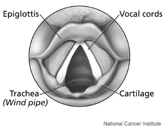
A very important function of the larynx is protecting the trachea from aspirated food. When swallowing occurs, the backward motion of the tongue forces a flap called the epiglottis to close over the entrance to the larynx. You can see the epiglottis in Figure \(\PageIndex{3}\). This prevents swallowed material from entering the larynx and moving deeper into the respiratory tract. If swallowed material does start to enter the larynx, it irritates the larynx and stimulates a strong cough reflex. This generally expels the material out of the larynx and into the throat.
Lower Respiratory Tract
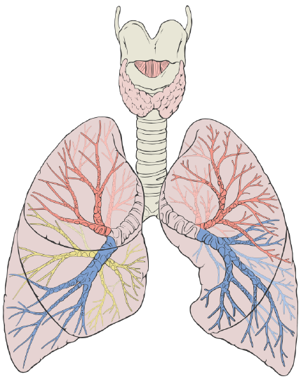
The trachea and other passages of the lower respiratory tract conduct air between the upper respiratory tract and the lungs. These passages form an inverted tree-like shape (Figure \(\PageIndex{4}\)), with repeated branching as they move deeper into the lungs. All told, there are an astonishing 1,500 miles of airways conducting air through the human respiratory tract! It is only in the lungs, however, that gas exchange occurs between the air and the bloodstream.
The trachea, or windpipe, is the widest passageway in the respiratory tract. It is about 2.5 cm (1 in.) wide and 10-15 cm (4-6 in.) long. It is formed by rings of cartilage, which make it relatively strong and resilient. The trachea connects the larynx to the lungs for the passage of air through the respiratory tract. The trachea branches at the bottom to form two bronchial tubes.
Bronchi and Bronchioles
There are two main bronchial tubes, or bronchi (singular, bronchus) , called the right and left bronchi. The bronchi carry air between the trachea and lungs. Each bronchus branches into smaller, secondary bronchi; and secondary bronchi branch into still smaller tertiary bronchi. The smallest bronchi branch into very small tubules called bronchioles. The tiniest bronchioles end in alveolar ducts, which terminate in clusters of minuscule air sacs, called alveoli (singular, alveolus), in the lungs.
The lungs are the largest organs of the respiratory tract. They are suspended within the pleural cavity of the thorax. In Figure \(\PageIndex{5}\), you can see that each of the two lungs is divided into sections. These are called lobes, and they are separated from each other by connective tissues. The right lung is larger and contains three lobes. The left lung is smaller and contains only two lobes. The smaller left lung allows room for the heart, which is just left of the center of the chest.
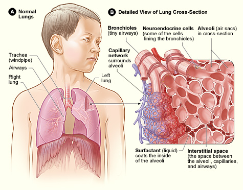
Lung tissue consists mainly of alveoli (Figure \(\PageIndex{6}\)). These tiny air sacs are the functional units of the lungs where gas exchange takes place. The two lungs may contain as many as 700 million alveoli, providing a huge total surface area for gas exchange to take place. In fact, alveoli in the two lungs provide as much surface area as half a tennis court! Each time you breathe in, the alveoli fill with air, making the lungs expand. Oxygen in the air inside the alveoli is absorbed by the blood in the mesh-like network of tiny capillaries that surrounds each alveolus. The blood in these capillaries also releases carbon dioxide into the air inside the alveoli. Each time you breathe out, air leaves the alveoli and rushes into the outside atmosphere, carrying waste gases with it.
The lungs receive blood from two major sources. They receive deoxygenated blood from the heart. This blood absorbs oxygen in the lungs and carries it back to the heart to be pumped to cells throughout the body. The lungs also receive oxygenated blood from the heart that provides oxygen to the cells of the lungs for cellular respiration.
Protecting the Respiratory System
You may be able to survive for weeks without food and for days without water, but you can survive without oxygen for only a matter of minutes except under exceptional circumstances. Therefore, protecting the respiratory system is vital. That’s why making sure a patient has an open airway is the first step in treating many medical emergencies. Fortunately, the respiratory system is well protected by the ribcage of the skeletal system. However, the extensive surface area of the respiratory system is directly exposed to the outside world and all its potential dangers in inhaled air. Therefore, it should come as no surprise that the respiratory system has a variety of ways to protect itself from harmful substances such as dust and pathogens in the air.
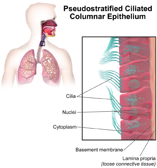
The main way the respiratory system protects itself is called the mucociliary escalator. From the nose through the bronchi, the respiratory tract is covered in the epithelium that contains mucus-secreting goblet cells. The mucus traps particles and pathogens in the incoming air. The epithelium of the respiratory tract is also covered with tiny cell projections called cilia (singular, cilium), as shown in Figure \(\PageIndex{7}\). The cilia constantly move in a sweeping motion upward toward the throat, moving the mucus and trapped particles and pathogens away from the lungs and toward the outside of the body.
What happens to the material that moves up the mucociliary escalator to the throat? It is generally removed from the respiratory tract by clearing the throat or coughing. Coughing is a largely involuntary response of the respiratory system that occurs when nerves lining the airways are irritated. The response causes air to be expelled forcefully from the trachea, helping to remove mucus and any debris it contains (called phlegm) from the upper respiratory tract to the mouth. The phlegm may spit out (expectorated), or it may be swallowed and destroyed by stomach acids.
Sneezing is a similar involuntary response that occurs when nerves lining the nasal passage are irritated. It results in forceful expulsion of air from the mouth, which sprays millions of tiny droplets of mucus and other debris out of the mouth and into the air, as shown in Figure \(\PageIndex{8}\). This explains why it is so important to sneeze into a sleeve rather than the air to help prevent the transmission of respiratory pathogens.
How the Respiratory System Works with Other Organ Systems
The amount of oxygen and carbon dioxide in the blood must be maintained within a limited range for the survival of the organism. Cells cannot survive for long without oxygen, and if there is too much carbon dioxide in the blood, the blood becomes dangerously acidic (pH is too low). Conversely, if there is too little carbon dioxide in the blood, the blood becomes too basic (pH is too high). The respiratory system works hand-in-hand with the nervous and cardiovascular systems to maintain homeostasis in blood gases and pH.
It is the level of carbon dioxide rather than the level of oxygen that is most closely monitored to maintain blood gas and pH homeostasis. The level of carbon dioxide in the blood is detected by cells in the brain, which speed up or slow down the rate of breathing through the autonomic nervous system as needed to bring the carbon dioxide level within the normal range. Faster breathing lowers the carbon dioxide level (and raises the oxygen level and pH); slower breathing has the opposite effects. In this way, the levels of carbon dioxide and oxygen, as well as pH, are maintained within normal limits.
The respiratory system also works closely with the cardiovascular system to maintain homeostasis. The respiratory system exchanges gases between the blood and the outside air, but it needs the cardiovascular system to carry them to and from body cells. Oxygen is absorbed by the blood in the lungs and then transported through a vast network of blood vessels to cells throughout the body where it is needed for aerobic cellular respiration. The same system absorbs carbon dioxide from cells and carries it to the respiratory system for removal from the body.
Feature: My Human Body
Choking is the mechanical obstruction of the flow of air from the atmosphere into the lungs. It prevents breathing and may be partial or complete. Partial choking allows some though inadequate airflow into the lung—prolonged or complete choking results in asphyxia, or suffocation, which is potentially fatal.
Obstruction of the airway typically occurs in the pharynx or trachea. Young children are more prone to choking than are older people, in part because they often put small objects in their mouths and do not appreciate the risk of choking that they pose. Young children may choke on small toys or parts of toys or on household objects in addition to food. Foods that can adapt their shape to that of the pharynx, such as bananas and marshmallows, are especially dangerous and may cause choking in adults as well as children.
How can you tell if a loved one is choking? The person cannot speak or cry out or has great difficulty doing so. Breathing, if possible, is labored, producing gasping or wheezing. The person may desperately clutch at his or her throat or mouth. If breathing is not soon restored, the person’s face will start to turn blue from lack of oxygen. This will be followed by unconsciousness if oxygen deprivation continues beyond a few minutes.
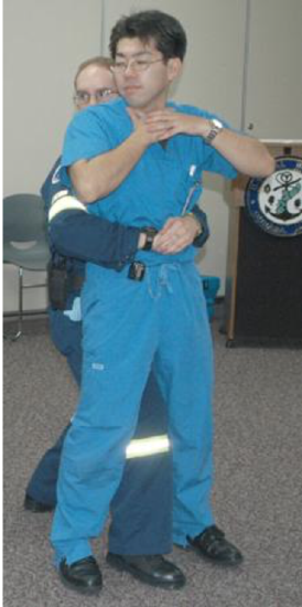
If an infant is choking, turning the baby upside down and slapping on the back may dislodge the obstructing object. To help an older person who is choking, first, encourage the person to cough. Give them a few hardback slaps to help force the lodged object out of the airway. If these steps fail, perform the Heimlich maneuver on the person. You can easily find instructional videos online to learn how to do it. If the Heimlich maneuver also fails, call for emergency medical care immediately.
- What is respiration, as carried out by the respiratory system? Name the two subsidiary processes it involves.
- Describe the respiratory tract.
- Identify the organs of the upper respiratory tract, and state their functions.
- List the organs of the lower respiratory tract. Which organs are involved only in conduction?
- Where does gas exchange take place?
- How does the respiratory system protect itself from potentially harmful substances in the air?
- Explain how the rate of breathing is controlled.
- Why does the respiratory system need the cardiovascular system to help it perform its main function of gas exchange?
trachea; nasal cavity; alveoli; bronchioles; larynx; bronchi; pharynx
D. Bronchus
- Describe two ways in which the body prevents food from entering the lungs.
- True or False. The lungs receive some oxygenated blood.
- True or False. Gas exchange occurs in both the upper and lower respiratory tracts.
B. food particles
D. All of the above
- What is the relationship between respiration and cellular respiration?
Explore More
Attributions.
- Snowboarders breath on a cold day by Alain Wong via Unsplash License
- Conducting Passages by Lord Akryl , Jmarchn, public domain via Wikimedia Commons
- Larynx by Alan Hoofring , National Cancer Institute, public domain via Wikimedia Commons
- Lung Diagram by Patrick J. Lynch ; CC BY 2.5 via Wikimedia Commons
- Lung Structure by National Heart Lung and Blood Institute, public domain via Wikimedia Commons
- Alveoli by helix84 licensed CC BY 2.5 , via Wikimedia Commons
- Ciliated Epithelium by Blausen.com staff (2014). " Medical gallery of Blausen Medical 2014 ". WikiJournal of Medicine 1 (2). DOI : 10.15347/wjm/2014.010 . ISSN 2002-4436 . licensed CC BY 3.0 via Wikimedia Commons
- Sneeze by James Gathany, CDC , public domain via Wikimedia Commons
- Abdominal Thrusts by Amanda M. Woodhead, public domain via Wikimedia Commons
- Text adapted from Human Biology by CK-12 licensed CC BY-NC 3.0

- school Campus Bookshelves
- menu_book Bookshelves
- perm_media Learning Objects
- login Login
- how_to_reg Request Instructor Account
- hub Instructor Commons
- Download Page (PDF)
- Download Full Book (PDF)
- Periodic Table
- Physics Constants
- Scientific Calculator
- Reference & Cite
- Tools expand_more
- Readability
selected template will load here
This action is not available.

14.2: Organs and Structures of the Respiratory System
- Last updated
- Save as PDF
- Page ID 57551
Learning Objectives
- List the structures that make up the respiratory system
- Describe how the respiratory system processes oxygen and CO 2
- Compare and contrast the functions of upper respiratory tract with the lower respiratory tract
The major organs of the respiratory system function primarily to provide oxygen to body tissues for cellular respiration, remove the waste product carbon dioxide, and help to maintain acid-base balance. Portions of the respiratory system are also used for non-vital functions, such as sensing odors, speech production, and for straining, such as during childbirth or coughing (Figure \(\PageIndex{1}\)).

Functionally, the respiratory system can be divided into a conducting zone and a respiratory zone. The conducting zone of the respiratory system includes the organs and structures not directly involved in gas exchange. The gas exchange occurs in the respiratory zone .
Conducting Zone
The major functions of the conducting zone are to provide a route for incoming and outgoing air, remove debris and pathogens from the incoming air, and warm and humidify the incoming air. Several structures within the conducting zone perform other functions as well. The epithelium of the nasal passages, for example, is essential to sensing odors, and the bronchial epithelium that lines the lungs can metabolize some airborne carcinogens.
The Nose and Its Adjacent Structures
The major entrance and exit for the respiratory system is through the nose. When discussing the nose, it is helpful to divide it into two major sections: the external nose, and the nasal cavity or internal nose.
The external nose consists of the surface and skeletal structures that result in the outward appearance of the nose and contribute to its numerous functions (Figure \(\PageIndex{2}\)). The root is the region of the nose located between the eyebrows. The bridge is the part of the nose that connects the root to the rest of the nose. The dorsum nasi is the length of the nose. The apex is the tip of the nose. On either side of the apex, the nostrils are formed by the alae (singular = ala). An ala is a cartilaginous structure that forms the lateral side of each naris (plural = nares), or nostril opening. The philtrum is the concave surface that connects the apex of the nose to the upper lip.

Underneath the thin skin of the nose are its skeletal features (see Figure \(\PageIndex{2}\), lower illustration). While the root and bridge of the nose consist of bone, the protruding portion of the nose is composed of cartilage. As a result, when looking at a skull, the nose is missing. The nasal bone is one of a pair of bones that lies under the root and bridge of the nose. The nasal bone articulates superiorly with the frontal bone and laterally with the maxillary bones. Septal cartilage is flexible hyaline cartilage connected to the nasal bone, forming the dorsum nasi. The alar cartilage consists of the apex of the nose; it surrounds the naris.
The nares open into the nasal cavity, which is separated into left and right sections by the nasal septum (Figure \(\PageIndex{3}\)). The nasal septum is formed anteriorly by a portion of the septal cartilage (the flexible portion you can touch with your fingers) and posteriorly by the perpendicular plate of the ethmoid bone (a cranial bone located just posterior to the nasal bones) and the thin vomer bones (whose name refers to its plough shape). Each lateral wall of the nasal cavity has three bony projections, called the superior, middle, and inferior nasal conchae. The inferior conchae are separate bones, whereas the superior and middle conchae are portions of the ethmoid bone. Conchae serve to increase the surface area of the nasal cavity and to disrupt the flow of air as it enters the nose, causing air to bounce along the epithelium, where it is cleaned and warmed. The conchae and meatuses also conserve water and prevent dehydration of the nasal epithelium by trapping water during exhalation. The floor of the nasal cavity is composed of the palate. The hard palate at the anterior region of the nasal cavity is composed of bone. The soft palate at the posterior portion of the nasal cavity consists of muscle tissue. Air exits the nasal cavities via the internal nares and moves into the pharynx.

Several bones that help form the walls of the nasal cavity have air-containing spaces called the paranasal sinuses, which serve to warm and humidify incoming air. Sinuses are lined with a mucosa. Each paranasal sinus is named for its associated bone: frontal sinus, maxillary sinus, sphenoidal sinus, and ethmoidal sinus. The sinuses produce mucus and lighten the weight of the skull.
The nares and anterior portion of the nasal cavities are lined with mucous membranes, containing sebaceous glands and hair follicles that serve to prevent the passage of large debris, such as dirt, through the nasal cavity. An olfactory epithelium used to detect odors is found deeper in the nasal cavity.
The conchae, meatuses, and paranasal sinuses are lined by respiratory epithelium composed of pseudostratified ciliated columnar epithelium (Figure \(\PageIndex{4}\)). The epithelium contains goblet cells, one of the specialized, columnar epithelial cells that produce mucus to trap debris. The cilia of the respiratory epithelium help remove the mucus and debris from the nasal cavity with a constant beating motion, sweeping materials towards the throat to be swallowed. Interestingly, cold air slows the movement of the cilia, resulting in accumulation of mucus that may in turn lead to a runny nose during cold weather. This moist epithelium functions to warm and humidify incoming air. Capillaries located just beneath the nasal epithelium warm the air by convection. Serous and mucus-producing cells also secrete the lysozyme enzyme and proteins called defensins, which have antibacterial properties. Immune cells that patrol the connective tissue deep to the respiratory epithelium provide additional protection.

The pharynx is a tube formed by skeletal muscle and lined by mucous membrane that is continuous with that of the nasal cavities (see Figure \(\PageIndex{3}\)). The pharynx is divided into three major regions: the nasopharynx, the oropharynx, and the laryngopharynx (Figure \(\PageIndex{5}\)).

The nasopharynx is flanked by the conchae of the nasal cavity, and it serves only as an airway. At the top of the nasopharynx are the pharyngeal tonsils. A pharyngeal tonsil , also called an adenoid, is an aggregate of lymphoid reticular tissue similar to a lymph node that lies at the superior portion of the nasopharynx. The function of the pharyngeal tonsil is not well understood, but it contains a rich supply of lymphocytes and is covered with ciliated epithelium that traps and destroys invading pathogens that enter during inhalation. The pharyngeal tonsils are large in children, but interestingly, tend to regress with age and may even disappear. The uvula is a small bulbous, teardrop-shaped structure located at the apex of the soft palate. Both the uvula and soft palate move like a pendulum during swallowing, swinging upward to close off the nasopharynx to prevent ingested materials from entering the nasal cavity. In addition, auditory (Eustachian) tubes that connect to each middle ear cavity open into the nasopharynx. This connection is why colds often lead to ear infections.
The oropharynx is a passageway for both air and food. The oropharynx is bordered superiorly by the nasopharynx and anteriorly by the oral cavity. The fauces is the opening at the connection between the oral cavity and the oropharynx. As the nasopharynx becomes the oropharynx, the epithelium changes from pseudostratified ciliated columnar epithelium to stratified squamous epithelium. The oropharynx contains two distinct sets of tonsils, the palatine and lingual tonsils. A palatine tonsil is one of a pair of structures located laterally in the oropharynx in the area of the fauces. The lingual tonsil is located at the base of the tongue. Similar to the pharyngeal tonsil, the palatine and lingual tonsils are composed of lymphoid tissue, and trap and destroy pathogens entering the body through the oral or nasal cavities.
The laryngopharynx is inferior to the oropharynx and posterior to the larynx. It continues the route for ingested material and air until its inferior end, where the digestive and respiratory systems diverge. The stratified squamous epithelium of the oropharynx is continuous with the laryngopharynx. Anteriorly, the laryngopharynx opens into the larynx, whereas posteriorly, it enters the esophagus.
The larynx is a cartilaginous structure inferior to the laryngopharynx that connects the pharynx to the trachea and helps regulate the volume of air that enters and leaves the lungs (Figure \(\PageIndex{6}\)). The structure of the larynx is formed by several pieces of cartilage. Three large cartilage pieces—the thyroid cartilage (anterior), epiglottis (superior), and cricoid cartilage (inferior)—form the major structure of the larynx. The thyroid cartilage is the largest piece of cartilage that makes up the larynx. The thyroid cartilage consists of the laryngeal prominence , or “Adam’s apple,” which tends to be more prominent in males. The thick cricoid cartilage forms a ring, with a wide posterior region and a thinner anterior region. Three smaller, paired cartilages—the arytenoids, corniculates, and cuneiforms—attach to the epiglottis and the vocal cords and muscle that help move the vocal cords to produce speech.

The epiglottis , attached to the thyroid cartilage, is a very flexible piece of elastic cartilage that covers the opening of the trachea (see Figure \(\PageIndex{3}\)). When in the “closed” position, the unattached end of the epiglottis rests on the glottis. The glottis is composed of the vestibular folds, the true vocal cords, and the space between these folds (Figure \(\PageIndex{7}\)). A vestibular fold , or false vocal cord, is one of a pair of folded sections of mucous membrane. A true vocal cord is one of the white, membranous folds attached by muscle to the thyroid and arytenoid cartilages of the larynx on their outer edges. The inner edges of the true vocal cords are free, allowing oscillation to produce sound. The size of the membranous folds of the true vocal cords differs between individuals, producing voices with different pitch ranges. Folds in males tend to be larger than those in females, which create a deeper voice. The act of swallowing causes the pharynx and larynx to lift upward, allowing the pharynx to expand and the epiglottis of the larynx to swing downward, closing the opening to the trachea. These movements produce a larger area for food to pass through, while preventing food and beverages from entering the trachea.

Continuous with the laryngopharynx, the superior portion of the larynx is lined with stratified squamous epithelium, transitioning into pseudostratified ciliated columnar epithelium that contains goblet cells. Similar to the nasal cavity and nasopharynx, this specialized epithelium produces mucus to trap debris and pathogens as they enter the trachea. The cilia beat the mucus upward towards the laryngopharynx, where it can be swallowed down the esophagus.
The trachea (windpipe) extends from the larynx toward the lungs (Figure \(\PageIndex{8}\).a). The trachea is formed by 16 to 20 stacked, C-shaped pieces of hyaline cartilage that are connected by dense connective tissue. The trachealis muscle and elastic connective tissue together form the fibroelastic membrane , a flexible membrane that closes the posterior surface of the trachea, connecting the C-shaped cartilages. The fibroelastic membrane allows the trachea to stretch and expand slightly during inhalation and exhalation, whereas the rings of cartilage provide structural support and prevent the trachea from collapsing. In addition, the trachealis muscle can be contracted to force air through the trachea during exhalation. The trachea is lined with pseudostratified ciliated columnar epithelium, which is continuous with the larynx. The esophagus borders the trachea posteriorly.

Bronchial Tree
The trachea branches into the right and left primary bronchi at the carina. These bronchi are also lined by pseudostratified ciliated columnar epithelium containing mucus-producing goblet cells (Figure \(\PageIndex{8}\).b). The carina is a raised structure that contains specialized nervous tissue that induces violent coughing if a foreign body, such as food, is present. Rings of cartilage, similar to those of the trachea, support the structure of the bronchi and prevent their collapse. The primary bronchi enter the lungs at the hilum, a concave region where blood vessels, lymphatic vessels, and nerves also enter the lungs. The bronchi continue to branch into bronchial a tree. A bronchial tree (or respiratory tree) is the collective term used for these multiple-branched bronchi. The main function of the bronchi, like other conducting zone structures, is to provide a passageway for air to move into and out of each lung. In addition, the mucous membrane traps debris and pathogens.
A bronchiole branches from the tertiary bronchi. Bronchioles, which are about 1 mm in diameter, further branch until they become the tiny terminal bronchioles, which lead to the structures of gas exchange. There are more than 1000 terminal bronchioles in each lung. The muscular walls of the bronchioles do not contain cartilage like those of the bronchi. This muscular wall can change the size of the tubing to increase or decrease airflow through the tube.

Respiratory Zone
In contrast to the conducting zone, the respiratory zone includes structures that are directly involved in gas exchange. The respiratory zone begins where the terminal bronchioles join a respiratory bronchiole , the smallest type of bronchiole (Figure \(\PageIndex{9}\)), which then leads to an alveolar duct, opening into a cluster of alveoli.

An alveolar duct is a tube composed of smooth muscle and connective tissue, which opens into a cluster of alveoli. An alveolus is one of the many small, grape-like sacs that are attached to the alveolar ducts.
An alveolar sac is a cluster of many individual alveoli that are responsible for gas exchange. An alveolus is approximately 200 μm in diameter with elastic walls that allow the alveolus to stretch during air intake, which greatly increases the surface area available for gas exchange. Alveoli are connected to their neighbors by alveolar pores , which help maintain equal air pressure throughout the alveoli and lung (Figure \(\PageIndex{10}\)).

The alveolar wall consists of three major cell types: type I alveolar cells, type II alveolar cells, and alveolar macrophages. A type I alveolar cell is a squamous epithelial cell of the alveoli, which constitute up to 97 percent of the alveolar surface area. These cells are about 25 nm thick and are highly permeable to gases. A type II alveolar cell is interspersed among the type I cells and secretes pulmonary surfactant , a substance composed of phospholipids and proteins that reduces the surface tension of the alveoli. Roaming around the alveolar wall is the alveolar macrophage , a phagocytic cell of the immune system that removes debris and pathogens that have reached the alveoli.
The simple squamous epithelium formed by type I alveolar cells is attached to a thin, elastic basement membrane. This epithelium is extremely thin and borders the endothelial membrane of capillaries. Taken together, the alveoli and capillary membranes form a respiratory membrane that is approximately 0.5 mm thick. The respiratory membrane allows gases to cross by simple diffusion, allowing oxygen to be picked up by the blood for transport and CO 2 to be released into the air of the alveoli.
Diseases of the Respiratory System: Asthma
Asthma is common condition that affects the lungs in both adults and children. Approximately 8.2 percent of adults (18.7 million) and 9.4 percent of children (7 million) in the United States suffer from asthma. In addition, asthma is the most frequent cause of hospitalization in children.
Asthma is a chronic disease characterized by inflammation and edema of the airway, and bronchospasms (that is, constriction of the bronchioles), which can inhibit air from entering the lungs. In addition, excessive mucus secretion can occur, which further contributes to airway occlusion (Figure \(\PageIndex{11}\)). Cells of the immune system, such as eosinophils and mononuclear cells, may also be involved in infiltrating the walls of the bronchi and bronchioles.
Bronchospasms occur periodically and lead to an “asthma attack.” An attack may be triggered by environmental factors such as dust, pollen, pet hair, or dander, changes in the weather, mold, tobacco smoke, and respiratory infections, or by exercise and stress.

Symptoms of an asthma attack involve coughing, shortness of breath, wheezing, and tightness of the chest. Symptoms of a severe asthma attack that requires immediate medical attention would include difficulty breathing that results in blue (cyanotic) lips or face, confusion, drowsiness, a rapid pulse, sweating, and severe anxiety. The severity of the condition, frequency of attacks, and identified triggers influence the type of medication that an individual may require. Longer-term treatments are used for those with more severe asthma. Short-term, fast-acting drugs that are used to treat an asthma attack are typically administered via an inhaler. For young children or individuals who have difficulty using an inhaler, asthma medications can be administered via a nebulizer.
In many cases, the underlying cause of the condition is unknown. However, recent research has demonstrated that certain viruses, such as human rhinovirus C (HRVC), and the bacteria Mycoplasma pneumoniae and Chlamydia pneumoniae that are contracted in infancy or early childhood, may contribute to the development of many cases of asthma.
Chapter Review
The respiratory system is responsible for obtaining oxygen and getting rid of carbon dioxide, and aiding in speech production and in sensing odors. From a functional perspective, the respiratory system can be divided into two major areas: the conducting zone and the respiratory zone. The conducting zone consists of all of the structures that provide passageways for air to travel into and out of the lungs: the nasal cavity, pharynx, trachea, bronchi, and most bronchioles. The nasal passages contain the conchae and meatuses that expand the surface area of the cavity, which helps to warm and humidify incoming air, while removing debris and pathogens. The pharynx is composed of three major sections: the nasopharynx, which is continuous with the nasal cavity; the oropharynx, which borders the nasopharynx and the oral cavity; and the laryngopharynx, which borders the oropharynx, trachea, and esophagus. The respiratory zone includes the structures of the lung that are directly involved in gas exchange: the terminal bronchioles and alveoli.
The lining of the conducting zone is composed mostly of pseudostratified ciliated columnar epithelium with goblet cells. The mucus traps pathogens and debris, whereas beating cilia move the mucus superiorly toward the throat, where it is swallowed. As the bronchioles become smaller and smaller, and nearer the alveoli, the epithelium thins and is simple squamous epithelium in the alveoli. The endothelium of the surrounding capillaries, together with the alveolar epithelium, forms the respiratory membrane. This is a blood-air barrier through which gas exchange occurs by simple diffusion.
Essay Questions
Q. What happens during an asthma attack. What are the three changes that occur inside the airways during an asthma attack?
A. Inflammation and the production of a thick mucus; constriction of the airway muscles, or bronchospasm; and an increased sensitivity to allergens.
Review Questions
Q. Which of the following anatomical structures is not part of the conducting zone?
B. nasal cavity
Q. What is the function of the conchae in the nasal cavity?
A. increase surface area
B. exchange gases
C. maintain surface tension
D. maintain air pressure
Q. The fauces connects which of the following structures to the oropharynx?
A. nasopharynx
B. laryngopharynx
C. nasal cavity
D. oral cavity
Q. Which of the following are structural features of the trachea?
A. C-shaped cartilage
B. smooth muscle fibers
D. all of the above
Q. Which of the following structures is not part of the bronchial tree?
C. terminal bronchioles
D. respiratory bronchioles
Q. What is the role of alveolar macrophages?
A. to secrete pulmonary surfactant
B. to secrete antimicrobial proteins
C. to remove pathogens and debris
D. to facilitate gas exchange
Critical Thinking Questions
Q. Describe the three regions of the pharynx and their functions.
A. The pharynx has three major regions. The first region is the nasopharynx, which is connected to the posterior nasal cavity and functions as an airway. The second region is the oropharynx, which is continuous with the nasopharynx and is connected to the oral cavity at the fauces. The laryngopharynx is connected to the oropharynx and the esophagus and trachea. Both the oropharynx and laryngopharynx are passageways for air and food and drink.
Q. If a person sustains an injury to the epiglottis, what would be the physiological result?
A. The epiglottis is a region of the larynx that is important during the swallowing of food or drink. As a person swallows, the pharynx moves upward and the epiglottis closes over the trachea, preventing food or drink from entering the trachea. If a person’s epiglottis were injured, this mechanism would be impaired. As a result, the person may have problems with food or drink entering the trachea, and possibly, the lungs. Over time, this may cause infections such as pneumonia to set in.
Q. Compare and contrast the conducting and respiratory zones.
A. The conducting zone of the respiratory system includes the organs and structures that are not directly involved in gas exchange, but perform other duties such as providing a passageway for air, trapping and removing debris and pathogens, and warming and humidifying incoming air. Such structures include the nasal cavity, pharynx, larynx, trachea, and most of the bronchial tree. The respiratory zone includes all the organs and structures that are directly involved in gas exchange, including the respiratory bronchioles, alveolar ducts, and alveoli.
Bizzintino J, Lee WM, Laing IA, Vang F, Pappas T, Zhang G, Martin AC, Khoo SK, Cox DW, Geelhoed GC, et al. Association between human rhinovirus C and severity of acute asthma in children. Eur Respir J [Internet]. 2010 [cited 2013 Mar 22]; 37(5):1037–1042.
Kumar V, Ramzi S, Robbins SL. Robbins Basic Pathology. 7th ed. Philadelphia (PA): Elsevier Ltd; 2005.
Martin RJ, Kraft M, Chu HW, Berns, EA, Cassell GH. A link between chronic asthma and chronic infection. J Allergy Clin Immunol [Internet]. 2001 [cited 2013 Mar 22]; 107(4):595-601.
Contributors and Attributions
OpenStax Anatomy & Physiology (CC BY 4.0). Access for free at https://openstax.org/books/anatomy-and-physiology
- Health Science
Essay Questions: Respiratory/Circulatory System
Related documents
Add this document to collection(s)
You can add this document to your study collection(s)
Add this document to saved
You can add this document to your saved list
Suggest us how to improve StudyLib
(For complaints, use another form )
Input it if you want to receive answer
Home — Essay Samples — Nursing & Health — Respiratory System — The Process of Respiration
The Process of Respiration
- Categories: Respiratory System
About this sample

Words: 1389 |
Published: Mar 1, 2019
Words: 1389 | Pages: 3 | 7 min read
Works Cited
- Hearing and Speech Science. (2017). Respiratory Physiology: Neural and Chemical Control of Respiration. Plural Publishing.
- Seikel, J. A., Drumright, D. G., & King, D. W. (2015). Anatomy & Physiology for Speech, Language, and Hearing (5th ed.). Delmar Cengage Learning.
- SOPHIA Learning. (2016). The Upper Respiratory Tract. Retrieved from https://www.sophia.org/tutorials/the-upper-respiratory-tract
- Berne, R. M., & Levy, M. N. (2017). Physiology (7th ed.). Elsevier.
- West, J. B. (2016). Respiratory Physiology: The Essentials (10th ed.). Wolters Kluwer.
- Khardori, N. (2019). Physiology, Respiration. In StatPearls [Internet]. StatPearls Publishing.
- Santrock, J. W. (2017). Life-Span Development (16th ed.). McGraw-Hill Education.
- West, J. B. (2020). Pulmonary Physiology and Pathophysiology: An Integrated, Case-Based Approach (3rd ed.). CRC Press.
- Widdicombe, J. (2016). Airway Receptors. In Comprehensive Physiology. John Wiley & Sons, Inc.
- Zaporozhanov, V. (2017). Physiology of respiration. In Basics of Anesthesia (2nd ed.). Springer.

Cite this Essay
Let us write you an essay from scratch
- 450+ experts on 30 subjects ready to help
- Custom essay delivered in as few as 3 hours
Get high-quality help

Dr Jacklynne
Verified writer
- Expert in: Nursing & Health

+ 120 experts online
By clicking “Check Writers’ Offers”, you agree to our terms of service and privacy policy . We’ll occasionally send you promo and account related email
No need to pay just yet!
Related Essays
2 pages / 858 words
2 pages / 774 words
2 pages / 1178 words
1 pages / 668 words
Remember! This is just a sample.
You can get your custom paper by one of our expert writers.
121 writers online
Still can’t find what you need?
Browse our vast selection of original essay samples, each expertly formatted and styled
Related Essays on Respiratory System
Oxygen is inhaled into alveoli and is passed into the capillaries and carbon dioxide is passed from the capillaries into the alveoli to be exhaled. The second step is called the internal respiration where the exchange of gases [...]
Cystic fibrosis (CF) is a genetic, recessive disorder which affects the lungs and the liver. CF develops when there is a mutation in the gene because it's autosomal recessive it means you would have inherited two copies of the [...]
Our body is made up of different systems. All of these systems collaborate together to make our human body function properly. Without all of these we wouldn’t be able to go through life normally. If you take just one away, the [...]
Hemostasis is the process that occurs when a blood vessel ruptures and large amounts of plasma and formed elements may escape (Bostwick and Wingerd, 2013). It can be divided into primary and secondary hemostasis. Primary [...]
Throughout history, the discovery of anatomy through the process of dissection has always elicited curiosity in numerous scholars scattered through the empires of the world. Of interest is how dissection was incorporated in the [...]
Abstract This paper includes the studies of techniques used to prevent, treat and nutritional or surgical therapies used for Coronary artery disease. This paper also outlines some of the causes and possible threats of having [...]
Related Topics
By clicking “Send”, you agree to our Terms of service and Privacy statement . We will occasionally send you account related emails.
Where do you want us to send this sample?
By clicking “Continue”, you agree to our terms of service and privacy policy.
Be careful. This essay is not unique
This essay was donated by a student and is likely to have been used and submitted before
Download this Sample
Free samples may contain mistakes and not unique parts
Sorry, we could not paraphrase this essay. Our professional writers can rewrite it and get you a unique paper.
Please check your inbox.
We can write you a custom essay that will follow your exact instructions and meet the deadlines. Let's fix your grades together!
Get Your Personalized Essay in 3 Hours or Less!
We use cookies to personalyze your web-site experience. By continuing we’ll assume you board with our cookie policy .
- Instructions Followed To The Letter
- Deadlines Met At Every Stage
- Unique And Plagiarism Free
Critical Thinking Questions
Describe the three regions of the pharynx and their functions.
If a person sustains an injury to the epiglottis, what would be the physiological result?
Compare and contrast the conducting and respiratory zones.
Compare and contrast the right and left lungs.
Why are the pleurae not damaged during normal breathing?
Describe what is meant by the term “lung compliance.”
Outline the steps involved in quiet breathing.
What is respiratory rate and how is it controlled?
Compare and contrast Dalton’s law and Henry’s law.
A smoker develops damage to several alveoli that then can no longer function. How does this affect gas exchange?
Compare and contrast adult hemoglobin and fetal hemoglobin.
Describe the relationship between the partial pressure of oxygen and the binding of oxygen to hemoglobin.
Describe three ways in which carbon dioxide can be transported.
Describe the neural factors involved in increasing ventilation during exercise.
What is the major mechanism that results in acclimatization?
During what timeframe does a fetus have enough mature structures to breathe on its own if born prematurely? Describe the other structures that develop during this phase.
Describe fetal breathing movements and their purpose.
As an Amazon Associate we earn from qualifying purchases.
This book may not be used in the training of large language models or otherwise be ingested into large language models or generative AI offerings without OpenStax's permission.
Want to cite, share, or modify this book? This book uses the Creative Commons Attribution License and you must attribute OpenStax.
Access for free at https://openstax.org/books/anatomy-and-physiology/pages/1-introduction
- Authors: J. Gordon Betts, Kelly A. Young, James A. Wise, Eddie Johnson, Brandon Poe, Dean H. Kruse, Oksana Korol, Jody E. Johnson, Mark Womble, Peter DeSaix
- Publisher/website: OpenStax
- Book title: Anatomy and Physiology
- Publication date: Apr 25, 2013
- Location: Houston, Texas
- Book URL: https://openstax.org/books/anatomy-and-physiology/pages/1-introduction
- Section URL: https://openstax.org/books/anatomy-and-physiology/pages/22-critical-thinking-questions
© Jan 27, 2022 OpenStax. Textbook content produced by OpenStax is licensed under a Creative Commons Attribution License . The OpenStax name, OpenStax logo, OpenStax book covers, OpenStax CNX name, and OpenStax CNX logo are not subject to the Creative Commons license and may not be reproduced without the prior and express written consent of Rice University.
Short / long answer type questions. Write an essay on the respiratory system of a human being.
In human beings, many organs take part in the process of respiration. these organs are called organs of the respiratory system. the main organs of the human respiratory system are nose, nasal passage, trachea (windpipe), bronchi, lungs, and diaphragm. the human respiratory system begins from the nose. the air then goes into the nasal passage. the nasal passage is lined is lined with fine hair and mucus. when air passes through the nasal passage, the dust particles and other impurities present in it are trapped by nasal hair and mucus so that clean air goes into the lungs. the part of throat between the mouth and windpipe is called pharynx. from the nasal passage, air enters into pharynx and then goes into the windpipe. trachea does not collapse even when there is no air in it because it is supported by rings of soft bones called cartilage. the trachea runs down the neck and divides into two smaller tubes called bronchi at its lower end. the bronchi are connected to the two lungs. the lungs lie in the chest cavity or thoracic cavity which is separated from the abdominal cavity by a muscular partition called diaphragm. each bronchus divides in the lungs to form a large number of still smaller tubes called ‘bronchioles’. the pouch-like air sacs at the ends of the smallest bronchioles are called alveoli. the walls of alveoli are very thin and they are surrounded by very thin blood capillaries. it is in the alveoli that gaseous exchange takes place..

IMAGES
VIDEO
COMMENTS
Get original essay. The major organs that make up the respiratory system consist of the three major parts: the airway, the lungs, and the muscles of respiration. Within those three major parts, there are organs that aid and pave the way for a healthy respiratory system. The airway, which includes the nose (Nasal cavity), mouth (Oral cavity ...
The main function of the respiratory system is to get oxygen in the body and to get carbon dioxide and water out of the body. The body needs oxygen to be able to release carbon dioxide. The air you breathe is mostly made of what gas? The air you breathe in is made up of 21% oxygen, 78% nitrogen, and 1% carbon dioxide, helium, and other gases ...
Respiratory system questions. Google Classroom. Bronchodilators are a class of drug often used in the treatment of asthma and COPD, which act on β-adrenergic receptors of the airways to induce smooth muscle relaxation. The anatomic distribution of these receptors is closely correlated to the function of each structural component of the lungs.
The respiratory system. The process of physiological respiration includes two major parts: external respiration and internal respiration. External respiration, also known as breathing, involves both bringing air into the lungs (inhalation) and releasing air to the atmosphere (exhalation). During internal respiration, oxygen and carbon dioxide ...
The organs of the respiratory system form a continuous system of passages called the respiratory tract, through which air flows into and out of the body. The respiratory tract has two major divisions: the upper respiratory tract and the lower respiratory tract. The organs in each division are shown in Figure 16.2.2 16.2.
The respiratory tract conveys air from the mouth and nose to the lungs, where oxygen and carbon dioxide are exchanged between the alveoli and the capillaries. Sagittal view of the human nasal cavity. The human gas-exchanging organ, the lung, is located in the thorax, where its delicate tissues are protected by the bony and muscular thoracic cage.
Study with Quizlet and memorize flashcards containing terms like 1.) Distinguish between the upper and lower respiratory tracts., 2.) Name and describe the locations of the major sinuses, explain function, and explain how a sinus headache may occur., 3.) List the successive branches of the bronchial tree from primary bronchi to alveoli. and more.
On either side of the apex, the nostrils are formed by the alae (singular = ala). An ala is a cartilaginous structure that forms the lateral side of each naris (plural = nares), or nostril opening. The philtrum is the concave surface that connects the apex of the nose to the upper lip. Figure 14.2.2 14.2. 2: Nose.
Respiratory System Essay Questions. Differentiate between atmospheric pressure, intrapulmonary pressure, and intrapleural pressure.. Atmospheric pressure is the pressure exerted on us on the earth due to the weight of air molecules. Humans are not overwhelmed with this because of Intrapulmonary pressure, the pressure within our lungs.
Free essays, homework help, flashcards, research papers, book reports, term papers, history, science, politics
The respiratory system is the body system responsible for breathing. It consists of the nose, mouth, pharynx, larynx, trachea, bronchi, and lungs. The main function of the respiratory system is to exchange gases, specifically oxygen and carbon dioxide, between the atmosphere and the body's cells.
The upper respiratory tract, which includes the nasal cavity, nose, oral cavity, pharynx, and larynx. The upper respiratory tract takes care of inhaling air, warming it, and moving it down into the lower respiratory tract. The lower respiratory tract consists of the trachea, lungs. Within the lungs are the bronchi, bronchioles, and alveoli.
Respiratory System. Your respiratory system is made up of your lungs, airways (trachea, bronchi and bronchioles), diaphragm, voice box, throat, nose and mouth. Its main function is to breathe in oxygen and breathe out carbon dioxide. It also helps protect you from harmful particles and germs and allows you to smell and speak.
The main organs of the human respiratory system are nose, nasal passage, Trachea (windpipe), Bronchi, Lungs, and Diaphragm. The human respiratory system begins from the nose. The air then goes into the nasal passage. The nasal passage is lined is lined with fine hair and mucus. When air passes through the nasal passage, the dust particles and ...
95% squamous (type 1 cells) and have a thin membrane (rapid diffusion) and a large surface area. 5% are Great (type 2 cells) which perform repair and produce surfactant that coats ... Lab 4 - The Respiratory System; Essay questions Exam 1; BIOH 211 Unit 3 Potential Essay Prompts; BIOH 211- Lab 6: packet; Preview text.
22.1 Organs and Structures of the Respiratory System ; 22.2 The Lungs ; 22.3 The Process of Breathing ; 22.4 Gas Exchange ; 22.5 Transport of Gases ; 22.6 Modifications in Respiratory Functions ; 22.7 Embryonic Development of the Respiratory System ; Key Terms; Chapter Review; Interactive Link Questions; Review Questions; Critical Thinking ...
Question. 4 answers. Mar 24, 2022. Erythromycin is used to prevent and treat infections in many different parts of the body, including respiratory tract infections, skin infections, diphtheria ...
The main function of the breathing system is to supply the blood with oxygen. The breathing system does this through breathing. Breathing is a natural process that we utilize to acquire oxygen, unlike consuming or drinking to get energy. When we breathe, we inhale oxygen and exhale co2. This exchange of gases is the respiratory system's means ...
Discover FREE essays on Respiratory System to understand writing styles, structures, and find new ideas. ... 🎓 Good Research Topics about Respiratory System; Questions and Answers; Essay examples. Essay Topic. guide. references. FAQ. 1. ... Bad effect of junk foodsJunk food most common and most known type of food now these days. It is ready ...
Short / long answer type questions. Write an essay on the respiratory system of a human being. Medium. Open in App. Solution. Verified by Toppr. In human beings, many organs take part in the process of respiration. These organs are called organs of the respiratory system. ... The human respiratory system begins from the nose. The air then goes ...
The respiratory system takes in oxygen from the atmosphere and then passes it into the bloodstream in order for it to be delivered to all the cells in the body. The circulatory system delivers the oxygen to all the cells but is also picks up the carbon dioxide and waste from the cells.…
Essay # 1. Organs of Digestive System: Digestion means simplification of complex foods. It is the process of breaking various foodstuff into simple products. The complex foods like carbohydrates, proteins and fats are converted into glucose, amino acids and fatly acids respectively by the action of digestive enzymes.