
Want to create or adapt books like this? Learn more about how Pressbooks supports open publishing practices.

Critical Thinking Questions
28 . Compare and contrast a human somatic cell to a human gamete.
29 . What is the relationship between a genome, chromosomes, and genes?
30 . Eukaryotic chromosomes are thousands of times longer than a typical cell. Explain how chromosomes can fit inside a eukaryotic nucleus.
31 . Briefly describe the events that occur in each phase of interphase.
32 . Chemotherapy drugs such as vincristine (derived from Madagascar periwinkle plants) and colchicine (derived from autumn crocus plants) disrupt mitosis by binding to tubulin (the subunit of microtubules) and interfering with microtubule assembly and disassembly. Exactly what mitotic structure is targeted by these drugs and what effect would that have on cell division?
33 . Describe the similarities and differences between the cytokinesis mechanisms found in animal cells versus those in plant cells.
34 . List some reasons why a cell that has just completed cytokinesis might enter the G 0 phase instead of the G 1 phase.
35 . What cell-cycle events will be affected in a cell that produces mutated (non-functional) cohesin protein?
36 . Describe the general conditions that must be met at each of the three main cell-cycle checkpoints.
37 . Compare and contrast the roles of the positive cell-cycle regulators negative regulators.
38 . What steps are necessary for Cdk to become fully active?
39 . Rb is a negative regulator that blocks the cell cycle at the G 1 checkpoint until the cell achieves a requisite size. What molecular mechanism does Rb employ to halt the cell cycle?
40 . Outline the steps that lead to a cell becoming cancerous.
41 . Explain the difference between a proto-oncogene and a tumor-suppressor gene.
42 . List the regulatory mechanisms that might be lost in a cell producing faulty p53.
43 . p53 can trigger apoptosis if certain cell-cycle events fail. How does this regulatory outcome benefit a multicellular organism?
44 . Name the common components of eukaryotic cell division and binary fission.
45 . Describe how the duplicated bacterial chromosomes are distributed into new daughter cells without the direction of the mitotic spindle.
Biology 2e for Biol 111 and Biol 112 Copyright © by Mary Ann Clark; Jung Choi; and Matthew Douglas is licensed under a Creative Commons Attribution 4.0 International License , except where otherwise noted.
Share This Book

Want to create or adapt books like this? Learn more about how Pressbooks supports open publishing practices.
18 3.3 The Nucleus and DNA Replication
Learning objectives.
By the end of this section, you will be able to:
- Describe the structure and features of the nuclear membrane
- List the contents of the nucleus
- Explain the organization of the DNA molecule within the nucleus
- Describe the process of DNA replication
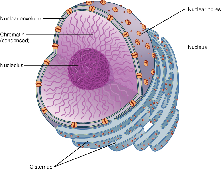
The nucleus is the largest and most prominent of a cell’s organelles ( Figure 1 ). The nucleus is generally considered the control center of the cell because it stores all of the genetic instructions for manufacturing proteins. Interestingly, some cells in the body, such as muscle cells, contain more than one nucleus ( Figure 2 ), which is known as multinucleated. Other cells, such as mammalian red blood cells (RBCs), do not contain nuclei at all. RBCs eject their nuclei as they mature, making space for the large numbers of hemoglobin molecules that carry oxygen throughout the body ( Figure 3 ). Without nuclei, the life span of RBCs is short, and so the body must produce new ones constantly.
View the University of Michigan WebScope at http://141.214.65.171/Histology/Basic%20Tissues/Muscle/058thin_HISTO_83X.svs/view.apml to explore the tissue sample in greater detail.
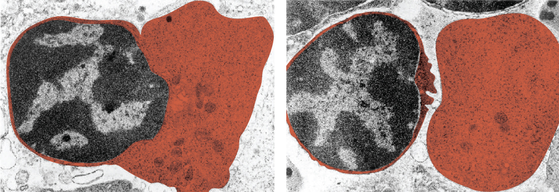
View the University of Michigan WebScope at http://virtualslides.med.umich.edu/Histology/EMsmallCharts/3%20Image%20Scope%20finals/139%20-%20Erythroblast_001.svs/view.apml to explore the tissue sample in greater detail.
Inside the nucleus lies the blueprint that dictates everything a cell will do and all of the products it will make. This information is stored within DNA. The nucleus sends “commands” to the cell via molecular messengers that translate the information from DNA. Each cell in your body (with the exception of germ cells) contains the complete set of your DNA. When a cell divides, the DNA must be duplicated so that the each new cell receives a full complement of DNA. The following section will explore the structure of the nucleus and its contents, as well as the process of DNA replication.
Organization of the Nucleus and Its DNA
Like most other cellular organelles, the nucleus is surrounded by a membrane called the nuclear envelope . This membranous covering consists of two adjacent lipid bilayers with a thin fluid space in between them. Spanning these two bilayers are nuclear pores. A nuclear pore is a tiny passageway for the passage of proteins, RNA, and solutes between the nucleus and the cytoplasm. Proteins called pore complexes lining the nuclear pores regulate the passage of materials into and out of the nucleus.
Inside the nuclear envelope is a gel-like nucleoplasm with solutes that include the building blocks of nucleic acids. There also can be a dark-staining mass often visible under a simple light microscope, called a nucleolus (plural = nucleoli). The nucleolus is a region of the nucleus that is responsible for manufacturing the RNA necessary for construction of ribosomes. Once synthesized, newly made ribosomal subunits exit the cell’s nucleus through the nuclear pores.
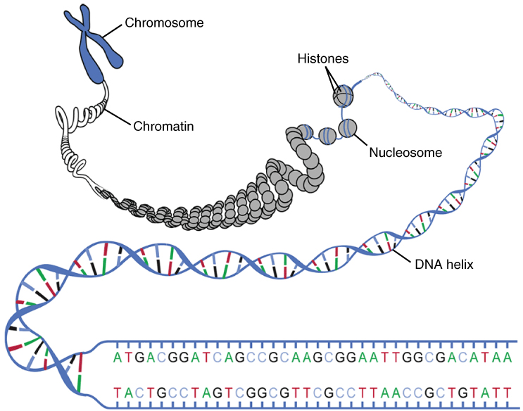
The genetic instructions that are used to build and maintain an organism are arranged in an orderly manner in strands of DNA. Within the nucleus are threads of chromatin composed of DNA and associated proteins ( Figure 4 ). Along the chromatin threads, the DNA is wrapped around a set of histone proteins. A nucleosome is a single, wrapped DNA-histone complex. Multiple nucleosomes along the entire molecule of DNA appear like a beaded necklace, in which the string is the DNA and the beads are the associated histones. When a cell is in the process of division, the chromatin condenses into chromosomes, so that the DNA can be safely transported to the “daughter cells.” The chromosome is composed of DNA and proteins; it is the condensed form of chromatin. It is estimated that humans have almost 22,000 genes distributed on 46 chromosomes.
DNA Replication
In order for an organism to grow, develop, and maintain its health, cells must reproduce themselves by dividing to produce two new daughter cells, each with the full complement of DNA as found in the original cell. Billions of new cells are produced in an adult human every day. Only very few cell types in the body do not divide, including nerve cells, skeletal muscle fibers, and cardiac muscle cells. The division time of different cell types varies. Epithelial cells of the skin and gastrointestinal lining, for instance, divide very frequently to replace those that are constantly being rubbed off of the surface by friction.
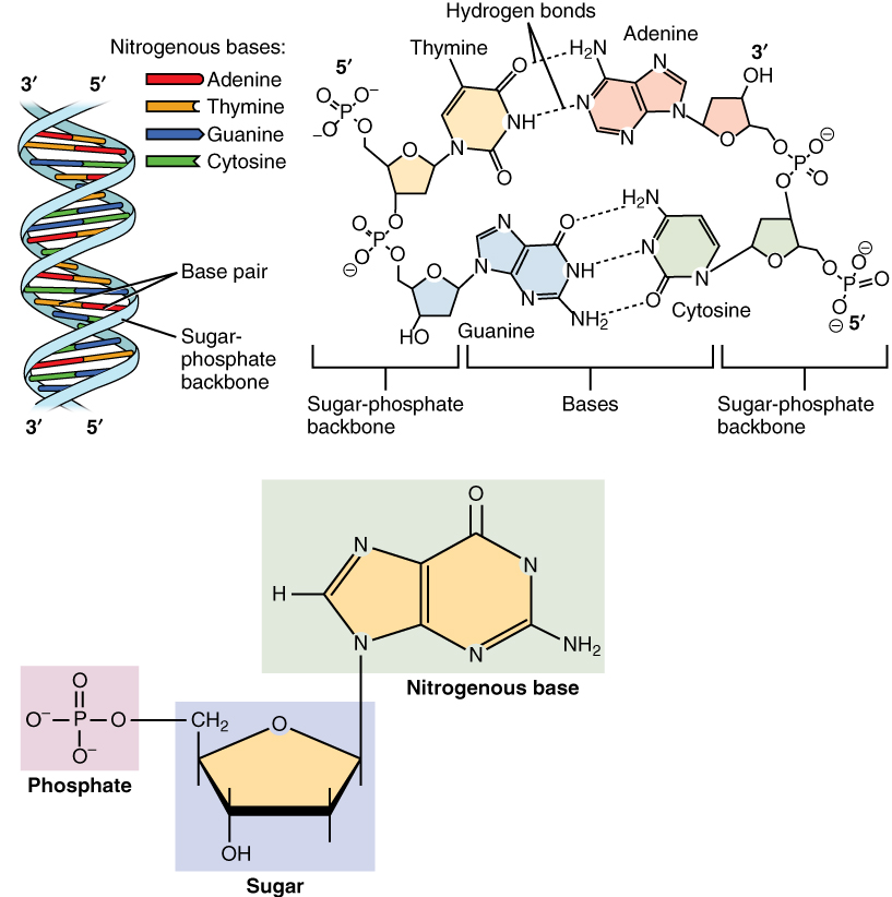
A DNA molecule is made of two strands that “complement” each other in the sense that the molecules that compose the strands fit together and bind to each other, creating a double-stranded molecule that looks much like a long, twisted ladder. Each side rail of the DNA ladder is composed of alternating sugar and phosphate groups ( Figure 5 ). The two sides of the ladder are not identical, but are complementary. These two backbones are bonded to each other across pairs of protruding bases, each bonded pair forming one “rung,” or cross member. The four DNA bases are adenine (A), thymine (T), cytosine (C), and guanine (G). Because of their shape and charge, the two bases that compose a pair always bond together. Adenine always binds with thymine, and cytosine always binds with guanine. The particular sequence of bases along the DNA molecule determines the genetic code. Therefore, if the two complementary strands of DNA were pulled apart, you could infer the order of the bases in one strand from the bases in the other, complementary strand. For example, if one strand has a region with the sequence AGTGCCT, then the sequence of the complementary strand would be TCACGGA.
DNA replication is the copying of DNA that occurs before cell division can take place. After a great deal of debate and experimentation, the general method of DNA replication was deduced in 1958 by two scientists in California, Matthew Meselson and Franklin Stahl. This method is illustrated in Figure 6 and described below.
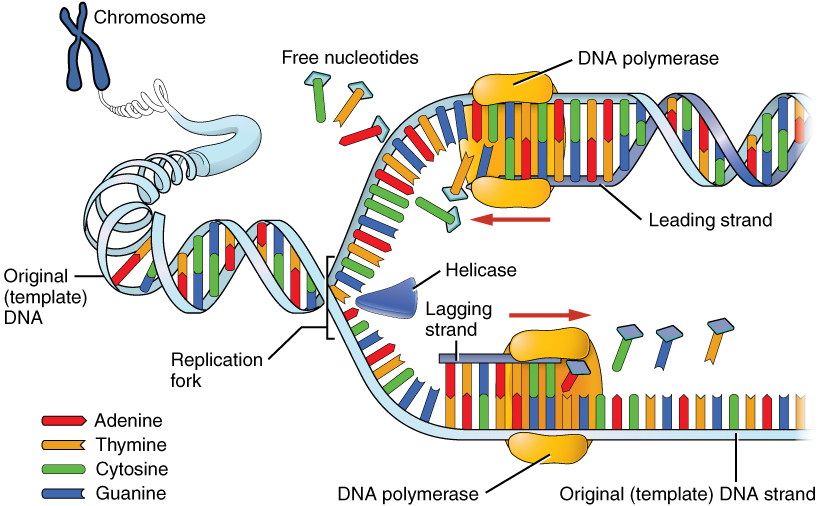
Stage 1: Initiation. The two complementary strands are separated, much like unzipping a zipper. Special enzymes, including helicase , untwist and separate the two strands of DNA.
Stage 2: Elongation. Each strand becomes a template along which a new complementary strand is built. DNA polymerase brings in the correct bases to complement the template strand, synthesizing a new strand base by base. A DNA polymerase is an enzyme that adds free nucleotides to the end of a chain of DNA, making a new double strand. This growing strand continues to be built until it has fully complemented the template strand.
Stage 3: Termination. Once the two original strands are bound to their own, finished, complementary strands, DNA replication is stopped and the two new identical DNA molecules are complete.
Each new DNA molecule contains one strand from the original molecule and one newly synthesized strand. The term for this mode of replication is “semiconservative,” because half of the original DNA molecule is conserved in each new DNA molecule. This process continues until the cell’s entire genome , the entire complement of an organism’s DNA, is replicated. As you might imagine, it is very important that DNA replication take place precisely so that new cells in the body contain the exact same genetic material as their parent cells. Mistakes made during DNA replication, such as the accidental addition of an inappropriate nucleotide, have the potential to render a gene dysfunctional or useless. Fortunately, there are mechanisms in place to minimize such mistakes. A DNA proofreading process enlists the help of special enzymes that scan the newly synthesized molecule for mistakes and corrects them. Once the process of DNA replication is complete, the cell is ready to divide. You will explore the process of cell division later in the chapter.

Watch this video to learn about DNA replication. DNA replication proceeds simultaneously at several sites on the same molecule. What separates the base pair at the start of DNA replication?
Chapter Review
The nucleus is the command center of the cell, containing the genetic instructions for all of the materials a cell will make (and thus all of its functions it can perform). The nucleus is encased within a membrane of two interconnected lipid bilayers, side-by-side. This nuclear envelope is studded with protein-lined pores that allow materials to be trafficked into and out of the nucleus. The nucleus contains one or more nucleoli, which serve as sites for ribosome synthesis. The nucleus houses the genetic material of the cell: DNA. DNA is normally found as a loosely contained structure called chromatin within the nucleus, where it is wound up and associated with a variety of histone proteins. When a cell is about to divide, the chromatin coils tightly and condenses to form chromosomes.
There is a pool of cells constantly dividing within your body. The result is billions of new cells being created each day. Before any cell is ready to divide, it must replicate its DNA so that each new daughter cell will receive an exact copy of the organism’s genome. A variety of enzymes are enlisted during DNA replication. These enzymes unwind the DNA molecule, separate the two strands, and assist with the building of complementary strands along each parent strand. The original DNA strands serve as templates from which the nucleotide sequence of the new strands are determined and synthesized. When replication is completed, two identical DNA molecules exist. Each one contains one original strand and one newly synthesized complementary strand.
Interactive Link Questions
Review questions.
1. The nucleus and mitochondria share which of the following features?
- protein-lined membrane pores
- a double cell membrane
- the synthesis of ribosomes
- the production of cellular energy
2. Which of the following structures could be found within the nucleolus?
- nucleosomes
3. Which of the following sequences on a DNA molecule would be complementary to GCTTATAT?
4. Place the following structures in order from least to most complex organization: chromatin, nucleosome, DNA, chromosome
- DNA, nucleosome, chromatin, chromosome
- nucleosome, DNA, chromosome, chromatin
- DNA, chromatin, nucleosome, chromosome
- nucleosome, chromatin, DNA, chromosome
5. Which of the following is part of the elongation step of DNA synthesis?
- pulling apart the two DNA strands
- attaching complementary nucleotides to the template strand
- untwisting the DNA helix
- none of the above
Critical Thinking Questions
1. Explain in your own words why DNA replication is said to be “semiconservative”?
2. Why is it important that DNA replication take place before cell division? What would happen if cell division of a body cell took place without DNA replication, or when DNA replication was incomplete?
Answers for Review Questions
Answers for Critical Thinking Questions
- DNA replication is said to be semiconservative because, after replication is complete, one of the two parent DNA strands makes up half of each new DNA molecule. The other half is a newly synthesized strand. Therefore, half (“semi”) of each daughter DNA molecule is from the parent molecule and half is a new molecule.
- During cell division, one cell divides to produce two new cells. In order for all of the cells in your body to maintain a full genome, each cell must replicate its DNA before it divides so that a full genome can be allotted to each of its offspring cells. If DNA replication did not take place fully, or at all, the offspring cells would be missing some or all of the genome. This could be disastrous if a cell was missing genes necessary for its function and health.
Anatomy and Physiology Copyright © 1999-2016 by Rice University is licensed under a Creative Commons Attribution 4.0 International License , except where otherwise noted.
Share This Book
Feedback/errata, leave a reply cancel reply.
Your email address will not be published. Required fields are marked *
Save my name, email, and website in this browser for the next time I comment.
If you're seeing this message, it means we're having trouble loading external resources on our website.
If you're behind a web filter, please make sure that the domains *.kastatic.org and *.kasandbox.org are unblocked.
To log in and use all the features of Khan Academy, please enable JavaScript in your browser.
AP®︎/College Biology
Course: ap®︎/college biology > unit 6.
- Antiparallel structure of DNA strands
- Leading and lagging strands in DNA replication
- Speed and precision of DNA replication
- Semi conservative replication
Molecular mechanism of DNA replication
- DNA structure and replication review
- Replication
Key points:
- DNA replication is semiconservative . Each strand in the double helix acts as a template for synthesis of a new, complementary strand.
- New DNA is made by enzymes called DNA polymerases , which require a template and a primer (starter) and synthesize DNA in the 5' to 3' direction.
- During DNA replication, one new strand (the leading strand ) is made as a continuous piece. The other (the lagging strand ) is made in small pieces.
- DNA replication requires other enzymes in addition to DNA polymerase, including DNA primase , DNA helicase , DNA ligase , and topoisomerase .
Introduction
The basic idea.
- DNA double helix.
- Hydrogen bonds break and helix opens.
- Each strand of DNA acts as a template for synthesis of a new, complementary strand.
- Replication produces two identical DNA double helices, each with one new and one old strand.
DNA polymerase
- They always need a template
- They can only add nucleotides to the 3' end of a DNA strand
- They can't start making a DNA chain from scratch, but require a pre-existing chain or short stretch of nucleotides called a primer
- They proofread , or check their work, removing the vast majority of "wrong" nucleotides that are accidentally added to the chain
Starting DNA replication
Primers and primase, leading and lagging strands, the maintenance and cleanup crew, summary of dna replication in e. coli.
- Helicase opens up the DNA at the replication fork.
- Single-strand binding proteins coat the DNA around the replication fork to prevent rewinding of the DNA.
- Topoisomerase works at the region ahead of the replication fork to prevent supercoiling.
- Primase synthesizes RNA primers complementary to the DNA strand.
- DNA polymerase III extends the primers, adding on to the 3' end, to make the bulk of the new DNA.
- RNA primers are removed and replaced with DNA by DNA polymerase I .
- The gaps between DNA fragments are sealed by DNA ligase .
DNA replication in eukaryotes
- Eukaryotes usually have multiple linear chromosomes, each with multiple origins of replication. Humans can have up to 100 , 000 origins of replication 5 !
- Most of the E. coli enzymes have counterparts in eukaryotic DNA replication, but a single enzyme in E. coli may be represented by multiple enzymes in eukaryotes. For instance, there are five human DNA polymerases with important roles in replication 5 .
- Most eukaryotic chromosomes are linear. Because of the way the lagging strand is made, some DNA is lost from the ends of linear chromosomes (the telomeres ) in each round of replication.
Explore outside of Khan Academy
Attribution:, works cited:.
- National Human Genome Research Institute. (2010, October 30). The human genome project completion: Frequently asked questions. In News release archives 2003 . Retrieved from https://www.genome.gov/11006943 .
- Reece, J. B., Urry, L. A., Cain, M. L., Wasserman, S. A., Minorsky, P. V., and Jackson, R. B. (2011). Origins of replication in E. coli and eukaryotes. In Campbell biology (10th ed., p. 321). San Francisco, CA: Pearson.
- Bell, S. P. and Kaguni, J. M. (2013). Helicase loading at chromosomal origins of replication. Cold Spring Harb. Perspect. Biol. , 5 (6), a010124. http://dx.doi.org/10.1101/cshperspect.a010124 .
- Yao, N. Y. and O'Donnell, M. (2008). Replisome dynamics and use of DNA trombone loops to bypass replication blocks. Mol. Biosyst. , 4 (11), 1075-1084. http://dx.doi.org/10.1039/b811097b . Retrieved from http://www.ncbi.nlm.nih.gov/pmc/articles/PMC4011192/ .
- OpenStax College, Biology. (2016, May 27). DNA replication in eukaryotes. In OpenStax CNX . Retrieved from http://cnx.org/contents/[email protected]:2l3nsfJK@5/DNA-Replication-in-Eukaryotes .
Additional references:
Want to join the conversation.
- Upvote Button navigates to signup page
- Downvote Button navigates to signup page
- Flag Button navigates to signup page


152 Critical Thinking Questions
19. If mRNA is complementary to the DNA template strand and the DNA template strand is complementary to the DNA non-template strand, then why are base sequences of mRNA and the DNA non-template strand not identical? Could they ever be?
20. A scientist observes that a cell has an RNA polymerase deficiency that prevents it from making proteins. Describe three additional observations that would together support the conclusion that a defect in RNA polymerase I activity, and not problems with the other polymerases, causes the defect.
21. Describe how transcription in prokaryotic cells can be altered by external stimulation such as excess lactose in the environment
22. Describe how controlling gene expression will alter the overall protein levels in the cell.
Biology Part I Copyright © 2022 by LOUIS: The Louisiana Library Network is licensed under a Creative Commons Attribution 4.0 International License , except where otherwise noted.
Share This Book
Critical Thinking Questions
Explain Griffith’s transformation experiments. What did he conclude from them?
- Two strains of S. pneumoniae were used for the experiment. Griffith injected a mouse with heat-inactivated S strain (pathogenic) and R strain (non-pathogenic). The mouse died and S strain was recovered from the dead mouse. He concluded that external DNA is taken up by a cell that changed morphology and physiology.
- Two strains of Vibrio cholerae were used for the experiment. Griffith injected a mouse with heat-inactivated S strain (pathogenic) and R strain (non-pathogenic). The mouse died and S strain was recovered from the dead mouse. He concluded that external DNA is taken up by a cell that changed morphology and physiology.
- Two strains of S. pneumoniae were used for the experiment. Griffith injected a mouse with heat-inactivated S strain (pathogenic) and R strain (non-pathogenic). The mouse died and R strain was recovered from the dead mouse. He concluded that external DNA is taken up by a cell that changed morphology and physiology.
- Two strains of S. pneumoniae were used for the experiment. Griffith injected a mouse with heat-inactivated S strain (pathogenic) and R strain (non-pathogenic). The mouse died and S strain was recovered from the dead mouse. He concluded that mutation occurred in the DNA of the cell that changed morphology and physiology.
Explain why radioactive sulfur and phosphorous were used to label bacteriophages in the Hershey and Chase experiments.
- Protein was labeled with radioactive sulfur and DNA was labeled with radioactive phosphorous. Phosphorous is found in DNA, so it will be tagged by radioactive phosphorous.
- Protein was labeled with radioactive phosphorous and DNA was labeled with radioactive sulfur. Phosphorous is found in DNA, so it will be tagged by radioactive phosphorous.
- Protein was labeled with radioactive sulfur and DNA was labeled with radioactive phosphorous. Phosphorous is found in DNA, so DNA will be tagged by radioactive sulfur.
- Protein was labeled with radioactive phosphorous and DNA was labeled with radioactive sulfur. Phosphorous is found in DNA, so DNA will be tagged by radioactive sulfur.
How can Chargaff’s rules be used to identify different species?
- The amount of adenine, thymine, guanine, and cytosine varies from species to species and is not found in equal quantities. They do not vary between individuals of the same species and can be used to identify different species.
- The amount of adenine, thymine, guanine, and cytosine varies from species to species and is found in equal quantities. They do not vary between individuals of the same species and can be used to identify different species.
- The amount of adenine and thymine is equal to guanine and cytosine and is found in equal quantities. They do not vary between individuals of the same species and can be used to identify different species.
- The amount of adenine, thymine, guanine, and cytosine varies from species to species and is not found in equal quantities. They vary between individuals of the same species and can be used to identify different species.
In the Hershey-Chase experiments, what conclusion would the scientists have drawn if bacteria containing both radioactive phosphorus and sulfur were found in the final pellets?
Describe the structure and complementary base pairing of DNA.
- DNA is made up of two strands that are twisted around each other to form a helix. Adenine pairs up with thymine and cytosine pairs with guanine. The two strands are anti-parallel in nature; that is, the 3' end of one strand faces the 5' end of other strand. Sugar, phosphate and nitrogenous bases contribute to the DNA structure.
- DNA is made up of two strands that are twisted around each other to form a helix. Adenine pairs up with cytosine and thymine pairs with guanine. The two strands are anti-parallel in nature; that is, the 3' end of one strand faces the 5' end of other strand. Sugar, phosphate and nitrogenous bases contribute to the DNA structure.
- DNA is made up of two strands that are twisted around each other to form a helix. Adenine pairs up with thymine and cytosine pairs with guanine. The two strands are parallel in nature; that is, the 3' end of one strand faces the 3' end of other strand. Sugar, phosphate and nitrogenous bases contribute to the DNA structure.
- DNA is made up of two strands that are twisted around each other to form a helix. Adenine pairs up with thymine and cytosine pairs with guanine. The two strands are anti-parallel in nature; that is, the 3' end of one strand faces the 5' end of other strand. Only sugar contributes to the DNA structure.
- Frederick Sanger’s sequencing is a chain termination method that is used to generate DNA fragments that terminate at different points using dye-labeled dideoxynucleotides. DNA is separated by electrophoresis on the basis of size. The DNA sequence can be read out on an electropherogram generated by a laser scanner.
- Frederick Sanger’s sequencing is a chain elongation method that is used to generate DNA fragments that elongate at different points using dye-labeled dideoxynucleotides. DNA is separated by electrophoresis on the basis of size. The DNA sequence can be read out on an electropherogram generated by a laser scanner.
- Frederick Sanger’s sequencing is a chain termination method that is used to generate DNA fragments that terminate at different points using dye-labeled dideoxynucleotides. DNA is joined together by electrophoresis on the basis of size. The DNA sequence can be read out on an electropherogram generated by a laser scanner.
- Frederick Sanger’s sequencing is a chain termination method that is used to generate DNA fragments that terminate at different points using dye-labeled dideoxynucleotides. DNA is separated by electrophoresis on the basis of size. The DNA sequence can be read out on an electropherogram generated by a magnetic scanner.
- Eukaryotes have a single, circular chromosome, while prokaryotes have multiple, linear chromosomes. Prokaryotes pack their chromosomes by super coiling, managed by DNA gyrase. Eukaryote chromosomes are wrapped around histone proteins that create heterochromatin and euchromatin, which is not present in prokaryotes.
- Prokaryotes have a single, circular chromosome, while eukaryotes have multiple, linear chromosomes. Prokaryotes pack their chromosomes by super coiling, managed by DNA gyrase. Eukaryote chromosomes are wrapped around histone proteins that create heterochromatin and euchromatin, which is not present in prokaryotes.
- Prokaryotes have a single, circular chromosome, while eukaryotes have multiple, linear chromosomes. Eukaryotes pack their chromosomes by super coiling, managed by DNA gyrase. Prokaryotes chromosomes are wrapped around histone proteins that create heterochromatin and euchromatin, which is not present in eukaryotes.
- Prokaryotes have a single, circular chromosome, while eukaryotes have multiple, linear chromosomes. Prokaryotes pack their chromosomes by super coiling, managed by DNA gyrase. Eukaryote chromosomes are wrapped around histone proteins that create heterochromatin and euchromatin, which is present in prokaryotes.
DNA replication is bidirectional and discontinuous; explain your understanding of those concepts.
- DNA polymerase reads the template strand in the 3' to 5' direction and adds nucleotides only in the 5' to 3' direction. The leading strand is synthesized in the direction of the replication fork. Replication on the lagging strand occurs in the direction away from the replication fork in short stretches of DNA called Okazaki fragments.
- DNA polymerase reads the template strand in the 5' to 3' direction and adds nucleotides only in the 5' to 3' direction. The leading strand is synthesized in the direction of the replication fork. Replication on the lagging strand occurs in the direction away from the replication fork in short stretches of DNA called Okazaki fragments.
- DNA polymerase reads the template strand in the 3' to 5' direction and adds nucleotides only in the 5' to 3' direction. The leading strand is synthesized in the direction away from the replication fork. Replication on the lagging strand occurs in the direction of the replication fork in short stretches of DNA called Okazaki fragments.
- DNA polymerase reads the template strand in the 5' to 3' direction and adds nucleotides only in the 3' to 5' direction. The leading strand is synthesized in the direction of the replication fork. Replication on the lagging strand occurs in the direction away from the replication fork in long stretches of DNA called Okazaki fragments.
Discuss how the scientific community learned that DNA replication takes place in a semi- conservative fashion.
- Meselson and Stahl experimented with E. coli. DNA grown in 15 N was heavier than DNA grown in 14 N . When DNA in 15 N was switched to 14 N media, DNA sedimented halfway between the 15 N and 14 N levels after one round of cell division, indicating 50 percent presence of 14 N . This supports the semiconservative replication model.
- Meselson and Stahl experimented with S. pneumonia . DNA grown in 15 N was heavier than DNA grown in 14 N . When DNA in 15 N was switched to 14 N media, DNA sedimented halfway between the 15 N and 14 N levels after one round of cell division, indicating 50 percent presence of 14 N . This supports the semiconservative replication model.
- Meselson and Stahl experimented with E. coli. DNA grown in 14 N was heavier than DNA grown in 15 N . When DNA in 15 N was switched to 14 N media, DNA sedimented halfway between the 15 N and 14 N levels after one round of cell division, indicating 50 percent presence of 14 N . This supports the semiconservative replication model.
- Meselson and Stahl experimented with S. pneumonia . DNA grown in 15 N was heavier than DNA grown in 14 N . When DNA in 15 N was switched to 14 N media, DNA sedimented halfway between the 15 N and 14 N levels after one round of cell division, indicating complete presence of 14 N . This supports the semiconservative replication model.
Explain why half of DNA is replicated in a discontinuous fashion.
- Replication of the lagging strand occurs in the direction away from the replication fork in short stretches of DNA, since access to the DNA is always from the 5' end. This results in pieces of DNA being replicated in a discontinuous fashion.
- Replication of the leading strand occurs in the direction away from the replication fork in short stretches of DNA, since access to the DNA is always from the 5' end. This results in pieces of DNA being replicated in a discontinuous fashion.
- Replication of the lagging strand occurs in the direction of the replication fork in short stretches of DNA, since access to the DNA is always from the 5' end. This results in pieces of DNA being replicated in a discontinuous fashion.
- Replication of the lagging strand occurs in the direction away from the replication fork in short stretches of DNA, since access to the DNA is always from the 3' end. This results in pieces of DNA being replicated in a discontinuous fashion.
- Helicase separates the DNA strands at the origin of replication. Topoisomerase breaks and reforms DNA’s phosphate backbone ahead of the replication fork, thereby relieving the pressure. Single-stranded binding proteins prevent reforming of DNA. Primase synthesizes RNA primer which is used by DNA polymerase to form a daughter strand. If helicase is mutated, the DNA strands will not be separated at the beginning of replication.
- Helicase joins the DNA strands together at the origin of replication. Topoisomerase breaks and reforms DNA’s phosphate backbone after the replication fork, thereby relieving the pressure. Single-stranded binding proteins prevent reforming of DNA. Primase synthesizes RNA primer which is used by DNA polymerase to form a daughter strand. If helicase is mutated, the DNA strands will not be joined together at the beginning of replication.
- Helicase separates the DNA strands at the origin of replication. Topoisomerase breaks and reforms DNA’s sugar backbone ahead of the replication fork, thereby increasing the pressure. Single-stranded binding proteins prevent reforming of DNA. Primase synthesizes DNA primer which is used by DNA polymerase to form a daughter strand. If helicase is mutated, the DNA strands will be separated at the beginning of replication.
- Helicase separates the DNA strands at the origin of replication. Topoisomerase breaks and reforms DNA’s sugar backbone ahead of the replication fork, thereby relieving the pressure. Single-stranded binding proteins prevent reforming of DNA. Primase synthesizes DNA primer which is used by RNA polymerase to form a parent strand. If helicase is mutated, the DNA strands will be separated at the beginning of replication.
- Okazaki fragments are short stretches of DNA on the lagging strand, which is synthesized in the direction away from the replication fork.
- Okazaki fragments are long stretches of DNA on the lagging strand, which is synthesized in the direction of the replication fork.
- Okazaki fragments are long stretches of DNA on the leading strand, which is synthesized in the direction away from the replication fork.
- Okazaki fragments are short stretches of DNA on the leading strand, which is synthesized in the direction of the replication fork.
- DNA polymerase I removes the RNA primers from the developing copy of DNA. DNA ligase seals the ends of the new segment, especially the Okazaki fragments.
- DNA polymerase I adds the RNA primers to the already developing copy of DNA. DNA ligase separates the ends of the new segment, especially the Okazaki fragments.
- DNA polymerase I seals the ends of the new segment, especially the Okazaki fragments. DNA ligase removes the RNA primers from the developing copy of DNA.
- DNA polymerase I removes the enzyme primase from the developing copy of DNA. DNA ligase seals the ends of the old segment, especially the Okazaki fragments.
If the rate of replication in a particular prokaryote is 900 nucleotides per second, how long would it take to make two copies of a 1.2 million base pair genome?
- 22.2 minutes
- 44.4 minutes
- 45.4 minutes
- 54.4 minutes
- The ends of the linear chromosomes are maintained by the activity of the telomerase enzyme.
- The ends of the linear chromosomes are maintained by the formation of a replication fork.
- The ends of the linear chromosomes are maintained by the continuous joining of Okazaki fragments.
- The ends of the linear chromosomes are maintained by the action of the polymerase enzyme.
- A prokaryotic organism’s rate of replication is ten times faster than that of eukaryotes. Prokaryotes have a single origin of replication and use five types of polymerases, while eukaryotes have multiple sites of origin and use fourteen polymerases. Telomerase is absent in prokaryotes. DNA pol I is the primer remover in prokaryotes, while in eukaryotes it is RNase H. DNA pol III performs strand elongation in prokaryotes and pol δ and pol ε do the same in eukaryotes.
- A prokaryotic organism’s rate of replication is ten times slower than that of eukaryotes. Prokaryotes have a single origin of replication and use five types of polymerases, while eukaryotes have multiple sites of origin and use fourteen polymerases. Telomerase is absent in eukaryotes. DNA pol I is the primer remover in prokaryotes, while in eukaryotes it is RNase H. DNA pol III performs strand elongation in prokaryotes and pol δ and pol ε do the same in eukaryotes.
- A prokaryotic organism’s rate of replication is ten times faster than that of eukaryotes. Prokaryotes have five origins of replication and use a single type of polymerase, while eukaryotes have a single site of origin and use fourteen polymerases. Telomerase is absent in prokaryotes. DNA pol I is the primer remover in prokaryotes, while in eukaryotes it is RNase H. DNA pol III performs strand elongation in prokaryotes and pol δ and pol ε do the same in eukaryotes.
- A prokaryotic organism’s rate of replication is ten times slower than that of eukaryotes. Prokaryotes have a single origin of replication and use five types of polymerases, while eukaryotes have multiple sites of origin and use fourteen polymerases. Telomerase is absent in prokaryotes. DNA pol I is the primer remover in eukaryotes, while in prokaryotes it is RNase H. DNA pol III performs strand elongation in prokaryotes and pol δ and pol ε do the same in eukaryotes.
- Mismatch repair corrects the errors after the replication is completed by excising the incorrectly added nucleotide and adding the correct base. Any mutation in a mismatch repair enzyme would lead to more permanent damage.
- Mismatch repair corrects the errors during the replication by excising the incorrectly added nucleotide and adding the correct base. Any mutation in the mismatch repair enzyme would lead to more permanent damage.
- Mismatch repair corrects the errors after the replication is completed by excising the added nucleotides and adding more bases. Any mutation in the mismatch repair enzyme would lead to more permanent damage.
- Mismatch repair corrects the errors after the replication is completed by excising the incorrectly added nucleotide and adding the correct base. Any mutation in the mismatch repair enzyme would lead to more temporary damage.
- Both will result in the production of defective proteins. The DNA mutation, if not corrected, is permanent, while the mRNA mutation will only affect proteins made from that mRNA strand. Production of defective protein ceases when the mRNA strand deteriorates.
- Both will result in the production of defective proteins. The DNA mutation, if not corrected, is permanent, while the mRNA mutation will not affect proteins made from that mRNA strand. Production of defective protein continues when the mRNA strand deteriorates.
- Only DNA will result in the production of defective proteins. The DNA mutation, if not corrected, is permanent. Production of defective protein ceases when the DNA strand deteriorates.
- Only mRNA will result in the production of defective proteins. The mRNA mutation will only affect proteins made from that mRNA strand. Production of defective protein ceases when the mRNA strand deteriorates.
Discuss the effects of point mutations on a DNA strand.
- Mutations can cause a single change in an amino acid. A nonsense mutation can stop the replication or reading of that strand. Insertion or deletion mutations can cause a frame shift. This can result in nonfunctional proteins.
- Mutations can cause a single change in amino acid. A missense mutation can stop the replication or reading of that strand. Insertion or deletion mutations can cause a frame shift. This can result in nonfunctional proteins.
- Mutations can cause a single change in amino acid. A nonsense mutation can stop the replication or reading of that strand. Substitution mutations can cause a frame shift. This can result in nonfunctional proteins.
- Mutations can cause a single change in amino acid. A nonsense mutation can stop the replication or reading of that strand. Insertion or deletion mutations can cause a frame shift. This can result in functional proteins.
- Mutations in tRNA and rRNA would lead to the production of defective proteins or no protein production.
- Mutations in tRNA and rRNA would lead to changes in the semi-conservative mode of replication of DNA.
- Mutations in tRNA and rRNA would lead to production of a DNA strand with a mutated single strand and normal other strand.
- Mutations in tRNA and rRNA would lead to skin cancer in patients of xeroderma pigmentosa.
Copy and paste the link code above.

Related Items

- school Campus Bookshelves
- menu_book Bookshelves
- perm_media Learning Objects
- login Login
- how_to_reg Request Instructor Account
- hub Instructor Commons
- Download Page (PDF)
- Download Full Book (PDF)
- Periodic Table
- Physics Constants
- Scientific Calculator
- Reference & Cite
- Tools expand_more
- Readability
selected template will load here
This action is not available.

9.2: DNA Replication
- Last updated
- Save as PDF
- Page ID 7023

When a cell divides, it is important that each daughter cell receives an identical copy of the DNA. This is accomplished by the process of DNA replication. The replication of DNA occurs during the synthesis phase, or S phase, of the cell cycle, before the cell enters mitosis or meiosis.
The elucidation of the structure of the double helix provided a hint as to how DNA is copied. Recall that adenine nucleotides pair with thymine nucleotides, and cytosine with guanine. This means that the two strands are complementary to each other. For example, a strand of DNA with a nucleotide sequence of AGTCATGA will have a complementary strand with the sequence TCAGTACT (Figure \(\PageIndex{1}\)).
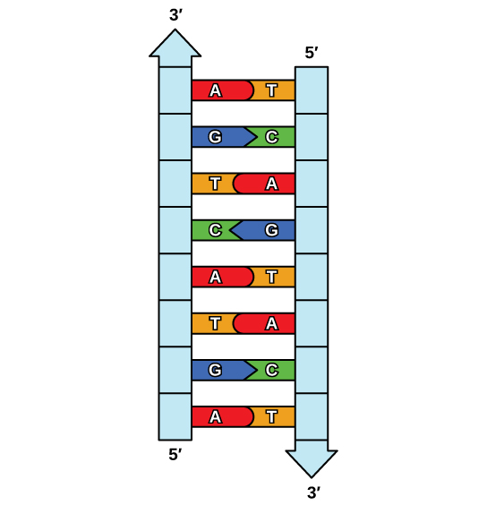
Because of the complementarity of the two strands, having one strand means that it is possible to recreate the other strand. This model for replication suggests that the two strands of the double helix separate during replication, and each strand serves as a template from which the new complementary strand is copied (Figure \(\PageIndex{2}\)).
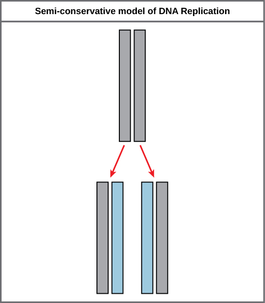
During DNA replication, each of the two strands that make up the double helix serves as a template from which new strands are copied. The new strand will be complementary to the parental or “old” strand. Each new double strand consists of one parental strand and one new daughter strand. This is known as semiconservative replication. When two DNA copies are formed, they have an identical sequence of nucleotide bases and are divided equally into two daughter cells.
DNA Replication in Eukaryotes
Because eukaryotic genomes are very complex, DNA replication is a very complicated process that involves several enzymes and other proteins. It occurs in three main stages: initiation, elongation, and termination.
Recall that eukaryotic DNA is bound to proteins known as histones to form structures called nucleosomes. During initiation, the DNA is made accessible to the proteins and enzymes involved in the replication process. How does the replication machinery know where on the DNA double helix to begin? It turns out that there are specific nucleotide sequences called origins of replication at which replication begins. Certain proteins bind to the origin of replication while an enzyme called helicase unwinds and opens up the DNA helix. As the DNA opens up, Y-shaped structures called replication forks are formed (Figure \(\PageIndex{3}\)). Two replication forks are formed at the origin of replication, and these get extended in both directions as replication proceeds. There are multiple origins of replication on the eukaryotic chromosome, such that replication can occur simultaneously from several places in the genome.
During elongation, an enzyme called DNA polymerase adds DNA nucleotides to the 3' end of the template. Because DNA polymerase can only add new nucleotides at the end of a backbone, a primer sequence, which provides this starting point, is added with complementary RNA nucleotides. This primer is removed later, and the nucleotides are replaced with DNA nucleotides. One strand, which is complementary to the parental DNA strand, is synthesized continuously toward the replication fork so the polymerase can add nucleotides in this direction. This continuously synthesized strand is known as the leading strand. Because DNA polymerase can only synthesize DNA in a 5' to 3' direction, the other new strand is put together in short pieces called Okazaki fragments. The Okazaki fragments each require a primer made of RNA to start the synthesis. The strand with the Okazaki fragments is known as the lagging strand. As synthesis proceeds, an enzyme removes the RNA primer, which is then replaced with DNA nucleotides, and the gaps between fragments are sealed by an enzyme called DNA ligase.
The process of DNA replication can be summarized as follows:
- DNA unwinds at the origin of replication.
- New bases are added to the complementary parental strands. One new strand is made continuously, while the other strand is made in pieces.
- Primers are removed, new DNA nucleotides are put in place of the primers and the backbone is sealed by DNA ligase.
ART CONNECTION
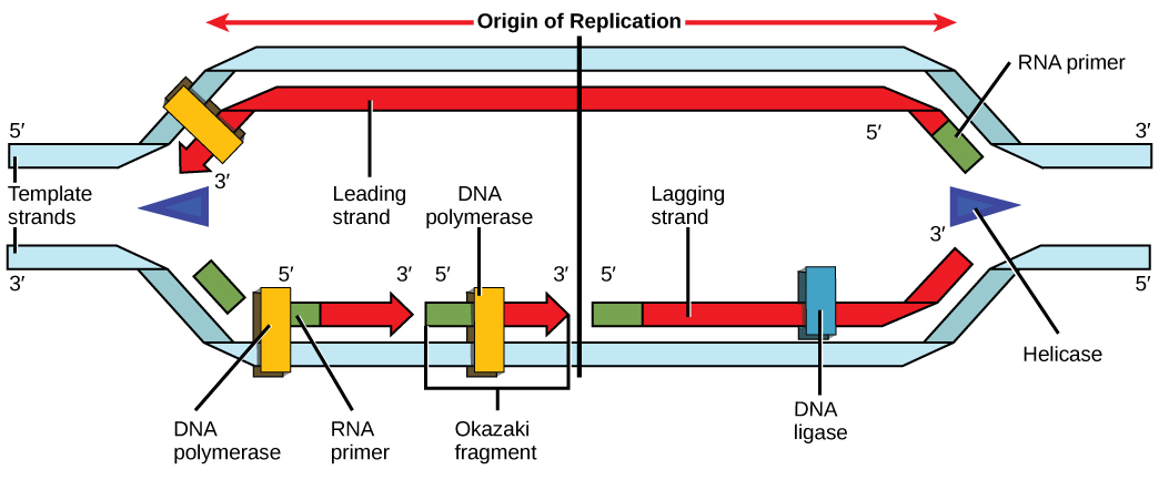
You isolate a cell strain in which the joining together of Okazaki fragments is impaired and suspect that a mutation has occurred in an enzyme found at the replication fork. Which enzyme is most likely to be mutated?
Telomere Replication
Because eukaryotic chromosomes are linear, DNA replication comes to the end of a line in eukaryotic chromosomes. As you have learned, the DNA polymerase enzyme can add nucleotides in only one direction. In the leading strand, synthesis continues until the end of the chromosome is reached; however, on the lagging strand there is no place for a primer to be made for the DNA fragment to be copied at the end of the chromosome. This presents a problem for the cell because the ends remain unpaired, and over time these ends get progressively shorter as cells continue to divide. The ends of the linear chromosomes are known as telomeres, which have repetitive sequences that do not code for a particular gene. As a consequence, it is telomeres that are shortened with each round of DNA replication instead of genes. For example, in humans, a six base-pair sequence, TTAGGG, is repeated 100 to 1000 times. The discovery of the enzyme telomerase (Figure \(\PageIndex{4}\)) helped in the understanding of how chromosome ends are maintained. The telomerase attaches to the end of the chromosome, and complementary bases to the RNA template are added on the end of the DNA strand. Once the lagging strand template is sufficiently elongated, DNA polymerase can now add nucleotides that are complementary to the ends of the chromosomes. Thus, the ends of the chromosomes are replicated.
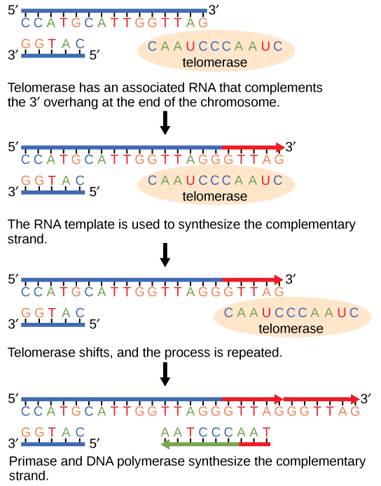
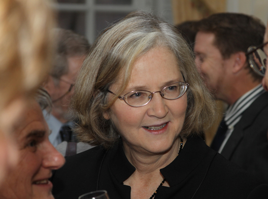
Telomerase is typically found to be active in germ cells, adult stem cells, and some cancer cells. For her discovery of telomerase and its action, Elizabeth Blackburn (Figure \(\PageIndex{5}\)) received the Nobel Prize for Medicine and Physiology in 2009.
Telomerase is not active in adult somatic cells. Adult somatic cells that undergo cell division continue to have their telomeres shortened. This essentially means that telomere shortening is associated with aging. In 2010, scientists found that telomerase can reverse some age-related conditions in mice, and this may have potential in regenerative medicine. 1 Telomerase-deficient mice were used in these studies; these mice have tissue atrophy, stem-cell depletion, organ system failure, and impaired tissue injury responses. Telomerase reactivation in these mice caused extension of telomeres, reduced DNA damage, reversed neurodegeneration, and improved functioning of the testes, spleen, and intestines. Thus, telomere reactivation may have potential for treating age-related diseases in humans.
DNA Replication in Prokaryotes
Recall that the prokaryotic chromosome is a circular molecule with a less extensive coiling structure than eukaryotic chromosomes. The eukaryotic chromosome is linear and highly coiled around proteins. While there are many similarities in the DNA replication process, these structural differences necessitate some differences in the DNA replication process in these two life forms.
DNA replication has been extremely well-studied in prokaryotes, primarily because of the small size of the genome and large number of variants available. Escherichia coli has 4.6 million base pairs in a single circular chromosome, and all of it gets replicated in approximately 42 minutes, starting from a single origin of replication and proceeding around the chromosome in both directions. This means that approximately 1000 nucleotides are added per second. The process is much more rapid than in eukaryotes. Table \(\PageIndex{1}\) summarizes the differences between prokaryotic and eukaryotic replications.
CONCEPT IN ACTION
Click through a tutorial on DNA replication.
DNA polymerase can make mistakes while adding nucleotides. It edits the DNA by proofreading every newly added base. Incorrect bases are removed and replaced by the correct base, and then polymerization continues (Figure \(\PageIndex{6}\) a ). Most mistakes are corrected during replication, although when this does not happen, the mismatch repair mechanism is employed. Mismatch repair enzymes recognize the wrongly incorporated base and excise it from the DNA, replacing it with the correct base (Figure \(\PageIndex{6}\) b ). In yet another type of repair, nucleotide excision repair, the DNA double strand is unwound and separated, the incorrect bases are removed along with a few bases on the 5' and 3' end, and these are replaced by copying the template with the help of DNA polymerase (Figure \(\PageIndex{6}\) c ). Nucleotide excision repair is particularly important in correcting thymine dimers, which are primarily caused by ultraviolet light. In a thymine dimer, two thymine nucleotides adjacent to each other on one strand are covalently bonded to each other rather than their complementary bases. If the dimer is not removed and repaired it will lead to a mutation. Individuals with flaws in their nucleotide excision repair genes show extreme sensitivity to sunlight and develop skin cancers early in life.
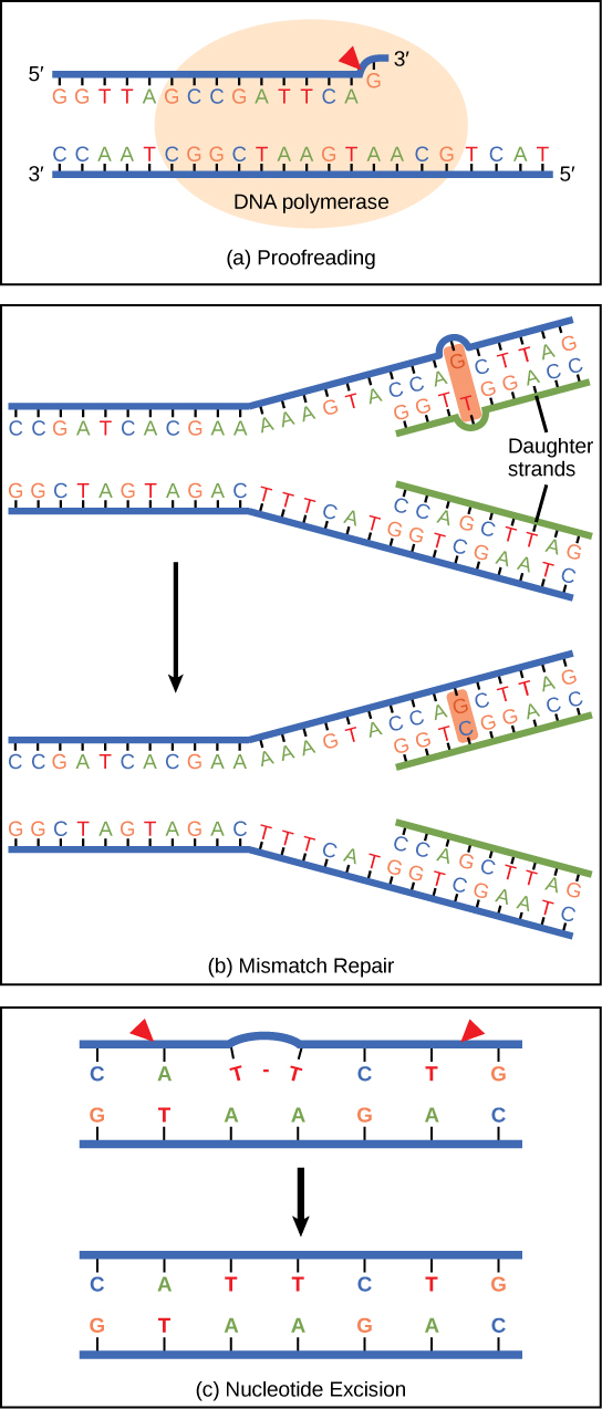
Most mistakes are corrected; if they are not, they may result in a mutation—defined as a permanent change in the DNA sequence. Mutations in repair genes may lead to serious consequences like cancer.
DNA replicates by a semi-conservative method in which each of the two parental DNA strands act as a template for new DNA to be synthesized. After replication, each DNA has one parental or “old” strand, and one daughter or “new” strand.
Replication in eukaryotes starts at multiple origins of replication, while replication in prokaryotes starts from a single origin of replication. The DNA is opened with enzymes, resulting in the formation of the replication fork. Primase synthesizes an RNA primer to initiate synthesis by DNA polymerase, which can add nucleotides in only one direction. One strand is synthesized continuously in the direction of the replication fork; this is called the leading strand. The other strand is synthesized in a direction away from the replication fork, in short stretches of DNA known as Okazaki fragments. This strand is known as the lagging strand. Once replication is completed, the RNA primers are replaced by DNA nucleotides and the DNA is sealed with DNA ligase.
The ends of eukaryotic chromosomes pose a problem, as polymerase is unable to extend them without a primer. Telomerase, an enzyme with an inbuilt RNA template, extends the ends by copying the RNA template and extending one end of the chromosome. DNA polymerase can then extend the DNA using the primer. In this way, the ends of the chromosomes are protected. Cells have mechanisms for repairing DNA when it becomes damaged or errors are made in replication. These mechanisms include mismatch repair to replace nucleotides that are paired with a non-complementary base and nucleotide excision repair, which removes bases that are damaged such as thymine dimers.
Art Connections
Figure \(\PageIndex{3}\): You isolate a cell strain in which the joining together of Okazaki fragments is impaired and suspect that a mutation has occurred in an enzyme found at the replication fork. Which enzyme is most likely to be mutated?
Ligase, as this enzyme joins together Okazaki fragments.
- 1 Mariella Jaskelioff, et al., “Telomerase reactivation reverses tissue degeneration in aged telomerase-deficient mice,” Nature , 469 (2011):102–7.
Contributors and Attributions
Samantha Fowler (Clayton State University), Rebecca Roush (Sandhills Community College), James Wise (Hampton University). Original content by OpenStax (CC BY 4.0; Access for free at https://cnx.org/contents/b3c1e1d2-83...4-e119a8aafbdd ).

3.3 The Nucleus and DNA Replication
Learning objectives.
By the end of this section, you will be able to:
- Describe the structure and features of the nuclear membrane
- List the contents of the nucleus
- Explain the organization of the DNA molecule within the nucleus
- Describe the process of DNA replication
The nucleus is the largest and most prominent of a cell’s organelles ( Figure 3.3.1 ). The nucleus is generally considered the control center of the cell because it stores all of the genetic instructions for manufacturing proteins. Interestingly, some cells in the body, such as muscle cells, contain more than one nucleus ( Figure 3.3.2 ), which is known as multinucleated. Other cells, such as mammalian red blood cells (RBCs), do not contain nuclei at all. RBCs eject their nuclei as they mature, making space for the large numbers of hemoglobin molecules that carry oxygen throughout the body ( Figure 3.3.3 ). Without nuclei, the life span of RBCs is short, and so the body must produce new ones constantly.

External Website

View the University of Michigan WebScope at http://141.214.65.171/Histology/Basic%20Tissues/Muscle/058thin_HISTO_83X.svs/view.apml to explore the tissue sample in greater detail.

View the University of Michigan WebScope at http://virtualslides.med.umich.edu/Histology/EMsmallCharts/3%20Image%20Scope%20finals/139%20-%20Erythroblast_001.svs/view.apml to explore the tissue sample in greater detail.
Inside the nucleus lies the blueprint that dictates everything a cell will do and all of the products it will make. This information is stored within DNA. The nucleus sends “commands” to the cell via molecular messengers that translate the information from the DNA. Each cell in your body (with the exception of germ cells) contains the complete set of your DNA. When a cell divides, the DNA must be duplicated so that each new cell receives a full complement of DNA. The following section will explore the structure of the nucleus and its contents, as well as the process of DNA replication.
Organization of the Nucleus and its DNA
Like most other cellular organelles, the nucleus is surrounded by a membrane called the nuclear envelope . This membranous covering consists of two adjacent lipid bilayers with a thin fluid space in between them. Spanning these two bilayers are nuclear pores. A nuclear pore is a tiny passageway for the passage of proteins, RNA, and solutes between the nucleus and the cytoplasm. Proteins called pore complexes lining the nuclear pores regulate the passage of materials into and out of the nucleus.
Inside the nuclear envelope is a gel-like nucleoplasm with solutes that include the building blocks of nucleic acids. There also can be a dark-staining mass often visible under a simple light microscope, called a nucleolus (plural = nucleoli). The nucleolus is a region of the nucleus that is responsible for manufacturing the RNA necessary for construction of ribosomes. Once synthesized, newly made ribosomal subunits exit the cell’s nucleus through the nuclear pores.
The genetic instructions that are used to build and maintain an organism are arranged in an orderly manner in strands of DNA. Within the nucleus are threads of chromatin composed of DNA and associated proteins ( Figure 3.3.4 ). Along the chromatin threads, the DNA is wrapped around a set of histone proteins. A nucleosome is a single, wrapped DNA-histone complex. Multiple nucleosomes along the entire molecule of DNA appear like a beaded necklace, in which the string is the DNA and the beads are the associated histones. When a cell is in the process of division, the chromatin condenses into chromosomes, so that the DNA can be safely transported to the “daughter cells.” The chromosome is composed of DNA and proteins; it is the condensed form of chromatin. It is estimated that humans have almost 22,000 genes distributed on 46 chromosomes.

DNA Replication
In order for an organism to grow, develop, and maintain its health, cells must reproduce themselves by dividing to produce two new daughter cells, each with the full complement of DNA as found in the original cell. Billions of new cells are produced in an adult human every day. Only very few cell types in the body do not divide, including nerve cells, skeletal muscle fibers, and cardiac muscle cells. The division time of different cell types varies. Epithelial cells of the skin and gastrointestinal lining, for instance, divide very frequently to replace those that are constantly being rubbed off of the surface by friction.
A DNA molecule is made of two strands that “complement” each other in the sense that the molecules that compose the strands fit together and bind to each other, creating a double-stranded molecule that looks much like a long, twisted ladder. Each side rail of the DNA ladder is composed of alternating sugar and phosphate groups ( Figure 3.3.5 ). The two sides of the ladder are not identical, but are complementary. These two backbones are bonded to each other across pairs of protruding bases, each bonded pair forming one “rung,” or cross member. The four DNA bases are adenine (A), thymine (T), cytosine (C), and guanine (G). Because of their shape and charge, the two bases that compose a pair always bond together. Adenine always binds with thymine, and cytosine always binds with guanine. The particular sequence of bases along the DNA molecule determines the genetic code. Therefore, if the two complementary strands of DNA were pulled apart, you could infer the order of the bases in one strand from the bases in the other, complementary strand. For example, if one strand has a region with the sequence AGTGCCT, then the sequence of the complementary strand would be TCACGGA.

DNA replication is the copying of DNA that occurs before cell division can take place. After a great deal of debate and experimentation, the general method of DNA replication was deduced in 1958 by two scientists in California, Matthew Meselson and Franklin Stahl. This method is illustrated in Figure 3.3.6 and described below.

Stage 1: Initiation. The two complementary strands are separated, much like unzipping a zipper. Special enzymes, including helicase , untwist and separate the two strands of DNA.
Stage 2: Elongation. Each strand becomes a template along which a new complementary strand is built. DNA polymerase brings in the correct bases to complement the template strand, synthesizing a new strand base by base. A DNA polymerase is an enzyme that adds free nucleotides to the end of a chain of DNA, making a new double strand. This growing strand continues to be built until it has fully complemented the template strand.
Stage 3: Termination. Once the two original strands are bound to their own, finished, complementary strands, DNA replication is stopped and the two new identical DNA molecules are complete.
Each new DNA molecule contains one strand from the original molecule and one newly synthesized strand. The term for this mode of replication is “semiconservative,” because half of the original DNA molecule is conserved in each new DNA molecule. This process continues until the cell’s entire genome , the entire complement of an organism’s DNA, is replicated. As you might imagine, it is very important that DNA replication take place precisely so that new cells in the body contain the exact same genetic material as their parent cells. Mistakes made during DNA replication, such as the accidental addition of an inappropriate nucleotide, have the potential to render a gene dysfunctional or useless. Fortunately, there are mechanisms in place to minimize such mistakes. A DNA proofreading process enlists the help of special enzymes that scan the newly synthesized molecule for mistakes and corrects them. Once the process of DNA replication is complete, the cell is ready to divide. You will explore the process of cell division later in the chapter.

Watch this video to learn about DNA replication. DNA replication proceeds simultaneously at several sites on the same molecule. What separates the base pair at the start of DNA replication?
Chapter Review
The nucleus is the command center of the cell, containing the genetic instructions for all of the materials a cell will make (and thus all of its functions it can perform). The nucleus is encased within a membrane of two interconnected lipid bilayers, side-by-side. This nuclear envelope is studded with protein-lined pores that allow materials to be trafficked into and out of the nucleus. The nucleus contains one or more nucleoli, which serve as sites for ribosome synthesis. The nucleus houses the genetic material of the cell: DNA. DNA is normally found as a loosely contained structure called chromatin within the nucleus, where it is wound up and associated with a variety of histone proteins. When a cell is about to divide, the chromatin coils tightly and condenses to form chromosomes.
There is a pool of cells constantly dividing within your body. The result is billions of new cells being created each day. Before any cell is ready to divide, it must replicate its DNA so that each new daughter cell will receive an exact copy of the organism’s genome. A variety of enzymes are enlisted during DNA replication. These enzymes unwind the DNA molecule, separate the two strands, and assist with the building of complementary strands along each parent strand. The original DNA strands serve as templates from which the nucleotide sequence of the new strands are determined and synthesized. When replication is completed, two identical DNA molecules exist. Each one contains one original strand and one newly synthesized complementary strand.
Interactive Link Questions
Review questions, critical thinking questions.
Explain in your own words why DNA replication is said to be “semiconservative”?
DNA replication is said to be semiconservative because, after replication is complete, one of the two parent DNA strands makes up half of each new DNA molecule. The other half is a newly synthesized strand. Therefore, half (“semi”) of each daughter DNA molecule is from the parent molecule and half is a new molecule.
Why is it important that DNA replication take place before cell division? What would happen if cell division of a body cell took place without DNA replication, or when DNA replication was incomplete?
During cell division, one cell divides to produce two new cells. In order for all of the cells in your body to maintain a full genome, each cell must replicate its DNA before it divides so that a full genome can be allotted to each of its offspring cells. If DNA replication did not take place fully, or at all, the offspring cells would be missing some or all of the genome. This could be disastrous if a cell was missing genes necessary for its function and health.
This work, Anatomy & Physiology, is adapted from Anatomy & Physiology by OpenStax , licensed under CC BY . This edition, with revised content and artwork, is licensed under CC BY-SA except where otherwise noted.
Images, from Anatomy & Physiology by OpenStax , are licensed under CC BY except where otherwise noted.
Access the original for free at https://openstax.org/books/anatomy-and-physiology/pages/1-introduction .
Anatomy & Physiology Copyright © 2019 by Lindsay M. Biga, Staci Bronson, Sierra Dawson, Amy Harwell, Robin Hopkins, Joel Kaufmann, Mike LeMaster, Philip Matern, Katie Morrison-Graham, Kristen Oja, Devon Quick, Jon Runyeon, OSU OERU, and OpenStax is licensed under a Creative Commons Attribution-ShareAlike 4.0 International License , except where otherwise noted.
DNA Replication and Mitosis Critical Thinking questions

- Word Document File
Description
Critical thinking questions about DNA replication (6 questions) and about Mitosis (7 questions) to give students after learning about each process. They provide new scenarios and ask students to predict how the process would be affected.
Great for a higher level review day, or to differentiate for higher achieving students, or to use as test questions!
Questions & Answers
The biobasement.
- We're hiring
- Help & FAQ
- Privacy policy
- Student privacy
- Terms of service
- Tell us what you think
11.2 DNA Replication
Learning objectives.
By the end of this section, you will be able to:
- Explain the meaning of semiconservative DNA replication
- Explain why DNA replication is bidirectional and includes both a leading and lagging strand
- Explain why Okazaki fragments are formed
- Describe the process of DNA replication and the functions of the enzymes involved
- Identify the differences between DNA replication in bacteria and eukaryotes
- Explain the process of rolling circle replication
The elucidation of the structure of the double helix by James Watson and Francis Crick in 1953 provided a hint as to how DNA is copied during the process of replication . Separating the strands of the double helix would provide two templates for the synthesis of new complementary strands, but exactly how new DNA molecules were constructed was still unclear. In one model, semiconservative replication , the two strands of the double helix separate during DNA replication, and each strand serves as a template from which the new complementary strand is copied; after replication, each double-stranded DNA includes one parental or “old” strand and one “new” strand. There were two competing models also suggested: conservative and dispersive, which are shown in Figure 11.4 .
Matthew Meselson (1930–) and Franklin Stahl (1929–) devised an experiment in 1958 to test which of these models correctly represents DNA replication ( Figure 11.5 ). They grew E. coli for several generations in a medium containing a “heavy” isotope of nitrogen ( 15 N) that was incorporated into nitrogenous bases and, eventually, into the DNA. This labeled the parental DNA. The E. coli culture was then shifted into a medium containing 14 N and allowed to grow for one generation. The cells were harvested and the DNA was isolated. The DNA was separated by ultracentrifugation, during which the DNA formed bands according to its density. DNA grown in 15 N would be expected to form a band at a higher density position than that grown in 14 N. Meselson and Stahl noted that after one generation of growth in 14 N, the single band observed was intermediate in position in between DNA of cells grown exclusively in 15 N or 14 N. This suggested either a semiconservative or dispersive mode of replication. Some cells were allowed to grow for one more generation in 14 N and spun again. The DNA harvested from cells grown for two generations in 14 N formed two bands: one DNA band was at the intermediate position between 15 N and 14 N, and the other corresponded to the band of 14 N DNA. These results could only be explained if DNA replicates in a semiconservative manner. If DNA replication was dispersive, a single purple band positioned closer to the red 14 14 would have been observed, as more 14 was added in a dispersive manner to replace 15 . Therefore, the other two models were ruled out. As a result of this experiment, we now know that during DNA replication, each of the two strands that make up the double helix serves as a template from which new strands are copied. The new strand will be complementary to the parental or “old” strand. The resulting DNA molecules have the same sequence and are divided equally into the two daughter cells.
Check Your Understanding
- What would have been the conclusion of Meselson and Stahl’s experiment if, after the first generation, they had found two bands of DNA?
DNA Replication in Bacteria
DNA replication has been well studied in bacteria primarily because of the small size of the genome and the mutants that are available. E. coli has 4.6 million base pairs (Mbp) in a single circular chromosome and all of it is replicated in approximately 42 minutes, starting from a single origin of replication and proceeding around the circle bidirectionally (i.e., in both directions). This means that approximately 1000 nucleotides are added per second. The process is quite rapid and occurs with few errors.
DNA replication uses a large number of proteins and enzymes ( Table 11.1 ). One of the key players is the enzyme DNA polymerase , also known as DNA pol. In bacteria, three main types of DNA polymerases are known: DNA pol I, DNA pol II, and DNA pol III. It is now known that DNA pol III is the enzyme required for DNA synthesis; DNA pol I and DNA pol II are primarily required for repair. DNA pol III adds deoxyribonucleotides each complementary to a nucleotide on the template strand, one by one to the 3’-OH group of the growing DNA chain. The addition of these nucleotides requires energy. This energy is present in the bonds of three phosphate groups attached to each nucleotide (a triphosphate nucleotide), similar to how energy is stored in the phosphate bonds of adenosine triphosphate (ATP) ( Figure 11.6 ). When the bond between the phosphates is broken and diphosphate is released, the energy released allows for the formation of a covalent phosphodiester bond by dehydration synthesis between the incoming nucleotide and the free 3’-OH group on the growing DNA strand.
The initiation of replication occurs at specific nucleotide sequence called the origin of replication , where various proteins bind to begin the replication process. E. coli has a single origin of replication (as do most prokaryotes), called oriC , on its one chromosome. The origin of replication is approximately 245 base pairs long and is rich in adenine-thymine (AT) sequences.
Some of the proteins that bind to the origin of replication are important in making single-stranded regions of DNA accessible for replication. Chromosomal DNA is typically wrapped around histones (in eukaryotes and archaea) or histone-like proteins (in bacteria), and is supercoiled , or extensively wrapped and twisted on itself. This packaging makes the information in the DNA molecule inaccessible. However, enzymes called topoisomerases change the shape and supercoiling of the chromosome. For bacterial DNA replication to begin, the supercoiled chromosome is relaxed by topoisomerase II , also called DNA gyrase . An enzyme called helicase then separates the DNA strands by breaking the hydrogen bonds between the nitrogenous base pairs. Recall that AT sequences have fewer hydrogen bonds and, hence, have weaker interactions than guanine-cytosine (GC) sequences. These enzymes require ATP hydrolysis. As the DNA opens up, Y-shaped structures called replication forks are formed. Two replication forks are formed at the origin of replication, allowing for bidirectional replication and formation of a structure that looks like a bubble when viewed with a transmission electron microscope; as a result, this structure is called a replication bubble . The DNA near each replication fork is coated with single-stranded binding proteins to prevent the single-stranded DNA from rewinding into a double helix.
Once single-stranded DNA is accessible at the origin of replication, DNA replication can begin. However, DNA pol III is able to add nucleotides only in the 5’ to 3’ direction (a new DNA strand can be only extended in this direction). This is because DNA polymerase requires a free 3’-OH group to which it can add nucleotides by forming a covalent phosphodiester bond between the 3’-OH end and the 5’ phosphate of the next nucleotide. This also means that it cannot add nucleotides if a free 3’-OH group is not available, which is the case for a single strand of DNA. The problem is solved with the help of an RNA sequence that provides the free 3’-OH end. Because this sequence allows the start of DNA synthesis, it is appropriately called the primer . The primer is five to 10 nucleotides long and complementary to the parental or template DNA. It is synthesized by RNA primase , which is an RNA polymerase . Unlike DNA polymerases, RNA polymerases do not need a free 3’-OH group to synthesize an RNA molecule. Now that the primer provides the free 3’-OH group, DNA polymerase III can now extend this RNA primer, adding DNA nucleotides one by one that are complementary to the template strand ( Figure 11.4 ).
During elongation in DNA replication , the addition of nucleotides occurs at its maximal rate of about 1000 nucleotides per second. DNA polymerase III can only extend in the 5’ to 3’ direction, which poses a problem at the replication fork. The DNA double helix is antiparallel; that is, one strand is oriented in the 5’ to 3’ direction and the other is oriented in the 3’ to 5’ direction (see Structure and Function of DNA ). During replication, one strand, which is complementary to the 3’ to 5’ parental DNA strand, is synthesized continuously toward the replication fork because polymerase can add nucleotides in this direction. This continuously synthesized strand is known as the leading strand . The other strand, complementary to the 5’ to 3’ parental DNA, grows away from the replication fork, so the polymerase must move back toward the replication fork to begin adding bases to a new primer, again in the direction away from the replication fork. It does so until it bumps into the previously synthesized strand and then it moves back again ( Figure 11.7 ). These steps produce small DNA sequence fragments known as Okazaki fragments , each separated by RNA primer. Okazaki fragments are named after the Japanese research team and married couple Reiji and Tsuneko Okazaki , who first discovered them in 1966. The strand with the Okazaki fragments is known as the lagging strand , and its synthesis is said to be discontinuous.
The leading strand can be extended from one primer alone, whereas the lagging strand needs a new primer for each of the short Okazaki fragments. The overall direction of the lagging strand will be 3’ to 5’, and that of the leading strand 5’ to 3’. A protein called the sliding clamp holds the DNA polymerase in place as it continues to add nucleotides. The sliding clamp is a ring-shaped protein that binds to the DNA and holds the polymerase in place. Beyond its role in initiation, topoisomerase also prevents the overwinding of the DNA double helix ahead of the replication fork as the DNA is opening up; it does so by causing temporary nicks in the DNA helix and then resealing it. As synthesis proceeds, the RNA primers are replaced by DNA. The primers are removed by the exonuclease activity of DNA polymerase I, and the gaps are filled in. The nicks that remain between the newly synthesized DNA (that replaced the RNA primer) and the previously synthesized DNA are sealed by the enzyme DNA ligase that catalyzes the formation of covalent phosphodiester linkage between the 3’-OH end of one DNA fragment and the 5’ phosphate end of the other fragment, stabilizing the sugar-phosphate backbone of the DNA molecule.
Termination
Once the complete chromosome has been replicated, termination of DNA replication must occur. Although much is known about initiation of replication, less is known about the termination process. Following replication, the resulting complete circular genomes of prokaryotes are concatenated, meaning that the circular DNA chromosomes are interlocked and must be separated from each other. This is accomplished through the activity of bacterial topoisomerase IV, which introduces double-stranded breaks into DNA molecules, allowing them to separate from each other; the enzyme then reseals the circular chromosomes. The resolution of concatemers is an issue unique to prokaryotic DNA replication because of their circular chromosomes. Because both bacterial DNA gyrase and topoisomerase IV are distinct from their eukaryotic counterparts, these enzymes serve as targets for a class of antimicrobial drugs called quinolones .
- Which enzyme breaks the hydrogen bonds holding the two strands of DNA together so that replication can occur?
- Is it the lagging strand or the leading strand that is synthesized in the direction toward the opening of the replication fork?
- Which enzyme is responsible for removing the RNA primers in newly replicated bacterial DNA?
DNA Replication in Eukaryotes
Eukaryotic genomes are much more complex and larger than prokaryotic genomes and are typically composed of multiple linear chromosomes ( Table 11.2 ). The human genome , for example, has 3 billion base pairs per haploid set of chromosomes, and 6 billion base pairs are inserted during replication. There are multiple origins of replication on each eukaryotic chromosome ( Figure 11.8 ); the human genome has 30,000 to 50,000 origins of replication. The rate of replication is approximately 100 nucleotides per second—10 times slower than prokaryotic replication.
The essential steps of replication in eukaryotes are the same as in prokaryotes. Before replication can start, the DNA has to be made available as a template. Eukaryotic DNA is highly supercoiled and packaged, which is facilitated by many proteins, including histone s (see Structure and Function of Cellular Genomes ). At the origin of replication , a prereplication complex composed of several proteins, including helicase , forms and recruits other enzymes involved in the initiation of replication, including topoisomerase to relax supercoiling, single-stranded binding protein, RNA primase , and DNA polymerase . Following initiation of replication, in a process similar to that found in prokaryotes, elongation is facilitated by eukaryotic DNA polymerases. The leading strand is continuously synthesized by the eukaryotic polymerase enzyme pol δ, while the lagging strand is synthesized by pol ε. A sliding clamp protein holds the DNA polymerase in place so that it does not fall off the DNA. The enzyme ribonuclease H ( RNase H ), instead of a DNA polymerase as in bacteria, removes the RNA primer, which is then replaced with DNA nucleotides. The gaps that remain are sealed by DNA ligase .
Because eukaryotic chromosomes are linear, one might expect that their replication would be more straightforward. As in prokaryotes, the eukaryotic DNA polymerase can add nucleotides only in the 5’ to 3’ direction. In the leading strand, synthesis continues until it reaches either the end of the chromosome or another replication fork progressing in the opposite direction. On the lagging strand, DNA is synthesized in short stretches, each of which is initiated by a separate primer. When the replication fork reaches the end of the linear chromosome, there is no place to make a primer for the DNA fragment to be copied at the end of the chromosome. These ends thus remain unpaired and, over time, they may get progressively shorter as cells continue to divide.
The ends of the linear chromosomes are known as telomere s and consist of noncoding repetitive sequences. The telomeres protect coding sequences from being lost as cells continue to divide. In humans, a six base-pair sequence, TTAGGG, is repeated 100 to 1000 times to form the telomere. The discovery of the enzyme telomerase ( Figure 11.9 ) clarified our understanding of how chromosome ends are maintained. Telomerase contains a catalytic part and a built-in RNA template. It attaches to the end of the chromosome, and complementary bases to the RNA template are added on the 3’ end of the DNA strand. Once the 3’ end of the lagging strand template is sufficiently elongated, DNA polymerase can add the nucleotides complementary to the ends of the chromosomes. In this way, the ends of the chromosomes are replicated. In humans, telomerase is typically active in germ cells and adult stem cells; it is not active in adult somatic cells and may be associated with the aging of these cells. Eukaryotic microbes including fungi and protozoans also produce telomerase to maintain chromosomal integrity. For her discovery of telomerase and its action, Elizabeth Blackburn (1948–) received the Nobel Prize for Medicine or Physiology in 2009.
Link to Learning
This animation compares the process of prokaryotic and eukaryotic DNA replication.
- How does the origin of replication differ between eukaryotes and prokaryotes?
- What polymerase enzymes are responsible for DNA synthesis during eukaryotic replication?
- What is found at the ends of the chromosomes in eukaryotes and why?
DNA Replication of Extrachromosomal Elements: Plasmids and Viruses
To copy their nucleic acids, plasmids and viruses frequently use variations on the pattern of DNA replication described for prokaryote genomes. For more information on the wide range of viral replication strategies, see The Viral Life Cycle .
Rolling Circle Replication
Whereas many bacterial plasmids (see Unique Characteristics of Prokaryotic Cells ) replicate by a process similar to that used to copy the bacterial chromosome, other plasmids, several bacteriophages , and some viruses of eukaryotes use rolling circle replication ( Figure 11.10 ). The circular nature of plasmids and the circularization of some viral genomes on infection make this possible. Rolling circle replication begins with the enzymatic nicking of one strand of the double-stranded circular molecule at the double-stranded origin (dso) site . In bacteria, DNA polymerase III binds to the 3’-OH group of the nicked strand and begins to unidirectionally replicate the DNA using the un-nicked strand as a template, displacing the nicked strand as it does so. Completion of DNA replication at the site of the original nick results in full displacement of the nicked strand, which may then recircularize into a single-stranded DNA molecule. RNA primase then synthesizes a primer to initiate DNA replication at the single-stranded origin (sso) site of the single-stranded DNA (ssDNA) molecule, resulting in a double-stranded DNA (dsDNA) molecule identical to the other circular DNA molecule.
- Is there a lagging strand in rolling circle replication? Why or why not?
As an Amazon Associate we earn from qualifying purchases.
This book may not be used in the training of large language models or otherwise be ingested into large language models or generative AI offerings without OpenStax's permission.
Want to cite, share, or modify this book? This book uses the Creative Commons Attribution License and you must attribute OpenStax.
Access for free at https://openstax.org/books/microbiology/pages/1-introduction
- Authors: Nina Parker, Mark Schneegurt, Anh-Hue Thi Tu, Philip Lister, Brian M. Forster
- Publisher/website: OpenStax
- Book title: Microbiology
- Publication date: Nov 1, 2016
- Location: Houston, Texas
- Book URL: https://openstax.org/books/microbiology/pages/1-introduction
- Section URL: https://openstax.org/books/microbiology/pages/11-2-dna-replication
© Jan 10, 2024 OpenStax. Textbook content produced by OpenStax is licensed under a Creative Commons Attribution License . The OpenStax name, OpenStax logo, OpenStax book covers, OpenStax CNX name, and OpenStax CNX logo are not subject to the Creative Commons license and may not be reproduced without the prior and express written consent of Rice University.

COMMENTS
24. Provide a brief summary of the Sanger sequencing method. 25. Describe the structure and complementary base pairing of DNA. 26. Prokaryotes have a single circular chromosome while eukaryotes have linear chromosomes. Describe one advantage and one disadvantage to the eukaryotic genome packaging compared to the prokaryotes.
Critical Thinking Questions; Test Prep for AP® Courses; Science Practice Challenge Questions; 22 Prokaryotes: Bacteria and Archaea. Introduction; ... The image shows electron microscope images of the DNA replication process. The strings in the image are DNA strands. The blob-like shapes are enzymes. The two DNA strands marked with arrows are ...
Critical Thinking Questions. 28. Compare and contrast a human somatic cell to a human gamete. 29. What is the relationship between a genome, chromosomes, and genes? 30. Eukaryotic chromosomes are thousands of times longer than a typical cell. Explain how chromosomes can fit inside a eukaryotic nucleus. 31.
14.3 Basics of DNA Replication; 14.4 DNA Replication in Prokaryotes; 14.5 DNA Replication in Eukaryotes; 14.6 DNA Repair; Key Terms; Chapter Summary; Review Questions; ... Critical Thinking Questions; Test Prep for AP® Courses; Science Practice Challenge Questions; 22 Prokaryotes: Bacteria and Archaea. Introduction;
Critical Thinking Questions; Evolution and the Diversity of Life. 11 Evolution and Its Processes. Introduction; 11.1 Discovering How Populations Change; ... DNA replication has been extremely well-studied in prokaryotes, primarily because of the small size of the genome and large number of variants available.
DNA Replication in Prokaryotes. 135. DNA Replication in Eukaryotes. 136. DNA Repair. 137. Key Terms. 138. Chapter Summary. 139. Visual Connection Questions. 140. ... 141 Critical Thinking Questions 21. If a purine were substituted for a pyrimidine at a single position in one strand of a DNA double helix, what would happen?
Answers for Critical Thinking Questions. DNA replication is said to be semiconservative because, after replication is complete, one of the two parent DNA strands makes up half of each new DNA molecule. The other half is a newly synthesized strand. Therefore, half ("semi") of each daughter DNA molecule is from the parent molecule and half is ...
DNA replication is semiconservative, meaning that each strand in the DNA double helix acts as a template for the synthesis of a new, complementary strand. This process takes us from one starting molecule to two "daughter" molecules, with each newly formed double helix containing one new and one old strand.
A list of student-submitted discussion questions for DNA Structure and Replication. Click Create Assignment to assign this modality to your LMS. We have a new and improved read on this topic.
which replication generates a double helix in which both strands are newly synthesized. Ergo, DNA replication is semi-conservative. DNA is synthesized by the repetitive addition of nucleotides to the 3' end of the growing polynucleotide chain DNA is synthesized by an iterative process in which nucleotides are added
Critical Thinking Questions; 22 Prokaryotes: Bacteria and Archaea. Introduction; 22.1 Prokaryotic Diversity; 22.2 Structure of Prokaryotes; ... During DNA replication, each of the two strands that make up the double helix serves as a template from which new strands are copied. The new strand will be complementary to the parental or "old ...
Basics of DNA Replication. 134. DNA Replication in Prokaryotes. 135. DNA Replication in Eukaryotes. 136. DNA Repair. 137. Key Terms. 138. Chapter Summary. 139. Visual Connection Questions. 140. Review Questions. 141. ... 152 Critical Thinking Questions 19. If mRNA is complementary to the DNA template strand and the DNA template strand is ...
DNA replication is bidirectional and discontinuous; explain your understanding of those concepts. DNA polymerase reads the template strand in the 3' to 5' direction and adds nucleotides only in the 5' to 3' direction. The leading strand is synthesized in the direction of the replication fork.
9.2: DNA Replication. When a cell divides, it is important that each daughter cell receives an identical copy of the DNA. This is accomplished by the process of DNA replication. The replication of DNA occurs during the synthesis phase, or S phase, of the cell cycle, before the cell enters mitosis or meiosis. The elucidation of the structure of ...
Critical Thinking Questions. Explain in your own words why DNA replication is said to be "semiconservative"? DNA replication is said to be semiconservative because, after replication is complete, one of the two parent DNA strands makes up half of each new DNA molecule. The other half is a newly synthesized strand.
DNA Critical Thinking Questions. Frederick Griffith. Click the card to flip 👆. Injected 2 strains of bacteria into mice and found bacteria can change their function/form. Click the card to flip 👆.
Our mission is to improve educational access and learning for everyone. OpenStax is part of Rice University, which is a 501 (c) (3) nonprofit. Give today and help us reach more students. This free textbook is an OpenStax resource written to increase student access to high-quality, peer-reviewed learning materials.
Critical thinking questions about DNA replication (6 questions) and about Mitosis (7 questions) to give students after learning about each process. They provide new scenarios and ask students to predict how the process would be affected. Great for a higher level review day, or to differentiate for higher achieving students, or to use as test ...
Question Set A (for ages 14 and older) What is a "strand" of DNA? How many strands make up a DNA double helix? Each strand is made up of two zones or regions. One zone of each strand is made up of identical repeating units, while another zone is made up of differing units. What are these zones of each strand called?
Matthew Meselson (1930-) and Franklin Stahl (1929-) devised an experiment in 1958 to test which of these models correctly represents DNA replication (Figure 11.5).They grew E. coli for several generations in a medium containing a "heavy" isotope of nitrogen (15 N) that was incorporated into nitrogenous bases and, eventually, into the DNA. This labeled the parental DNA.