
Weird, Unsocialized Homeschoolers
Honest. Quirky. Real. Stereotype-smashing humor for homeschooling families.

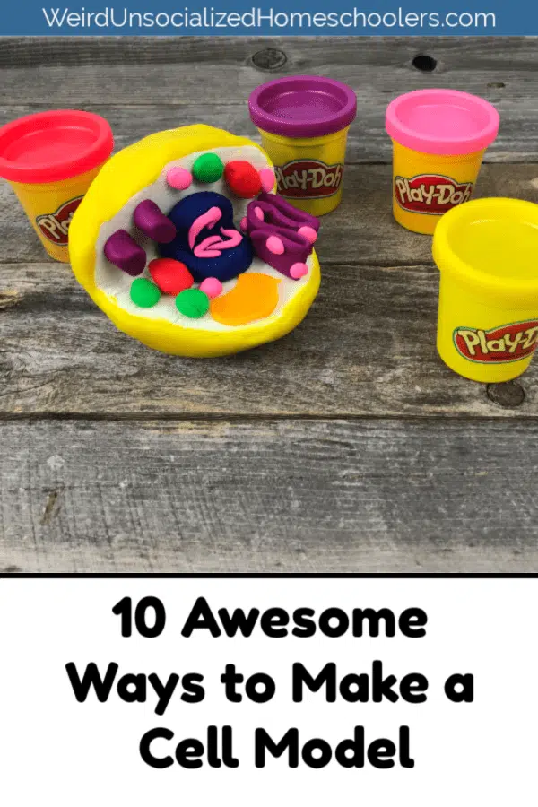
10+ Awesome Ways to Make a Cell Model

Did you like this article? If so, please help by sharing it!
- Facebook 802
- Pinterest 16.9K

Is your student studying biology? Choose any one of these awesome ways to make a cell model to create a colorful, hands-on project to help them learn all those sometimes hard-to-remember cell parts!
It doesn’t matter where they attend school – home, public, or private – at some point nearly every teen is going to take biology. When they do, the first thing they’ll likely study is the cell. And, chances are their instructor is going to assign them a cell model project.
Do you feel like you just read some weird, homeschooled teenager version of If You Give a Mouse a Cookie ? You’re welcome.
If you’re a homeschooling parent and your teen’s science curriculum doesn’t include building a cell model as one of their projects, do it anyway because it’s lots of fun!
There are lots of different ways to ways to make a cell model, whether you choose a plant or an animal cell. But, the following are some of our favorites. Some are edible, some are not, but they’re all super cool.
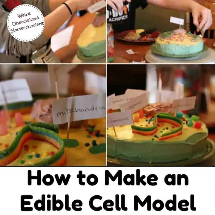
1. Cell Cake
Starting with my family’s all-time favorite, this cell cake uses a cake as its base with an icing cell wall, a cupcake nucleus, and a variety of candy organelles. We probably should have branched out a bit, but we made this model for a couple of kids’ biology lessons. Not only was it colorful and fun, but it made studying cells much more interesting because it was delicious!
Plus, it’s just funny to me imagining my kids at some future point in their lives thinking, “Mitochondria – was that the Mike and Ikes or the gummy worms??”
2. Jell-O Cell
Many, many eons ago, when my oldest was around twelve, we made our first Jell-O cell model . It was not pretty, y’all. And it was definitely not appetizing. As well as I can recall, we followed the directions and used what we had on hand, but I don’t care how great a slice of boiled egg represents a cell nucleus, eggs do not belong in Jell-O.
Even if I could find the obscure blog post featuring the tiny, grainy photo of that cell model, I wouldn’t subject y’all to that. This model from 123 Homeschool 4 Me is much prettier and more appetizing than the one we made.
My best tip if you make a cell model using Jell-O? Choose foods like fruits, marshmallows, gummies, and nuts to represent the organelles so that you’ll actually want to eat it when you’re finished. A banana slice makes a much better nucleus than an egg slice for a Jell-O cell.

photo credit 123 homeschool 4 me
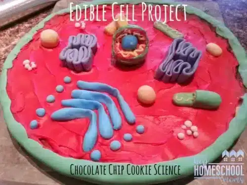
3. Cookie Cell
Another tasty edible option is a cookie cell model . This one at Hip Homeschool Moms starts with a large chocolate chip cookie as the base. Bake the cookie in a pizza pan for an animal cell model or in a jellyroll pan for a plant cell. Then use icing, fondant, and candies to represent the organelles. Yum! This multi-use project lets you study science, brush up on some home ec skills , and provide dessert for the family after dinner all in one tasty project.
4. Pizza Cell
For edible models, you can’t go wrong with pizza, either. You can make a regular pizza cell model or a fruit pizza cell model . Or, do both! Make an animal cell pizza and a plant cell fruit pizza like the one pictured. You can study both cell types, compare and contrast them, and then eat your projects for lunch and dessert!

photo credit proverbial homemaker
5. 3D Cell Model
This 3D animal cell made from Styrofoam is fantastic! If edible projects aren’t your thing or you just want something more permanent, this is perfect.
Unfortunately, the Crafts N Coffee site is no longer active, but I thought you could still use their photo as a guide to make your own Styrofoam cell model! It features a foam craft ball with a wedge cut out as the base with the orange paint representing the cell wall. Use the leftover foam wedge to form the nucleus. Then, use a variety of self-stick foam dots and shapes or cut your own from foam craft sheets to represent the organelles.

photo credit crafts n coffee
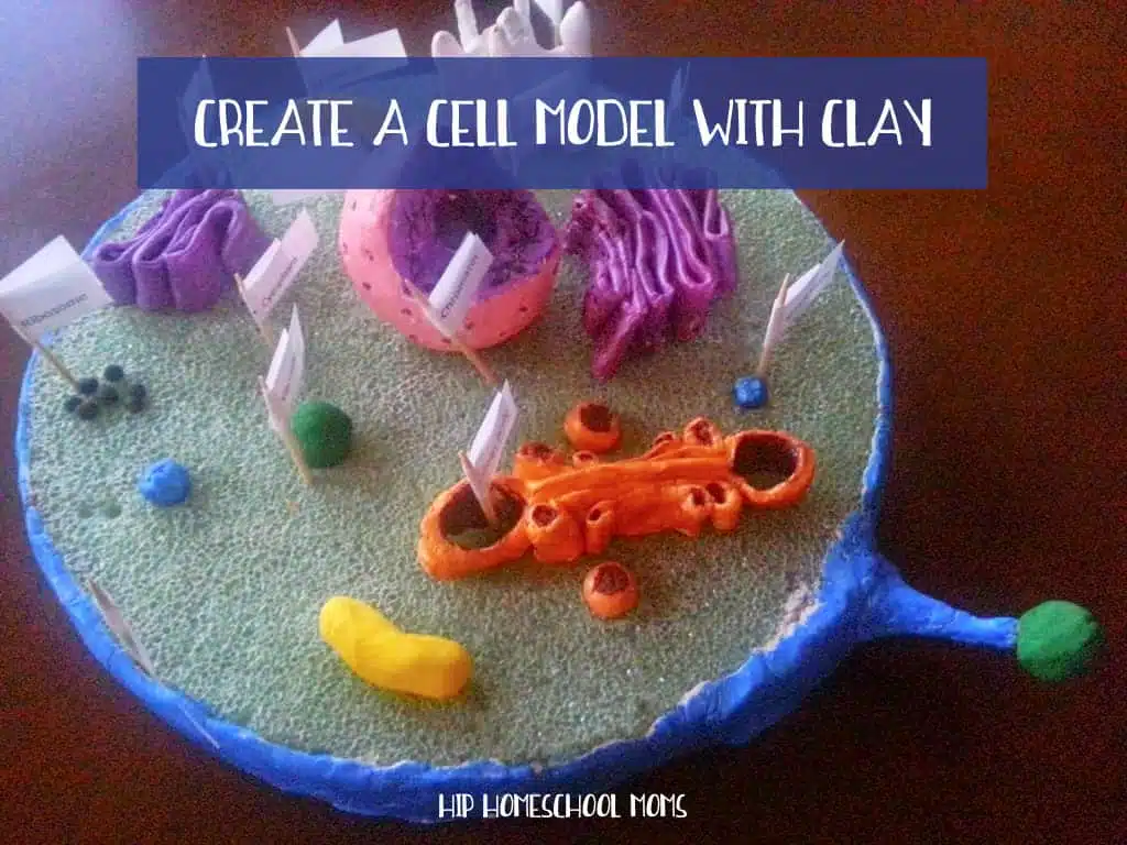
6. Clay Cell
If you like the idea of creating a foam model but want something a little more sophisticated than foam-shape organelles, check out this article from Hip Homeschool Moms on how to make a cell model from styrofoam and clay . It’s fantastic! I love the idea of forming the organelles from modeling clay ! What a great way to review the shape of each and its job!
7. Sandwich Cell
If you’re short on time or have younger siblings who want to get in on the cell model making fun, try this simple edible sandwich cell idea. If you want to get more detailed, try using a large round biscuit or cookie cutter to create an animal cell model, or leave the bread square for a simple plant cell option. Then choose your favorite toppings for the organelles.
This example uses a caramel candy for the nucleus, which is one of my favorites, but you could also use a banana slice with other fruits like apples and raisins for other organelles. I do suggest sticking with the butterscotch and chocolate chip organelles, though, because yum!

photo credit adventures in mommydom
8. Shrinky Dinks Cell
Because my oldest used to love Shrinky Dinks, this Shrinky Dink cell model idea is one of my favorites! I wish I’d found it when my kids were younger. The article includes a tutorial with printable templates that your students can color in and label for both a plant cell and an animal cell. And Amazon has Shrinky Dink paper if you can’t find it locally.

photo credit Teacher Thrive
9. Lego Cell
If you’ve got a Lego lover, this Lego cell model is the perfect fit! My son definitely would have loved this one! No, you won’t get the right cell shape for an animal cell using Lego blocks, but it might be better to let that detail slide if the result is a learning activity that engages your Lego-loving kid.
Let your Lego fans design their own organelles and create a chart or labels to indicate what each represents. I love the way the kid who made the example pictured even created his own label markers out of Lego parts.

photo credit homegrown learners
10. Rice Krispies Treat Cell
For another tasty edible cell model idea, try this Rice Krispies treat cell . The original article is no longer available, but the photo provides plenty of inspiration. I’m pretty sure that’s a Fruit Roll-up cell wall and I’m here to testify that Fruit Roll-ups make a delicious addition to Rice Krispies treats! When my kids were little, we paired the two for some sweet “sushi rolls” when we studied Japan. Delicious! (Though, you may be dealing with a bit of a sugar rush craziness after your kids consume this one.)

11. Spielgaban Cell Model
Have you seen the Speilgaban educational sets? They’re amazing, open-ended, hands-on learning tools featuring all kinds of kid-friendly manipulative parts. If you’ve got one, you’re going to love this idea for creating a 3D Model using the Spielgaban educational set.
And if you don’t have the set, I bet you can still use the inspiration to use similar items like blocks and craft supplies that you have at home to create your own cell model.
This is another opportunity to get younger students (or interested siblings) involved in the creative learning process.

Bonus: Cell Printable Packs!
For more hands-on learning fun and activity ideas, add this Animal Cell Printable Pack or Plant Cell Printable Pack to your cell study. Or check out the Animal Cell Interactive Notebook .

Have you made a cell model that isn’t represented here? Leave your idea in the comments!
Pin this post so you can find it when you get ready to make cell models!
You Might Also Like:
- 100 Hands-On Activities for Middle and High School
- 10 Hands-On History and Geography Activities
- 13 Hands-On Activities to Enhance Any Study

Kris Bales is a newly-retired homeschool mom and the quirky, Christ-following, painfully honest founder (and former owner) of Weird, Unsocialized Homeschoolers. She has a pretty serious addiction to sweet tea and Words with Friends. Kris and her husband of over 30 years are parents to three amazing homeschool grads. They share their home with three dogs, two cats, a ball python, a bearded dragon, and seven birds.
- Kris Bales https://www.weirdunsocializedhomeschoolers.com/author/kris_wuhsmom/ 8 Tips for Helping Kids Explore Their Interests (When You Don’t Have a Clue)
- Kris Bales https://www.weirdunsocializedhomeschoolers.com/author/kris_wuhsmom/ 10 Tips to Help Easily Distractible Teens Focus
- Kris Bales https://www.weirdunsocializedhomeschoolers.com/author/kris_wuhsmom/ 4 Practical Ways to Be Your Teen's Homeschool Guidance Counselor
- Kris Bales https://www.weirdunsocializedhomeschoolers.com/author/kris_wuhsmom/ 16+ Ways a Homeschool Homestead Provides the Ultimate Education
These all look so awesome! I hope to try a few of them while we’re on break.
Love these! We did a pizza and jello/candy– why didn’t I ever think of Legos??
These are totally awesome! I want to make the fruit pizza with my kids!
These look fantastic! Can’t wait to try them all! 😊
Doing a school project! Thanks for all of the great ideas!!!!!!!!!!!!
I am all about hands-on learning! Thank you for these great ideas that will inspire my students to achieve greatness! This follows the BJU curriculum and the assignment gives the assessment I need for those tacticle learners. Thanks for sharing!
Using play dough for 3 d is plant cell. Will the play dough get hard or change colors What can i.use on the play dough to keep the colors bright
theses are so cute i love them i am going to definety do one for my school project
my teacher is using these for our cell project and I saw the cake and was like yes thanks for the inspiration to my teacher
Leave a Reply Cancel reply
Your email address will not be published. Required fields are marked *
Notify me via e-mail if anyone answers my comment.
This site uses Akismet to reduce spam. Learn how your comment data is processed .

Build a Model of a Human Cell

Introduction: Build a Model of a Human Cell

In this project we will be making a model of a Animal (Human) cell and talking about the various structures within an animal cell. I made my cell out of just cardboard and paper, but you can use anything you have around the house to make your cell. Things like bits of fabric, buttons, and pipe cleaners could all be used.
A cardboard circle. I took a cardboard box and traced a plate on it to get my circle shape.
Colored Pencils
Glue (I used a glue stick, but forgot to include it in my picture.)
Step 1: Create Your Nucleus.

The first thing you’ll want to draw is a circle to be the Nucleolus for your cell. Then you’ll draw a bigger circle around that. This bigger circle is the Nucleus. The Nucleus of a cell has 2 main functions, it contains all the DNA of the cell and functions as a “brain” for the cell. The Nucleolus is the smaller circle within the nucleus, and it’s where Ribosomes are made. Ribosomes will be discussed in a later step.
Step 2: Create Your Endoplasmic Reticulum

The Endoplasmic Reticulum is connected to the nucleus by the Nuclear Membrane. Because of this you’ll want to start by drawing a circle to fit around your nucleus, before drawing the rest of the cisternae. Endoplasmic reticulum may be either rough or smooth, and both serve different functions. Rough endoplasmic reticulum has Ribosomes bound to it and helps modify and transport the proteins made by those ribosomes. Smooth endoplasmic reticulum modifies lipids.
Step 3: Create the Golgi Apparatus

The Golgi Apparatus is made of cisternae much like the Endoplasmic Reticulum, but the cisternae are not interconnected. The golgi apparatus’ job is to take in molecules produced by the cell, make any modifications, and then package them into Vesicles. These vesicles can then ship the proteins either within the cell itself, or can send the proteins outside the cell.
Step 4: Create Your Ribosomes.

Ribosomes are small organelles made of RNA. The way I made mine was to first make a square, before cutting the square in smaller and smaller bits of paper. I then glued these bits where they were needed. Make sure you glue some onto your endoplasmic reticulum, and to also glue them around the rest of your cell. Ribosomes help with protein synthesis (making proteins) within the cell.
Step 5: Create Your Mitochondria.

Mitochondria are larger organelles with an inner and outer membrane, and they also have their own DNA.
Mitochondria are where oxygen and glucose are taken into the cell, before being converted into energy.
Step 6: Create Your Cell Membrane

The Cell Membrane is the outer edge of the cell and forms the cell’s boundary. To create cell membrane I first traced my entire cell onto a sheet of paper. I then colored around the edge, before cutting the whole thing out. I then folded the paper in half and cut out another circle. You can then glue down the outer circle to form your membrane.
Step 7: Create Your Vacuole

Vacuoles are small pockets formed by a membrane layer. They contain the gas and fluids found in cells, such as oxygen or water.
Step 8: Futher Information and Learning
https://www.cellsalive.com/cells/cell_model.htm
https://www.khanacademy.org/science/biology/struct...

Recommendations

Text Contest

Paper and Cardboard Contest

Made with AI - Autodesk Design & Make - Student Contest

How Can You Make a Model of a Cell?
Purpose: The purpose of this project is to make a 3D model of a cell in order to better understand the parts and workings of a cell.

Designing a cell model using household objects can be a creative and engaging way for students to understand the structure and components of a cell. Here's a step-by-step guide they can follow:
1. Gather Materials:
Gather all the household objects and materials you'll need. These objects will represent the different organelles and structures within the cell.
2. Identify Cell Parts:
Review the different parts of a cell (organelles like nucleus, mitochondria, cytoplasm, cell membrane, etc.). Assign specific household objects to represent each part based on their shape or function.
- A small bead or marble could represent the nucleus.
- Pieces of pipe cleaners or wires can be shaped to represent the endoplasmic reticulum.
- Small candies or beads might be used for lysosomes.
- Play-doh or clay can also be used to represent structures
- Food items, like macaroni or beans can also represent structures
3. Prepare the Base:
The base can be cardboard (like a shoebox) or a styrofoam ball cut in half. You can be creative if you think other bases could work.
4. Labeling and Coloring:
Use paint, markers, or colored paper to paint or label the different parts of the cell. You could also attach a "key" that identifies each part.
Grading Rubric

Science By Sinai
Middle School Science Tips, Ideas, and Resources
Create a Cell Model with Recyclables
Do you want your students to create a creative cell model project, using recyclables, but you’re not sure where to start and how to structure it?

Karen Sinai

October 11, 2023
I put together a structured set of directions so that the students don’t get distracted and off task. I believe accountability and time management are the secret to any successful project.
Using Creativity to Select Organelle Materials
What I like about this project is that the students are using their creativity as they research the organelle’s shape and function. Using only recycled materials, students try to replicate the approximate shape of each plant or animal cell organelle. I don’t allow food because I don’t want ants and other critters crawling around my classroom as I put these on display.
I keep these projects in the back of the classroom for a while so we can refer to them throughout our life science unit.
The Cell Model Project Parameters
I learned the hard way to put size restraints on the cell projects. I usually say it shouldn’t be bigger than a large pizza or smaller than a dessert plate. You will need to determine whether the students are working on this in your classroom or at home. With my population, I have had success with them working on it at home because of the project check-ins. However, you could easily set up an area of your classroom with materials for them to work on it.
Students are required to represent each organelle and they should try their best to match the basic shape that they have learned through research. They will label the cell with typed labels or reference numbers for a typed key. They will also need to briefly describe what each organelle does for the cell.
After assembling the cell, students will present it to the class through a video or an in-person presentation.

The Project Specifics
Students are given a list of twelve organelles to include and two extra ones for plant cells, which are the chloroplasts and the cell wall. I tell students that they will get more credit if they tackle a plant cell.
We start with a brainstorming session, in school, where students will figure out what their base will be. I have found this to be super important because they may start building their cell and then they find that they can’t pick it up! Students have used cookie sheets, heavy cardboard, boxes etc. to support their projects.
One of the things I ask students at the beginning is how they will get the cell to school if they ride a bus. They need to think about how big their project will be and how stable. This is super important to talk about at the FRONT end of the project. I definitely learned THAT the hard way!
During the brainstorming session, they should also have ideas for what materials they will be using to represent the organelles. I’ve had students use pieces of old toys, batteries for the mitochondria, yarn, balloons, dental floss, etc. They have fun going through their house looking for materials to fit the shape of the organelles!
Cell Project Check-in Forms
Since I tend to have students do this project at home, I require project check-ins. The check-ins have students write one or two sentences about what they have done on the project so far. I ask them what they will do next and if they need any help. In my product I have included a rubric just for grading the check-ins. That makes a big difference in accountability, plus it’s extra grading data for you.
If you are doing this in your classroom, I recommend still having a project check-in that the students must fill out. They include what they have accomplished so far and what they plan to do next.
I am in the unique and wonderful situation that allows the students to bring their iPads home every night. I have them do PHOTO check-ins as well. Seeing a picture is worth 1000 words!
I tend to give the students at least a week to finish this project if it is at home. If I’m doing it in class, it is generally three 40 minute class periods. I let them know when the presentation date will be and the students will either create a video or do a standard classroom presentation. I have included a final project rubric to help grade it. I always show the students the final rubric somewhere near the beginning of the project so they know what they are working towards.
What I love about this project is that, year after year, the students are super creative and come up with completely different projects than I have seen before! Because they are working with the vocabulary, while creating the key with the organelle functions, student retention has increased.
You can find the project directions, check-ins and rubrics for this cell project at Teachers Pay Teachers in my store called Science by Sinai.

Similar Posts

How To Create STEM Electrical Circuit Games
How To Create STEM Electrical Circuit Games Are you looking for a STEM electricity project that solidifies student’s knowledge of series and parallel circuits? Have your students design and create electrical circuit arcade games that they can let younger students come in and play! It is amazing to watch students design and build projects from…

Science Activities to Do Outside with Middle School
Science Activities to Do Outside with Middle School Do you want to do science experiments, scavenger hunts and activities outside, with your middle school students, but you’re not sure about what lessons and structures to use? Even if your playground/schoolyard is limited, there are many activities that you can do outside as long as you…

Go Outdoors on an Exciting Schoolyard Ecosystem Scavenger Hunt!
Go Outdoors on an Exciting Schoolyard Ecosystem Scavenger Hunt! Are you looking for a way to get students observing their local ecosystem? Do you love taking classes outside? Here is a way to go on a fun scavenger hunt that involves students bringing their devices that have cameras outside! I put together this scavenger hunt…

Teaching Students About the Threat of Invasive Species
Teaching Students About the Threat of Invasive Species Do your students understand the threat of invasive species? Are you doing a unit on how biological components of an ecosystem affect populations(MS-LS2-4 or HS-LS2-7)? Students may be surprised to learn that some of the “local animals”, that they grew up with, are actually non native! I…

Build an Interactive Student Food Web!
Build an Interactive Student Food Web! Are you looking for a fun way to teach food webs that is guaranteed to be engaging? Grab some scissors, some yarn, and some plant and animal food chain pages, from your chosen ecosystem, and turn your class into a giant, interactive student food web! Prepare to Build Your…

Inspiring Middle School Students to Minimize Their Impact on Municipal Waste
Inspiring Middle School Students to Minimize Their Impact on Municipal Waste Do your students have any idea of their personal impact on municipal waste? Are you introducing a unit on human impact as part of the NGSS standard MS-ESS3? Let’s start the unit with relatable scenarios. Your mom asks you to clean your bedroom. You…
Click here to cancel reply.
Don't subscribe All new comments Replies to my comments Notify me of followup comments via e-mail. You can also subscribe without commenting.
How to make a model animal and plant cell
Part of Biology Living organisms
Cells make up all life on Earth.
Cells are the smallest unit of life.
All cells have specific structures, called components.
Models in science are used to help us understand complicated ideas in a simple and memorable way.
Slideshow - Model animal cell

A step-by-step guide to making a model animal cell.

YOU WILL NEED: A balloon and a grape.

Pop a grape into the deflated balloon.

Place the neck of the balloon over a tap and fill it with water.

Tie off the end of the balloon so it is sealed.

There you go! You've now made your very own model of an animal cell.
Animal cells are made up of four main components:
Nucleus close nucleus A cell component found in most cells which contains the genetic material (DNA) of the organism and controls the cell’s activities. : In the model, this is the grape.
Cell membrane close cell membrane This surrounds the outside of animal cells and controls what can enter and exit it. : In the model, this is the balloon.
Cytoplasm close cytoplasm The liquid that makes up most of the cell in which chemical reactions happen. This is mainly water. : In the model, this is the water.
Mitochondria close mitochondria Tiny parts of cells floating in the cytoplasm where energy is released from glucose. The glucose comes from food.
Video - Model plant cell
This video can not be played
To play this video you need to enable JavaScript in your browser.
Video transcript video transcript.
This is how you make a model plant cell.
There are a few things you'll need, a plastic box, a small sealable sandwich bag filled with water, cling film, some peas, frozen or otherwise, and some water with green food colouring if you've got any, and a trusty grape.
Line the plastic box with cling film. Pop the sealed plastic bag in. Spread the peas generously. Get the grape in there. And finally, top it up with the water. And then you'll have a model plant cell with all of its parts.
A cell wall, which keeps everything together, cell membrane, which surrounds the cytoplasm, vacuole, the space within the cytoplasm which contains the fluids and nutrients, and a nucleus, which contains the cell's genetic material, cytoplasm, where the cell's genetic reactions happen, and chloroplasts, these are where photosynthesis takes place.
So there you have it, the building blocks of plant life.
1. What items do you need to make this model?
2. What does the plastic box represent?
3. What do the peas and grape represent?
Show answer Hide answer
A plastic box, a small bag filled with water, clingfilm, peas, water with green food colouring, and a grape
The plastic box is the plant cell wall.
The peas represent chloroplasts and the grape represents the nucleus.
Plant cells have the same four components in animal cells but have these extra three as well:
Cell wall close cell wall The outside of the plant cell. It is made of cellulose. It is rigid and gives strength to the cell, allowing it to keep its shape. : In the model, this is the plastic box.
Vacuole close vacuole Filled with cell sap to store nutrients and help keep the cell turgid. : In the model, this is the sandwich bag of water.
Chloroplasts close chloroplast Structures found in plant cells which have a green pigment called chlorophyll in them. Photosynthesis occurs here. : In the model, these are the peas.
Test your knowledge
Quiz - multiple choice, quiz - label the parts of animal and plant cells, test questions.
Write a paragraph to answer the following questions. Tap 'Show answer' to see points you could have included.
1. Describe the components of animal cells and their functions.
2. Describe the components of plant cells and their functions.
- Your answer could include:
- Most animal cells possess a nucleus, cell membrane, cytoplasm and mitochondria.
- The nucleus contains the genetic material (DNA) of the organism and controls the cell’s activities.
- The cell membrane is a flexible outer layer that surrounds the cell and controls which substances can pass into and out from it.
- The cytoplasm is the liquid that makes up most of the cell in which chemical reactions happen. This is mainly water.
- Mitochondria are tiny parts of cells floating in the cytoplasm where energy is released from glucose from food.
Working safely in the lab
Find out how to spot risks, hazards and understand hazard symbols

More on Living organisms
Find out more by working through a topic
How to observe cells under a microscope
- count 15 of 15
Animal and plant cells
- count 1 of 15
Specialised animal cells
- count 2 of 15
Specialised plant cells
- count 3 of 15
- PRO Courses Guides New Tech Help Pro Expert Videos About wikiHow Pro Upgrade Sign In
- EDIT Edit this Article
- EXPLORE Tech Help Pro About Us Random Article Quizzes Request a New Article Community Dashboard This Or That Game Popular Categories Arts and Entertainment Artwork Books Movies Computers and Electronics Computers Phone Skills Technology Hacks Health Men's Health Mental Health Women's Health Relationships Dating Love Relationship Issues Hobbies and Crafts Crafts Drawing Games Education & Communication Communication Skills Personal Development Studying Personal Care and Style Fashion Hair Care Personal Hygiene Youth Personal Care School Stuff Dating All Categories Arts and Entertainment Finance and Business Home and Garden Relationship Quizzes Cars & Other Vehicles Food and Entertaining Personal Care and Style Sports and Fitness Computers and Electronics Health Pets and Animals Travel Education & Communication Hobbies and Crafts Philosophy and Religion Work World Family Life Holidays and Traditions Relationships Youth
- Browse Articles
- Learn Something New
- Quizzes Hot
- This Or That Game
- Train Your Brain
- Explore More
- Support wikiHow
- About wikiHow
- Log in / Sign up
- Education and Communications
How to Build 3D Models of Animal and Plant Cells
Last Updated: September 30, 2023 Fact Checked
This article was co-authored by Bess Ruff, MA . Bess Ruff is a Geography PhD student at Florida State University. She received her MA in Environmental Science and Management from the University of California, Santa Barbara in 2016. She has conducted survey work for marine spatial planning projects in the Caribbean and provided research support as a graduate fellow for the Sustainable Fisheries Group. This article has been fact-checked, ensuring the accuracy of any cited facts and confirming the authority of its sources. This article has been viewed 1,823,643 times.
Every student in a junior high or high school science class has had to learn about the structures of living cells at some time or another. Perhaps you have recently taken your turn, learning about the various organelles of plant and animal cells. If you have decided to show off your recently acquired knowledge by creating a 3D model of the cell and its structures (or have been assigned to do so by a teacher), this article can help guide you through the process.
Planning for Your Model

- You must know the different organelles if you are going to model them. Vitally, you must understand their shape. The colors usually given to the different cell components in textbooks are used for contrast and usually bear no resemblance to reality, so in that instance you can be creative. But you must develop the correct shapes in order to model them.
- It is also important to know how the various cell structures relate to one another. For example, the endoplasmic reticulum (ER) is always located close to the nucleus because it processes the proteins that are used in DNA replication. Consider where you will place your organelle as you are creating your model. [2] X Trustworthy Source Nature Respected Multidisciplinary Scientific Journal Go to source
- Know the differences between plant and animal cells. Most importantly, plant cells have an exterior cell wall made of cellulose, contain very large vacuoles (a membrane-bound collection of water and enzymes), and possess chloroplasts (the portions of the plant cell that convert sunlight into usable energy). [3] X Research source Centrosomes are only present in animal cells. Similarly, animal cells always have cilia, while plant cells often don't. Animal cells are typically round and irregular, while plant cells are rectangular and fixed in shape.

- The first option is a fully three-dimensional representation of a cell, with all of the organelles suspended in clear gelatin.
- The second option involves using craft materials to construct a cut-away model that shows a cell with a section removed to facilitate viewing.

- It is easiest to use items that already have the general shape of the object you are modeling--say, something roughly circular for a cell nucleus.
- Of course, many of the organelles are shaped so strangely that it may be impossible to find something that already has the same appearance. In this case you should think of materials that are flexible and can be fitted to whatever appearance you need.

Using Gelatin

- Clear gelatin will work as the cytoplasm, which is present in both animal and plant cells. If you are simply going for authenticity, non-flavored gelatin would work perfectly well. If you have decided to go edible, choose a variety that won't be so darkly colored as to obscure the model organelles you place inside.
- For the nucleus, nucleolus and nuclear membrane: Purchase a pitted fruit, such as a plum or peach. The pit is the nucleolus, the fruit is the nucleus, and the skin is the nuclear membrane. (If you are not expected to deliver this level of complexity, a simple round food item will do). You'll need this for either a plant or animal cell.
- Centrosomes, which are only present in animal cells, are supposed to be spiky, try putting bits of toothpick through a gumdrop or other small gummy item.
- Model the Golgi apparatus, which is present in both plant and animal cells, using cut-out pieces of cardboard, wafers, crackers, sliced bananas or, perhaps best yet, a fruit roll-up stacked like an accordion.
- For the Lysosomes in either type of cell, use small, round candies or chocolate chips.
- Mitochondrion, which are also present in both animal and plant cells, are somewhat oblong, so try using lima beans or perhaps certain types of un-shelled nuts.
- For ribosomes, you'll want something small. Try sprinkles, peppercorns, or plain pepper. These are present in both plant and animal cells.
- The rough endoplasmic reticulum, which is present in both animal and plant cells, looks much like the Golgi apparatus, in that it is a structure of flat, folded sections clumped together; though unlike the Golgi apparatus it has a rough-looking surface. You could use similar materials for it, but try to find a way to stick something rough or textured to it (perhaps sprinkles) in order to make the two distinct.
- The smooth endoplasmic reticulum looks more like a tangled and irregularly sized series of connected tubes. For this, you'll want something smooth and bendy. Use cooked spaghetti, gummy worms, or stretched-out taffy.
- When creating your vacuoles, you'll choose different shapes for animal versus plant cells. For an animal cell, use a few moderately sized gumballs--ideally uniform in color, but with some transparent quality (there are essentially just sacks of water and enzymes, after all). Vacuoles in plant cells are much, much larger. If you really want to get tricky here, you could make a separate gelatin (perhaps made with the concentrated formula for extra rigidity) earlier and attempt to insert it into the model plant cell.
- Microtubules can be modeled using uncooked pieces of spaghetti or, depending on the scale of your project, straws.
- For chloroplasts (plant cell only), use peas, green jelly beans, or green beans cut in half. Keep them green.

- If you're making a plant cell, the first thing you'll need is a rectangular baking dish, preferably made out of porcelain. The dish itself will be your cell wall and membrane, in your model.
- If you're making an animal cell, you'll want a round or oblong baking dish, like a casserole dish. This dish can be your cell membrane, or you can later remove the cell model from the dish and cover it in saran wrap cut to shape and size and call that the membrane.

- If you can't find clear gelatin, buy the lightest color possible, like yellow or orange. You can also make gelatin from scratch.

- Put the nucleus near the middle (unless you are modeling a plant cell).
- Place the centrosome near the nucleus. This is only present in animal cells.
- Put the smooth endoplasmic reticulum near the nucleus. This is present in both animal and plant cells.
- Place the Golgi body, which is present in both animal and plant cells, near the nucleus (though farther away than the endoplasmic reticulum).
- Add the rough endoplasmic reticulum onto the other side of the smooth endoplasmic reticulum (away from the nucleus). This is present in both plant and animal cells.
- Arrange everything else wherever you have room. Try not to crowd too much into one space. In a real cell, there are a few structures that float all around the cytoplasm. These can be mixed in almost randomly.

Using Craft Items

- You can use a styrofoam cell base. Craft or art stores and will have styrofoam balls (if making an animal cell) roughly the size of a basketball or a styrofoam rectangular cube (if making a plant cell).
- Cardstock can be used to form a number of cell structures, such as the Golgi apparatus or rough endoplasmic reticulum.
- Straws or small hoses can be used to form tube-like structures. The microtubules could be constructed out of stirring straws, while flexible straws or tubes can be used to model the smooth endoplasmic reticulum.
- Use beads of various sizes and shapes as other cell structures, such as mitochondria or chloroplasts. Try to keep them on an appropriate scale compared to the other structures in the model cell.
- Modeling clay can be used to create any structure that is difficult to replicate using preexisting materials.
- Paint can be used to fill in the cytoplasm and differentiate between it and the exterior of the cell. You can also paint any clay structures you have created.

- For the plant cell, do this by drawing the center line on any two adjoining sides and continue those lines all the way around until they circle back.
- If doing this for the animal cell, draw the lines like you were making the equator and the meridians on a globe.

- The trickiest of these will be the parts that you must model out of clay. Keep these structures as simple as possible while remaining true to the basic structure you are modeling. It may be best to only make the simplest of structures out of clay and leave more complex parts--say, the smooth endoplasmic reticulum--to be replicated using tubes or some other item.

- The Golgi apparatus and rough endoplasmic reticulum can be shaped out of cardstock using your hands. In this case, make slices into the styrofoam and slide pieces of cardstock in to form the folded shapes of these structures.

Community Q&A
- You'll be able to add parts more quickly if you have a friend or parent help. Thanks Helpful 2 Not Helpful 0
- Make sure the gelatin has enough time to solidify after you have added the "organelles." Try to keep it in the fridge overnight. Thanks Helpful 0 Not Helpful 2
- You might want to papier-mâché the styrofoam for safety reasons. Add extra layers for good measure. Thanks Helpful 0 Not Helpful 0
Tips from our Readers
- Double-check that all of the cell parts are in the right place before putting the gelatin in the fridge to solidify.
- If you're making an edible cell, make sure no one is allergic to the food you use as the cell parts.

You Might Also Like

- ↑ https://medlineplus.gov/genetics/understanding/basics/cell/
- ↑ http://www.nature.com/scitable/topicpage/endoplasmic-reticulum-golgi-apparatus-and-lysosomes-14053361
- ↑ https://www.khanacademy.org/science/high-school-biology/hs-cells/hs-plant-vs-animal-cells/a/hs-plant-vs-animal-cells-review
- ↑ https://www.education.com/science-fair/article/biology_building-blocks/
- ↑ https://sciencing.com/make-model-plant-cell-plastic-bag-12094635.html
- ↑ https://sciencing.com/make-3d-model-plant-cell-styrofoam-ball-12072121.html
About This Article

Before you build your 3D model of a cell, make sure to review cell structure and purchase the supplies you need. When you get home, measure and cut out 1/4 of the styrofoam ball so you can show the inside of the cell. Paint the styrofoam and build the simpler structures, like the nucleus, out of clay. Then, use tubes and cardstock to shape the Golgi apparatus and other more detailed parts. To learn how to make an edible 3D cell using gelatin, scroll down! Did this summary help you? Yes No
- Send fan mail to authors
Reader Success Stories
Lucia Madrigales
Oct 8, 2016
Did this article help you?
Jul 27, 2016
Dec 28, 2019
Maya Wicker
Nov 18, 2016

Featured Articles

Trending Articles

Watch Articles

- Terms of Use
- Privacy Policy
- Do Not Sell or Share My Info
- Not Selling Info
Get all the best how-tos!
Sign up for wikiHow's weekly email newsletter
Resources: Course Assignments
Module 4 assignment: cell builder.
Create a model of a eukaryotic cell using any material of your choice. In your model be sure to include all the organelles appropriate to your cell (either plant or animal). Once complete, take multiple photographs of your model from all angles. Include these images in a document that also contains the following in table format:
- A detailed key/legend that matches the model;
- Each organelle or part with its basic function;
- A disease or disorder that is associated with the malfunction of each cellular component
- How this organelle is visualized microscopically
Note for the disease information, you can list a disease in either animals or plants, regardless of what type of cell you are modeling. In other words, its okay to discuss a “human” disease even if you are making a plant model, provided the organelle is present in both types of cells.
Some suggestions for 3D models include Legos, a decorated cake with candy toppings, or standard Styrofoam base with appropriate pieces attached. You can also draw or illustrate a model. Here is an example of what you might make.
Basic Requirements (the assignment will not be accepted or assessed unless the follow criteria have been met):
- Assignment has been proofread and does not contain any major spelling or grammatical errors
- Assignment includes appropriate references
- Assignment includes photographs or images of created model from all angles.
- Assignment includes a key documenting how each organelle is represented in the model.
- Assignment includes a completed table such as the one illustrated in the example document.
- Assignment includes a disease caused by malfunction of each identified component in the model.
- Assignment includes at least 7 organelles in the model and table.
- Performance Assessments: Cell Builder. Authored by : Shelli Carter. Provided by : Columbia Basin College. Located at : https://www.columbiabasin.edu/ . License : CC BY: Attribution

- Free Resources
- Project Search
- Featured Projects
- Member Benefits
1059 Main Avenue, Clifton, NJ 07011
The most valuable resources for teachers and students
(973) 777 - 3113
1059 Main Avenue
Clifton, NJ 07011
07:30 - 19:00
Monday to Friday
123 456 789
Goldsmith Hall
New York, NY 90210
- Why We’re Unique
Make a Model of a Plant or Animal Cell
Introduction: (initial observation).
What are cells? Cells are structural units that make up plants and animals. There are also many single cell organisms such as bacteria and amebas. What all cells have in common is a membrane (small ‘sacks’) composed mostly of water. The ‘sacks’ are made from a phospholipid bilayer. The membrane is semi-permeable (allowing some things to pass in and out of the cell while blocking others).
Since cells are very small, we have to use microscopes in order to see their details. Another problem with cell observation is that many material forming a cell are clear. We often need to use colorants that can help us see cell membrane or other cell elements.

Cells may be studied for different purposes. The main purpose is usually to gain a good understanding of the cell’s biological structure as well as feeding, defense, growth and reproduction. Studying cells is a branch of life science known as cell biology.
Making a model of a cell is an educational activity that can help a student to learn about the cell structure and transfer such knowledge to others as well.
In this project you will make models of a plant cells or animal cells. Your display may be used to answer any of the following questions.
How are plant cells different from animal cells? What are the main parts of a plant cell?
I have a question about cells…I am doing a project on the white cell named Basophile and I need some ideas on how to do a model for school.. please give me some ideas on how to build it!
This project guide contains information that you need in order to start your project. If you have any questions or need more support about this project, click on the “Ask Question” button on the top of this page to send me a message.
If you are new in doing science project, click on “How to Start” in the main page. There you will find helpful links that describe different types of science projects, scientific method, variables, hypothesis, graph, abstract and all other general basics that you need to know.
Project advisor
Information Gathering:
Find out about cells, which are the smallest living organisms that may live by themselves or as a part of a larger organism. Read books, magazines or ask professionals who have knowledge in this area in order to learn about a variety of plant and animal cells and learn in what aspects they are different. Keep track of where you got your information.
Following are samples of information that you may find.
So what is in a cell? The cell is a fluid like membrane that surrounds the contents of the cell. Each component will be discussed in more detail later.
While studying about cells, we will frequently hear the terms Eukaryotic and Prokaryotic. Lets see what they mean (by looking at a dictionary or an encyclopedia).
Eukaryotic: A single-celled or multi-cellular organism whose cells contain a distinct membrane-bound nucleus.
Prokaryotic: An organism of the kingdom Monera (or Prokaryotae), comprising the bacteria and cyanobacteria, characterized by the absence of a distinct, membrane-bound nucleus or membrane-bound organelles, and by DNA that is not organized into chromosomes. Also called moneran.
Animal Cell Structure
Animal cells are typical of the eukaryotic cell, enclosed by a plasma membrane and containing a membrane-bound nucleus and organelles. Unlike the cells of the two other eukaryotic kingdoms, plants and fungi, animal cells don’t have a cell wall.

The lack of a rigid cell wall allows animals to develop a greater diversity of cell types, tissues, and organs. Specialized cells that formed nerves and muscles — tissues impossible for plants to evolve — gave these organisms mobility. The ability to move about by the use of specialized muscle tissues is the hallmark of the animal world. (Protozoans locomote, but by nonmuscular means, i.e. cilia, flagella, pseudopodia.)
The animal kingdom is unique amongst eukaryotic organisms because animal tissues are bound together by a triple helix of protein, called collagen. Plant and fungal cells are bound together in tissues or aggregations by other molecules, such as pectin. The fact that no other organisms utilize collagen in this manner is one of the indications that all animals arose from a common unicellular ancestor.
Animals are a large and incredibly diverse group of organisms. Making up about three-quarters of the species on Earth, they run the gamut from sponges and jellyfish to ants, whales, elephants, and — of course — human beings. Being mobile has given animals the flexibility to adopt many different modes of feeding, defense, and reproduction.
Animal cells include the following parts:

- Cilia and Flagella – For single-celled eukaryotes, cilia and flagella are essential for the locomotion of individual organisms. In multicellular organisms, cilia function to move fluid or materials past an immobile cell as well as moving a cell or group of cells.

- Lysosomes – The main function of these microbodies is digestion. Lysosomes break down cellular waste products and debris from outside the cell into simple compounds, which are transferred to the cytoplasm as new cell-building materials. Lysosomes are formed in golgi. You can see small spherical lysosomes around the golgi in the above image.
- Microfilaments – Microfilaments are solid rods made of globular proteins called actin. These filaments are primarily structural in function and are an important component of the cytoskeleton.
- Microtubules – These straight, hollow cylinders, composed of tubulin protein, are found throughout the cytoplasm of all eukaryotic cells and perform a number of functions.

- Nucleus – The nucleus is a highly specialized organelle that serves as the information and administrative center of the cell.
- Peroxisomes – Microbodies are a diverse group of organelles that are found in the cytoplasm, roughly spherical and bound by a single membrane. There are several types of microbodies but peroxisomes are the most common. Peroxisomes are organelles that contain oxidative enzymes. They may resemble a lysosome, however, they are not formed in the Golgi complex. Peroxisomes are distinguished by a crystalline structure inside a sac which also contains amorphous gray material. They are self replicating, like the mitochondria. Components accumulate at a given site and they can be assembled into a peroxisome.
- Plasma Membrane – All living cells have a plasma membrane that encloses their contents. In prokaryotes (like bacteria), the membrane is the inner layer of protection surrounded by a rigid cell wall. Eukaryotic animal cells have only the membrane to contain and protect their contents. These membranes also regulate the passage of molecules in and out of the cells.
- Ribosomes – All living cells contain ribosomes, tiny organelles composed of approximately 60 percent RNA and 40 percent protein. In eukaryotes, ribosomes are made of four strands of RNA. In prokaryotes, they consist of three strands of RNA.
The basic plant cell has a similar construction to the animal cell, but does not have centrioles, lysosomes, cilia, or flagella. It does have additional structures such as a rigid cell wall, central vacuole, plasmodesmata, and chloroplasts. Explore the structure of a plant cell with our three-dimensional graphics.
Plant Cell Structure
Plants are unique amongst the eukaryotes, organisms whose cells have membrane-enclosed nuclei and organelles, because they can manufacture their own food. Chlorophyll, which gives plants their green color, enables them to use sunlight to convert water and carbon dioxide into sugars and carbohydrates; chemicals the cell uses for fuel.
Like the fungi, another kingdom of eukaryotes, plant cells have retained the protective cell wall structure of their prokaryotic ancestors. The basic plant cell has the basic construction of a typical eukaryote cell, but does not have centrioles, lysosomes, cilia, or flagella as does the animal cell. Plant cells do have specialized structures: a rigid cell wall, central vacuole, plasmodesmata, and chloroplasts.
Plant cells include the following parts:
- Cell Wall – Like their prokaryotic ancestors, plant cells have a rigid wall surrounding the plasma membrane. However, it is a far more complex structure, and serves a variety of functions, from protecting the cell to regulating the life cycle of the plant organism.
- Chloroplast – The most important characteristic of plants is their ability to photosynthesize, i.e. make their own food by converting light energy into chemical energy. This process is carried out in specialized organelles called chloroplasts.
- Endoplasmic Reticulum – The endoplasmic reticulum is a network of sacs that manufactures, processes, and transports chemical compounds for use inside and outside of the cell. It is attached to the double-layered nuclear envelope, providing a connection between the nucleus and the cytoplasm. In plants, it also connects between cells via the plasmodesmata.
- Golgi Apparatus – The Golgi apparatus is the distribution and shipping department for the cell’s chemical products. It modifies proteins and fats built in the endoplasmic reticulum and prepares them for export as outside of the cell.
- Mitochondria – Mitochondria are oblong shaped organelles found in the cytoplasm of all eukaryotic cells. In plant cells, they break down carbohydrate and sugar molecules to provide energy, particularly when light isn’t available for the chloroplasts to produce energy.
- Peroxisomes – Microbodies are a diverse group of organelles that are found in the cytoplasm, roughly spherical and bound by a single membrane. There are several types of microbodies but peroxisomes are the most common.
- Plasmodesmata – Plasmodesmata are small tubes that connect plant cells to each other, providing living bridges between cells.
- Plasma Membrane – All living cells have a plasma membrane that encloses their contents. In prokaryotes and plants, the membrane is the inner layer of protection surrounded by a rigid cell wall. These membranes also regulate the passage of molecules in and out of the cells.
- Vacuole – Each plant cell has a large, single vacuole that stores compounds, helps in plant growth, and plays an important structural role for the plant.
Some Single Cell Organisms:
Amebas are tiny, one celled organisms in the Protista kingdom that can usually only be seen under a microscope. Amebas vary in size from about 1/100 inch (0.25 millimeter) to 1/10 inch (2.5 millimeters) across. Amebas can live in water, moist soil or in the bodies of animals and human beings. The single cell that makes up an ameba is a shapeless mass of protoplasm, the living, jelly like material found in the cells of all living things.

A thin, selectively permeable plasma membrane surrounds the protoplasm to hold the ameba together and allow water, gases, food, wastes, and other substances to pass in and out of the cell. The protoplasm extends and recoils in a pseudo pod , allowing the ameba to move about its environment. Cells that move in this way are called ameboid cells.
Amebas eat tiny living organisms and particles of dead and decaying matter. They engulf their food by slowly wrapping pseudo pods around a food particle. In this way, the food gets inside the cell. The section of the cell that contains the food is called a food vacuole. It floats in the protoplasm until the food is digested. All undigested food is forced out of the cell through the plasma membrane. Amebas in fresh water must remain hypertonic to their environment in order to survive, thus they contain contractile vacuoles to export excess water that enters the cell. Amebas reproduce by fission when they reach a certain size, and each new daughter cell is able to grow, feed, and divide. Most amebas are harmless to people, but some may cause diseases such amebic dysentery caused by infection in the large intestine.
Bacteria are prokaryotes, lacking well-defined nuclei and membrane-bound organelles, and with chromosomes composed of a single closed DNA circle. They come in many shapes and sizes, from minute spheres, cylinders and spiral threads, to flagellated rods, and filamentous chains. They are found practically everywhere on Earth and live in some of the most unusual and seemingly inhospitable places.

What is basophilic?
Tissue stains are usually classified as acids or bases and their targets as acidophilic (“acid-loving”) or basophilic (“base-loving”). Some cellular structures and tissues, referred to as chromophobic, will not absorb stains.
Basophils : Structures, cells, or other histologic elements that stain readily with basic dyes.
LEUKOCYTES (white cells)
Leukocytes, or white cells, are responsible for the defense of the organism. In the blood, they are much less numerous than red cells. The density of the leukocytes in the blood is 5000-7000 /mm3.
There are different types of Leukocyte.
Each type of leukocyte is present in the blood in different proportions:
neutrophil 50 – 70 % eosinophil 2 – 4 % basophil 0,5 – 1 % lymphocyte 20 – 40 % monocyte 3 – 8 %

Neutrophils are very active in phagocyting bacteria and are present in large amounts in the pus of wounds. Unfortunately, these cells are not able to renew the lysosomes used in digesting microbes and dead after having phagocyted a few of them.
Eosinophils attack parasites and phagocyte antigen-antibody complexes.
Basophil secrete anti-coagulant and vasodilatory substances as histamines and serotonin. Even if they have a phagocytory capability, their main function is secreting substances which mediate the hypersensitivity reaction.
Basophil Model
Basophils are the rarest leukocytes: less than 1%. They are quite small: 9-10 µm in diameter. Cytoplasm is very rich in granules which take a dark purple color. The nucleus is bi- or tri-lobed, but it is hard to see because of the number of granules which hide it.
Construction of a Basophil cell model is similar to any other animal cell. The only noticeable difference is the shape of nucleus and presence of dark purple granules. All other parts are the same.

Lymphocytes are cells which, besides being present in the blood, populate the lymphoid tissues and organs too, as well as the lymph circulating in the lymphatic vessel. The lymphoid organs include thymus, bone marrow (in birds bursa), spleen, lymphoid nodules, palatine tonsils, Peyer’s patches and lymphoid tissue of respiratory and gastrointestinal tracts.
Question/ Purpose:
What do you want to find out? Write a statement that describes what you want to do. Use your observations and questions to write the statement.
The purpose of this project is to construct a model of an animal or plant cell.
Identify Variables:
When you think you know what variables may be involved, think about ways to change one at a time. If you change more than one at a time, you will not know what variable is causing your observation. Sometimes variables are linked and work together to cause something. At first, try to choose variables that you think act independently of each other.
Variables and hypothesis are not required for display projects.
Hypothesis:
Based on your gathered information, make an educated guess about what types of things affect the system you are working with. Identifying variables is necessary before you can make a hypothesis.
Experiment Design:
Design an experiment to test each hypothesis. Make a step-by-step list of what you will do to answer each question. This list is called an experimental procedure. For an experiment to give answers you can trust, it must have a “control.” A control is an additional experimental trial or run. It is a separate experiment, done exactly like the others. The only difference is that no experimental variables are changed. A control is a neutral “reference point” for comparison that allows you to see what changing a variable does by comparing it to not changing anything. Dependable controls are sometimes very hard to develop. They can be the hardest part of a project. Without a control you cannot be sure that changing the variable causes your observations. A series of experiments that includes a control is called a “controlled experiment.”
Experiment 1: Make a model of an animal cell
Introduction:
Making a model of a cell’s cross section is science combined with arts and crafts. You may use different material in constructing your model. Everything that you need for your model may be found at home or purchased from a local craft store. Following is a sample procedure. You may change it as you need. Make sure you include Plasma Membrane, Ribosomes, Nucleus, Cilia and Flagella, Endoplasmic Reticulum, Peroxisomes, Mitochondria, Microtubules, Microfilaments, Lysosomes, Golgi Apparatus, Centrioles.

- Get a piece of rigid foam board and cut it into a round or oval shape the size of the cell model that you want to make. Small models are usually about 9 inches long; Larger models are about 18 inches long.
- Paint the board in skin color or pink with any water based paint such as latex paints or acrylic paints. This color represents the cytoplasm.
- Nucleus : Cut a Styrofoam ball in half and paint it in light purple. This represents half of a nucleus. Place it in the center of the cell and secure it there with some wood glue or hot-melt glue.
- Endoplasmic Reticulum : Get a few long strip of crepe paper (about 1 inch wide) and fold them a few times in a zigzag manner and place them around the nucleus.
- Ribosomes : Use a black marker to put dots on the strip of paper used as endoplasmic reticulum. These dots represent ribosomes. Most ribosomes are attached to the endoplasmic reticular, but there are also free ribosomes which float in the cytosol. ( What is cytosol? Cytosol is the fluid component of cytoplasm, excluding organelles and the insoluble, usually suspended, cytoplasmic components.)
- Plasma Membrane: Get a long strip of clear plastic (about 3 inches wide) and wrap it around the cell model. This will be the cell membrane.
- Mitochondria : Cut small pieces of soft foam and paint them like Mitochondria. Place a few of those in the cytoplasm. You may use peanuts for Mitochondria.
- Microtubules : Cut small pieces of thin straws and place them on the cytoplasm. These will be microtubules.
- Cilia : Cut short strands of yarn (2 or 3 inches long each) and connect them to the outside membrane of the cell. You may use a nail to push one end of these strands into the foam. If you don’t have access to yarn, cut some toothpicks in half and insert their sharper end in plasma membrane. About 1/2 inch of each toothpick remain outside to resemble the cilia.
- Centrioles : Cut small – same size pieces of thin straw and glue them side by side to resemble Centrioles. Place one or two Centrioles on the cell background (cytoplasm).
- Golgi Apparatus : Use light brown or yellow crepe paper to make a model of Golgi apparatus as shown in different images. Place that somewhere on the cell.
- Microfilaments : Use cotton thread to represent microfilaments. Cut about 20 strands of 8 inches long thread and glue them on the cell’s cross section, parallel and close to the cell membrane.
- Peroxisomes : Get a few more small foam balls and place them on the cell cross section. Color them different from lysosomes so you can distinguish them as peroxisomes.
- Label all cell parts in your model.
Additional notes:
- Almost all organelles can also be formed using modeling clay. If you don’t have access to crepe paper or Styrofoam balls, you may make models of different organelles using modeling clay.
- If you need to see more images of different organelles, search for them using google.com or yahoo.com. Click on images tap to see just the images.
- Cell components are only viewable by electronic microscopes in black and white. Most colors are personal choices of creators and are used for illustration purpose only.
Experiment 2: Make a model of a plant cell
Introduction : A model of a plant cell cross section is very similar to a model of an animal cell. Main differences are:
- In animal cells the nucleus is usually in the center of the cell, but in a plant cell because of the large vacuole, it is more to the side. It is colorless, clear, and more dense than cytoplasm.
- Plant cells have a rigid cell wall over the cell membrane. Cell wall in your model can be made of a long strip of cardboard.
- Plant cells have vacuole, chloroplast and plasmodesmata. Plant cells have no cilia or flagella.

- Get a piece of rigid foam board and cut it into a rounded corner rectangle shape the size of the cell model that you want to make. Small models are usually about 9 inches long; Larger models are about 18 inches long.
- Paint the board light green with any water based paint such as latex paints or acrylic paints. This color represents the cytoplasm.
- Vacuole : Cut a large kidney shape sponge and place it in the center of the model. Vacuole is the largest piece in your model. It occupies about 1/5th to 1/4th of the cell model area.
- Nucleus : Cut a Styrofoam ball in half and paint it in light purple. This represents half of a nucleus. Place it between the vacuole and membrane and secure it there with some wood glue or hot-melt glue. It must be touching the vacuole.
- Chloroplast : Cut some peanut shape foam or sponge and paint them green to represent chloroplast. You may paint unshelled peanuts and use them as chloroplast. (Some schools do not allow using food items in display models)
- Plasma Membrane : Get a long strip of clear plastic (about 3 inches wide) and wrap it around the cell model. (or mount it like a wall surrounding the entire cell) This will be the cell membrane.
- Cell Wall : Get a long strip of cardboard about 3 inches wide. Paint it green or brown and wrap it around the cell model, right over the cell membrane. (or mount it like a second wall or outer wall). This will be the cell wall.
- plasmodesmata : Connect about 5 small buttons to the outside of cell wall to represent plasmodesmata.
- Cytoskeleton : Use cotton thread to represent cytoskeleton. Cut about 20 strands of 8 inches long thread and glue them on the cell’s cross section, parallel and close to the cell membrane.
- Colgi Vesicles or Lysosomes: Get 2 or 3 small foam balls and place them around the golgi on the cell’s cross section to represent the lysosomes. You may cut some of these balls in half and use a marker to place a few dots in their center to show enzymes that might be present in lysosomes.

Materials and Equipment:
List of material may vary based on your model design. You may make cell models using Styrofoam, paper, modeling clay or a combination of them. Following is a sample list of material.
- Block of Styrofoam (18″ x 12″ x 1″)
- One 3″ Styrofoam ball
- Six 1′ Styrofoam balls
- Set of acrylic paints (main colors)
- One sheet of soft foam
- Modeling clay
- Papers and cardboard (or foam board)
- cutting tools
Results of Experiment (Observation):
Experiments are often done in series. A series of experiments can be done by changing one variable a different amount each time. A series of experiments is made up of separate experimental “runs.” During each run you make a measurement of how much the variable affected the system under study. For each run, a different amount of change in the variable is used. This produces a different amount of response in the system. You measure this response, or record data, in a table for this purpose. This is considered “raw data” since it has not been processed or interpreted yet. When raw data gets processed mathematically, for example, it becomes results.
Following are some cell model samples.
Calculations:
No calculation is required.
Summary of Results:
Summarize what happened. This can be in the form of a table of processed numerical data, or graphs. It could also be a written statement of what occurred during experiments.
It is from calculations using recorded data that tables and graphs are made. Studying tables and graphs, we can see trends that tell us how different variables cause our observations. Based on these trends, we can draw conclusions about the system under study. These conclusions help us confirm or deny our original hypothesis. Often, mathematical equations can be made from graphs. These equations allow us to predict how a change will affect the system without the need to do additional experiments. Advanced levels of experimental science rely heavily on graphical and mathematical analysis of data. At this level, science becomes even more interesting and powerful.
Conclusion:
Using the trends in your experimental data and your experimental observations, try to answer your original questions. Is your hypothesis correct? Now is the time to pull together what happened, and assess the experiments you did.
Related Questions & Answers:
What you have learned may allow you to answer other questions. Many questions are related. Several new questions may have occurred to you while doing experiments. You may now be able to understand or verify things that you discovered when gathering information for the project. Questions lead to more questions, which lead to additional hypothesis that need to be tested.
Possible Errors:
If you did not observe anything different than what happened with your control, the variable you changed may not affect the system you are investigating. If you did not observe a consistent, reproducible trend in your series of experimental runs there may be experimental errors affecting your results. The first thing to check is how you are making your measurements. Is the measurement method questionable or unreliable? Maybe you are reading a scale incorrectly, or maybe the measuring instrument is working erratically.
If you determine that experimental errors are influencing your results, carefully rethink the design of your experiments. Review each step of the procedure to find sources of potential errors. If possible, have a scientist review the procedure with you. Sometimes the designer of an experiment can miss the obvious.
References:
List of References
http://academic.wsc.edu/faculty/jatodd1/ap1/ch3.html
http://www.tedankara.k12.tr/lise/zumreler/bioted/notes/CELL_dosyalar2/CELL2.htm
http://www.nigms.nih.gov/news/science_ed/lysme2.html
http://sun.menloschool.org/~cweaver/cells/e/lysosomes/
http://sun.menloschool.org/~cweaver/cells/e/nucleus/
It is always important for students, parents and teachers to know a good source for science related equipment and supplies they need for their science activities. Please note that many online stores for science supplies are managed by MiniScience.
Testimonials
" I called School Time and my husband and son came with me for the tour. We felt the magic immediately."
- Robby Robinson
" My husband and son came with me for the tour. We felt the magic immediately."
- Zoe Ranson
Contact Info
Our address, working hours.
Week Days: 07:00-19:00
Saturday: 09:00-15:00
Sunday: Closed
Science Project

- school Campus Bookshelves
- menu_book Bookshelves
- perm_media Learning Objects
- login Login
- how_to_reg Request Instructor Account
- hub Instructor Commons
Margin Size
- Download Page (PDF)
- Download Full Book (PDF)
- Periodic Table
- Physics Constants
- Scientific Calculator
- Reference & Cite
- Tools expand_more
- Readability
selected template will load here
This action is not available.
3.1: Assignment- Build a Cell
- Last updated
- Save as PDF
- Page ID 44540
\( \newcommand{\vecs}[1]{\overset { \scriptstyle \rightharpoonup} {\mathbf{#1}} } \)
\( \newcommand{\vecd}[1]{\overset{-\!-\!\rightharpoonup}{\vphantom{a}\smash {#1}}} \)
\( \newcommand{\id}{\mathrm{id}}\) \( \newcommand{\Span}{\mathrm{span}}\)
( \newcommand{\kernel}{\mathrm{null}\,}\) \( \newcommand{\range}{\mathrm{range}\,}\)
\( \newcommand{\RealPart}{\mathrm{Re}}\) \( \newcommand{\ImaginaryPart}{\mathrm{Im}}\)
\( \newcommand{\Argument}{\mathrm{Arg}}\) \( \newcommand{\norm}[1]{\| #1 \|}\)
\( \newcommand{\inner}[2]{\langle #1, #2 \rangle}\)
\( \newcommand{\Span}{\mathrm{span}}\)
\( \newcommand{\id}{\mathrm{id}}\)
\( \newcommand{\kernel}{\mathrm{null}\,}\)
\( \newcommand{\range}{\mathrm{range}\,}\)
\( \newcommand{\RealPart}{\mathrm{Re}}\)
\( \newcommand{\ImaginaryPart}{\mathrm{Im}}\)
\( \newcommand{\Argument}{\mathrm{Arg}}\)
\( \newcommand{\norm}[1]{\| #1 \|}\)
\( \newcommand{\Span}{\mathrm{span}}\) \( \newcommand{\AA}{\unicode[.8,0]{x212B}}\)
\( \newcommand{\vectorA}[1]{\vec{#1}} % arrow\)
\( \newcommand{\vectorAt}[1]{\vec{\text{#1}}} % arrow\)
\( \newcommand{\vectorB}[1]{\overset { \scriptstyle \rightharpoonup} {\mathbf{#1}} } \)
\( \newcommand{\vectorC}[1]{\textbf{#1}} \)
\( \newcommand{\vectorD}[1]{\overrightarrow{#1}} \)
\( \newcommand{\vectorDt}[1]{\overrightarrow{\text{#1}}} \)
\( \newcommand{\vectE}[1]{\overset{-\!-\!\rightharpoonup}{\vphantom{a}\smash{\mathbf {#1}}}} \)
Build a model of a prokaryotic cell using any material of your choice. In your model be sure to include all the structures appropriate to your cell. You may model a bacterial cell or an archaea. You will model at least 4 structures in any model.
Next, build a model of a virus using the same type of materials. You may choose any type of virus but be sure you can illustrate the key features of a virus (head shape for example).
Once complete, take multiple photographs of your models from all angles. Include these images in a document that also contains the following completed table format. You may need to add rows to your table depending on the type of prokaryote you model.
Once your model is complete, write a brief description of your cell and your virus. Emphasize features that are absent in viruses that classify them as non-living. You can refer to specific structures in your table in this explanation.
Some suggestions for 3D models include Legos, a decorated cake with candy toppings, or standard Styrofoam base with appropriate pieces attached. You can also draw or illustrate a model.
Rubric: Build a Cell
Contributors and attributions.
- Assignment: Build a Cell. Authored by : Shelli Carter and Lumen Learning. Provided by : Lumen Learning. License : CC BY: Attribution
BIOLOGY JUNCTION
Test And Quizzes for Biology, Pre-AP, Or AP Biology For Teachers And Students
Cell Model Instructions
Construct a 3-dimensional eukaryotic plant or animal cell that includes the organelles listed in the table below. Your cell must show all of the cellular organelles listed on the table. These organelles should be LABELED with straight pin “flags” with the FUNCTION of each organelle written on the back Make sure that your cell has a FLAT NOT round bottom so it will sit on a table or shelf.
- NO FOOD ITEMS MAY BE USED IN THE CONSTRUCTION OF YOUR CELL MODEL!
- DO NOT USE CLAY OR PLAY DOUGH AS THE CELL WILL BE TOO HEAVY OR NOT STAY TOGETHER!

- Grades 6-12
- School Leaders
Don't Miss the Grand Prize: A $2,500 Office Depot/OfficeMax Card!
17 Creative Plant Cell Project Ideas To Try This Year
These ideas won’t leaf you disappointed!

Making science come alive in the classroom is important because it helps keep students engaged. By fifth grade, most students begin to learn some biology basics, including what a plant cell is and how it’s structured. While many plant cell project ideas and lessons are geared toward upper elementary school students, the simpler concepts can be taught to younger students using supplies like play dough.
Whether you have your students create 3D plant cell projects in school or as part of a take-home assignment, they can really help kids better understand cells and their organelles. A plant cell project can be complicated (stitching a cell), but many are fairly easy and require little more than the supplies you likely already have on hand.
3D Plant Cell Projects
1. jelly plant cell model.

First, you’ll need to make Jell-O in a lightly greased container. Then, you’ll add candies to represent each organelle. Finally, use toothpicks and stickers to label everything. Bonus: Once you’re finished, you get to eat the leftover candy!
Learn more: Jell-O Plant Cell Model at Science Sparks
2. Clay Model

Grab some Air-Dry Modeling Clay and then get building! Print out the various names of the parts of the cell, including the cell wall and membrane, and then create little flags out of them with toothpicks.
3. Altoids Model

These Altoids tins make for the perfect and oh-so adorably pocket-sized home for a mini 3D plant cell model. You can use card stock to make the various parts of the model and then use two layers of mounting tape or craft foam to make it pop.
Learn more: 3D Mint Tin Cell Model at Teacher Thrive
4. Cardboard Plant Cell Model
This one is somewhat time-consuming, but it requires little more than some recycled cardboard and construction paper or card stock. If a younger child is doing this project, you’ll want an adult to handle the X-Acto knife.
5. Plant Cell Model From Seeds
This 3D plant cell project will take a while, but the results will be well worth it. We especially love the idea of using seeds to create the various parts of the plant cell!
6. LEGO Plant Cell
Kids love LEGO so why not incorporate them into your science unit on plant and animal cells?
7. Plant Cell Cake

This idea is so creative and all you need is a cake pan, frosting, and some candy. Add some toothpicks with labels and your delicious cake just became educational!
8. Stitched Model

You’ll definitely want to have sewing experience before tackling this plant cell project. Since it is time-consuming and requires skill, we think it would be perfect for a handy teacher to create to use as a teaching tool.
Learn more: Stitched Plant Cell at Becky Button
9. Peanut Butter Cell

Another edible option! This one is so simple that it will be easy for young kids to recreate. Since some kids have peanut allergies, you can replace the peanut butter with a more allergy-friendly spread. And you’ll have a tasty treat once the learning is done!
Learn more: Edible Cell Model for Elementary School at Adventures in Mommydom
10. Play-Doh Model

Kids love playing with Play-Doh, so they will really enjoy creating an animal or plant cell in different colors. We especially love that supplies are minimal. Creating each individual part of the plant cell will help kids remember their names and purposes.
Learn more: Introducing Animal and Plant Cells to Kids at Spongy Kids
11. Whole-Class Plant Model

This idea takes a 3D plant cell project to the next level! Students are divided into groups by organelles and then they need to create a blueprint for and build their plant cell component to scale. The giant plant cell is created from clear painter’s drop cloths and then inflated using fans. This activity will engage all your students while also being fun and educational.
Learn more: Biology Students and the Giant Plant Cell at Teachers Network
Other Plant Cell Projects
12. plant cell drawing tutorial.
Kids love to follow drawing tutorials, and this one will be no exception. The muscle memory involved in actually drawing each part should help them with remembering the various components of the plant cell.
13. Rock ‘n Learn Video
This cute video uses relatable characters to teach about the different parts of a plant cell while also highlighting the differences between plant and animal cells.
14. Two-Minute Lesson
This is another video lesson, but this one is geared toward slightly older kids. It’s a great video for kids to bookmark so they can refresh their memories later.
15. Shrinky Dinks Model

Shrinky Dinks have been around for decades, so many teachers and parents probably remember crafting with them at some point in their childhood. They are essentially thin sheets of plastic that you cut and color and then bake in an oven. Once baked, you have a tiny version of what you created.
Learn more: Shrinky Dinks Cell Models at Teacher Thrive
16. Cut-and-Paste Worksheet

These worksheets are a great way to introduce the concept of a plant cell and the various organelles. Grab scissors and glue sticks and get to work learning about plant cells and their organelles!
Learn more: Free Build-Your-Own Animal and Plant Cell Worksheet at You’ve Got This Math
17. A Complete Lesson Plan

This is a complete lesson plan that has students travel to different stations while learning all about animal and plant cells. Students will learn different things when making their way through the four E’s in this plan—engagement, exploration, explanation, and elaboration.
Learn more: Plant and Animal Cell Lesson at Kesler Science
Continue the STEM learning with these 54 Fifth Grade Science Projects .
Plus, sign up for our newsletters to get all the latest teaching tips and ideas straight to your inbox..

You Might Also Like

Use Our FREE Guess Who Template To Make Your Own Game
Whether it's animals, shapes, or math, students will love switching up the game! Continue Reading
Copyright © 2024. All rights reserved. 5335 Gate Parkway, Jacksonville, FL 32256
- Skip to primary navigation
- Skip to main content
- Skip to footer
BJU Press Blog
4 Edible Cell Model Project Ideas
November 16, 2021 by Guest Writer

How to make a cell model
- Choose a plant or animal cell. A plant cell should include a cell wall, chloroplasts, and a central vacuole, but all other parts of a plant and animal cell are the same.
- Choose your favorite edible cell project idea. The simplest project is listed first.
- Gather the parts for your cell model.
- Follow the baking and preparation instructions
- Add candy cell parts and label them.
- Enjoy your tasty creation!
Plant cell model vs. animal cell model
Plant cells are surrounded by a cell wall which gives them a specific shape, usually cube-like. Plants are not mobile like animals, and so they have some specialized organelles to help them with energy needs. They create energy from sunlight in their chloroplasts and store nutrients in a large central vacuole in case they encounter unfavorable conditions. Animal and plant cells have other features in common, including a cell membrane, a nucleus, cytoplasm, and many organelles.
Parts of a cell
Choose your favorite topping that looks most like each part of the cell. Decide ahead of time which cell organelles you will include. A basic cell model for elementary students might only include the first 4-5 parts. For more advanced students, include the entire list. We’ve given some ideas for which toppings to use, but you’re welcome to choose your own. Involving students in the selection of toppings is a valuable learning opportunity and a way to encourage creativity. They might choose toppings that look most like the structure or toppings that remind them of the function of each organelle.
1. Cell wall
Cell walls are only found surrounding plant cells. The cell wall is found just outside the cell membrane. You might think of the cake pan or pizza crust as representing the cell wall and membrane for simplicity.
2. Cell membrane
The cell membrane is made of fats, and because it is permeable, it allows animal cells to share nutrients and enzymes. In the gelatin model, the cell membrane is represented by fruit strips.
3. Nucleus
The nucleus is surrounded by a membrane. It houses the genetic material, DNA, for each cell. The nucleus is usually a large component of the cell and is represented by the gumball in the gelatin model.
4. Cytoplasm
The cytosol is the goo surrounding all the organelles. Together the cytosol and organelles make up the cytoplasm. The gelatin, the icing on the cake, or the cheese on your pizza can represent the cytoplasm.
5. Chloroplast
Chloroplasts are unique to plant cells. They contain special structures that turn sunlight into energy. Their shape is like a jellybean or Mike & Ike. Make sure you use green ones!
6. Central vacuole
Plant cells have a large central vacuole that serves as a storage site for nutrients the plant cell can use in case of unfavorable conditions. A different color of icing could represent the central vacuole on a plant cell cake. Animal cells also have many small vacuoles but not one large central vacuole.
7. Mitochondria
Both plant and animal cells have mitochondria. They turn sugars into energy for the cell. The remaining colors of jellybeans or Mike & Ike will make great mitochondria.
8. Endoplasmic Reticulum (rough and smooth)
The endoplasmic reticulum (ER) is a membranous organelle that sits alongside the nuclear membrane. There is no real distinction between the three inter-connected membrane structures: nuclear membrane, rough ER, and smooth ER. In general, rough ER produces new proteins, and smooth ER produces new membranes. Rough ER is so-called because it is dotted with ribosomes. These ribosomes make proteins that will be embedded in membranes or that need to go to the outside of the cell. Fruit strips or deli meat look the most like this membranous organelle.
9. Ribosomes
Ribosomes are special proteins that read the instructions from the nucleus to make proteins. Free-floating ribosomes make proteins that will reside in the cytoplasm. Small round sprinkles would make great ribosomes on a cake. Bacon bits are the perfect pizza topping.
10. Golgi apparatus
The Golgi apparatus is another membranous organelle that resides near the nucleus. It is like a dispatch station for the membranes and proteins from the ER.
11. Cytoskeleton (optional)
The cytoskeleton is not strictly an organelle, but it has an important job to do. It gives cells structure and allows movement in animal cells like your body’s skeleton. The cytoskeleton is made of long tubules, so it might be represented by individual pieces of Twizzlers pull ‘n’ peel or by cookie sticks, like pocky.
Edible cell project ideas
Plant cell cake, you will need:.
- Boxed cake mix, plus ingredients listed on the box
- Frosting for the cake
- Food coloring, if desired
- Candies to represent different cellular organelles
Instructions:
Prepare the cake in a 9×13 pan, following directions on the box. After the cake cools, frost the cake all over with colored frosting for the cytoplasm. Use white frosting or another color for the central vacuole. Add candies to decorate the top of the cake to look like a plant cell model. Take a picture that you can digitally label. Enjoy your plant cell cake.
Candy cell model
- Rice Krispies, plus ingredients listed on the box for Rice Krispies treats
- prepared frosting
- A variety of candies to represent different cellular organelles
Prepare the Rice Krispies treats as directed on the box, and mold into a spherical shape with a wedge missing while the treats are still hot. You can be creative with the shape since animal cells lack a cell wall.
After the have cooled in the refrigerator, frost the entire shape with white or colored frosting as desired. Add candies to decorate the inside of the wedge so the edible cell model looks like an animal cell. Take a picture that you can digitally label. Enjoy your edible cell model.
Jello cell model
This edible cell model activity will work best for younger students and uses a few common ingredients. And, if you prepare your models in the morning, these will make an excellent dessert for after dinner. Your children can share what they learned about cells with the rest of the family and explain how the model works. You will need:
- Cell model printable
- Clear plastic cup
- Measuring cups (1 cup and ½ cup)
- Refrigerator
- Fruit strips
- Gelatin powder (light colored or clear)
- Gummy candies (optional)
- Prepare gelatin according to package instructions. Make sure gelatin is cool before adding it to the cups.
- Line the cup with the fruit strip to represent the cell membrane. It should touch the bottom of the cup all the way around. This will start our cross section of a cell. Have your child answer the questions in the printable as she goes.
- Add the gelatin to the cup with the fruit strip to represent the cytoplasm. Make sure the gelatin is cool or cold. Only add enough gelatin to go to the top of the strip. Since you may have enough gelatin to make more than one cup, feel free to make several models. Another model may come in handy if you decide to do the optional portion.
- Chill the gelatin cup in the refrigerator until the gelatin is no longer a liquid. This will take 4–6 hours. Putting it in the freezer for an hour before moving it to the refrigerator for an additional 2–3 hours will help it set more quickly.
- Push the gumball into the gelatin to represent the nucleus. Any round candy will also work.
- (Optional) Add additional gummy candies to represent a chloroplast or other parts of a cell.
- Leave the cell model in the cup. The mingling of sugars from the fruit strip and the gelatin may cause the fruit strip to liquefy, so leaving it in the cup will keep it from completely dissolving.
- On the printable, have your child draw a diagram of the cell model, label the parts, and color it. Then, ask your child to explain why models are useful and what each part represents.
Pizza cell model
- A pizza crust mix, plus the ingredients listed on the package
- pizza sauce
- Your favorite pizza cheese
- Another variety of cheese with a different color, if desired
- A variety of pizza toppings to represent different cellular organelles
Prepare the pizza crust as directed on the package. Spread onto a rectangular cookie sheet or round pizza pan. Add the sauce and your favorite pizza cheese all over the crust. You can use another variety of cheese to make large structures like the nucleus or central vacuole. Chop toppings into shapes that represent organelles. Bake the pizza according to the crust package directions. Take a picture that you can digitally label and enjoy your pizza cell model for dinner or a snack.
More advanced students may take the level of difficulty up a notch and create a meat and cheese board for their plant or animal cell model.
Once your child has finished his or her plant or animal cell project, set aside time to review the parts of the cell and assess what your child has learned. Ask how a plant cell is different from an animal cell and see if your child can label the parts from memory.
Let us know how your cells turn out. We’d love to see them! Please share photos on Facebook or Instagram and tag us with @bjupresshomeschool.
Valerie is a wife and a mother to a very busy toddler. In her free time she enjoys reading all kinds of books. She earned a B.S. in Biology from Bob Jones University, minoring in Mathematics, and a Ph.D. in Molecular Genetics from Ohio State University. Valerie has 15 years of experience working in research laboratories and has coauthored 8 original research articles. She has also taught several classes and laboratories at the high school and college levels. She currently works as a Data Analyst and a freelance writer.
About Guest Writer
This post was written by a guest writer for BJU Press. Learn more about the guest writer in the description above. If you have any questions regarding this post, please direct them to [email protected] .
Reader Interactions
Leave a reply.
Your email address will not be published. Required fields are marked *
Save my name, email, and website in this browser for the next time I comment.
- About BJU Press
- Conversation Guidelines
- Terms of Use & Copyright
Blood Model Project
Our blood is our life force, and for most kids it is scary to see when they cut themselves but also fascinating to learn what is inside their body. Today we are going to learn more about the body’s circulatory system and specifically the components of blood. This is an essential curriculum component that we are going to bring to life by making a Blood Model. This classroom project can provide a vivid, interactive learning experience that enhances students’ understanding of human biology and encourages their curiosity about how their own bodies work. So, grab your lab coats and prepare to embark on a microscopic adventure through the bloodstream!
Build a Blood Model
What you will discover in this article!

Disclaimer: This article may contain commission or affiliate links. As an Amazon Influencer I earn from qualifying purchases. Not seeing our videos? Turn off any adblockers to ensure our video feed can be seen. Or visit our YouTube channel to see if the video has been uploaded there. We are slowly uploading our archives. Thanks!
Today we are going to build a simple blood model where we can identify the different components that make up blood and how they could look in the body. I have a couple of ways you can do this depending on your teaching situation. First, let’s dive into a little blood biology lesson.
What is Blood?
Blood is a crucial fluid in our body, and the transportation of blood throughout our entire body is known as the Circulatory System. Blood performs functions like transporting oxygen and nutrients to cells, removing waste, and defending against infections. An adult has around 5 liters or 1.5 gallons of blood in their body. It makes up about 10% of your body weight.
Learn more about the Circulatory System with our printable lesson pack .

Parts of the Blood
There are several key components that in our blood:
Red Blood Cells (RBCs)
Red Blood cells make up about 40% of your blood volume and contain hemoglobin, which binds to oxygen and carries it throughout the body. RBCs are made in our bone marrow.
White Blood Cells (WBCs)
Also called leukocytes, these cells are part of the immune system and play a crucial role in defending the body against infections. There are a lot of different kinds of white blood cells, including neutrophils, lymphocytes, monocytes, eosinophils, and basophils.
These are small cell fragments involved in blood clotting (coagulation). When a blood vessel is damaged, platelets adhere to the site of injury and release substances that help form a clot to stop bleeding.
This is the liquid portion of blood, composed mainly of water, electrolytes, proteins, hormones, and waste products. Plasma serves as a medium for transporting blood cells, nutrients, hormones, and waste products throughout the body.
Antibodies, also known as immunoglobulins, are proteins produced by specialized white blood cells called B lymphocytes (B cells). They are a critical component of the immune system and help to identify, neutralize, and eliminate foreign pathogens such as bacteria, viruses, and other microorganisms. Antibodies recognize specific antigens (molecules on the surface of pathogens) and bind to them, marking the pathogens for destruction by other immune cells or processes.
All of these parts of your blood work together to keep your body working correctly, protect against infections, and facilitate all of the processes necessary for life.
Blood Types
Blood types are like labels that tell us what kind of blood someone has, and they’re determined by specific markers called antigens that are present on the surface of red blood cells. There are four main blood types which are determined by antigens. They are: A, B, AB, and O.
Each of these types can also be either “positive” or “negative,” based on whether there’s another marker called the Rh factor.
The most common blood type varies by population, but globally, Type O is considered the most prevalent, while AB negative is the rarest.
Something special about Type O negative (O-) is that it’s known as the “universal donor” type. This means that in emergencies, O negative blood can be given to anyone, making it extremely valuable for blood transfusions. On the other hand, people with AB positive (AB+) blood are called “universal recipients” because they can receive any type of blood.
Books about Blood
If you want a really cool book for younger kids that explores blood, check out A Drop of Blood, Let’s Read and Find Out Science or Cardiology for Kids – A fun picture book
Or for a more in depth exploration take a look at DK Knowledge Encyclopedias Human Body! or How the Body Works – The Facts Simply Explained
BLOOD VESSEL MODELS
Putting together a representation of what blood looks like in our body can help students have a better understanding of what all of these words mean. We are going to explore two different ways you can make a blood model.
Blood Model in a Jar

Red Hots or similar small red candy Mentos or similar white candy White sprinkles or similar Light Corn Syrup Colored Tiny Sprinkles A clear glass jar or vase
For this model we want to start with a clear glass vase or jar. The different kinds of candy are used to represent the parts of our blood. We are going to layer them into the jar and then stir them around to combine the blood.
Light Corn Syrup = Plasma Red Hots = Red Blood Cells Mentos = White Blood Cells Snowflake Sprinkles = Platelets Colored Tiny Sprinkles = Antibodies
Fill the jar about half way with the corn syrup, then layer in the red hots – approximately a half a cup, then the mentos (maybe an eighth of a cup), a palmful of platelet sprinkles and a tablespoon of the tiny sprinkles. Use a chopstick or pencil to stir the candy into the syrup and then take a look. You can clearly see all of the blood components suspended in the syrup and intermixed with one another to form the blood model.

The kids love picking out the different parts of the blood and seeing everything suspended in the plasma. The colors in the candy don’t run and you can leave this one up for several days for observation.
Blood Model in a Blood Vessel
This model is cool because it also creates the blood vessel walls, just like our veins and arteries.

Paper Towel Tube or similar cardboard tube Paint, Crayons or Markers (Optional) Hot Glue Gun and Glue Sticks Masking Tape Aluminum Foil Red Hots or similar small red candy Mentos or similar white candy White sprinkles or similar Colored Tiny Sprinkles
*If you choose to use a glue gun make sure there are adequate safety precautions and an adult is in charge of the gluing.
For this blood model you will need to cut a cardboard tube like a paper towel tube down the length on both sides so you end up with two equal half circle long tubes. If you want to you can paint or color the tube but that step is up to you.
Now for the fun part. Tape over each of the ends of your tube so nothing can seep out and then using hot glue* or clear school glue put a bead of glue along the length of the bottom of your tube.
Layer in some red hots and then add another layer of glue, place a few of the mentos candies down the tube and then a few more red hots and some snowflake sprinkles and rainbow sprinkles. Repeat the process until you’re happy with the way it looks and then let it dry completely.
Reminder in this model: Clear Glue = Plasma Red Hots = Red Blood Cells Mentos = White Blood Cells Snowflake Sprinkles = Platelets Colored Tiny Sprinkles = Antibodies
Remember when you’re placing the candies that the red blood cells are the most plentiful, there are only a few white blood cells in comparison to the red blood cells. Same with the platelets and antibodies.

These two blood models are very hands on and visual so the students get a nice representation of what the pieces are that make up our blood. Either would be great for a science fair because they are so bright and colorful.
Extension Ideas
Want to dive into Biology even more with your students? Make DNA Models or extract the DNA from a Strawberry .
Or perhaps you want to build cell models and learn the parts of the cells.
You can also build working, interactive models of the human lungs or the heart .
Have fun exploring and learning all about the human body!
If you're seeing this message, it means we're having trouble loading external resources on our website.
If you're behind a web filter, please make sure that the domains *.kastatic.org and *.kasandbox.org are unblocked.
To log in and use all the features of Khan Academy, please enable JavaScript in your browser.
Unit 2: Cell structure and function
About this unit.
Take your cellular knowledge to the next level! From organelles to membrane transport, this unit covers the facts you need to know about cells - the tiny building blocks of life.
Cell structures and their functions
- Introduction to the cell (Opens a modal)
- Organelles in eukaryotic cells (Opens a modal)
- Intro to eukaryotic cells (Opens a modal)
- Endoplasmic reticulum and Golgi bodies (Opens a modal)
- Endomembrane system (Opens a modal)
- The endomembrane system (Opens a modal)
- Mitochondria (Opens a modal)
- Mitochondria and chloroplasts (Opens a modal)
- Cell structures and their functions Get 5 of 7 questions to level up!
- Scale of cells (Opens a modal)
- Cell size (Opens a modal)
- Introduction to cilia, flagella and pseudopodia (Opens a modal)
- Surface area to volume ratio of cells (Opens a modal)
- Surface area of a box (cuboid) (Opens a modal)
- Volume of a sphere (Opens a modal)
- Cell size Get 3 of 4 questions to level up!
Plasma membranes
- Fluid mosaic model of cell membranes (Opens a modal)
- Cell membrane proteins (Opens a modal)
- Structure of the plasma membrane (Opens a modal)
- The cell membrane review (Opens a modal)
- Plasma membranes Get 3 of 4 questions to level up!
Membrane permeability
- Cell membrane introduction (Opens a modal)
- Cell membrane overview and fluid mosaic model (Opens a modal)
- Fluid mosaic model: cell membranes article (Opens a modal)
- Plant cell walls (Opens a modal)
- The extracellular matrix and cell wall (Opens a modal)
- Membrane permeability Get 3 of 4 questions to level up!
Membrane transport
- Introduction to passive and active transport (Opens a modal)
- Passive transport and selective permeability (Opens a modal)
- Passive transport review (Opens a modal)
- Active transport review (Opens a modal)
- Endocytosis, phagocytosis, and pinocytosis (Opens a modal)
- Exocytosis (Opens a modal)
- Bulk transport (Opens a modal)
- Membrane transport Get 3 of 4 questions to level up!
Facilitated diffusion
- Facilitated diffusion (Opens a modal)
- Diffusion and passive transport (Opens a modal)
- Electrochemical gradients and secondary active transport (Opens a modal)
- Uniporters, symporters and antiporters (Opens a modal)
- Active transport (Opens a modal)
- Sodium potassium pump (Opens a modal)
- Facilitated diffusion Get 3 of 4 questions to level up!
Mechanisms of transport: tonicity and osmoregulation
- Diffusion and osmosis (Opens a modal)
- Osmosis (Opens a modal)
- Hypotonic, isotonic, and hypertonic solutions (tonicity) (Opens a modal)
- Osmosis and tonicity (Opens a modal)
- Water potential example (Opens a modal)
- Mechanisms of transport: tonicity and osmoregulation Get 3 of 4 questions to level up!
Cell compartmentalization and its origins
- Prokaryotic and eukaryotic cells (Opens a modal)
- Endosymbiosis theory (Opens a modal)
- Prokaryotes and eukaryotes review (Opens a modal)
- Cell compartmentalization Get 3 of 4 questions to level up!
Phone Numbers
Routine and emergency care.
Companion Animal Hospital in Ithaca, NY for cats, dogs, exotics, and wildlife
Equine and Nemo Farm Animal Hospitals in Ithaca, NY for horses and farm animals
Cornell Ruffian Equine Specialists, on Long Island for every horse
Ambulatory and Production Medicine for service on farms within 30 miles of Ithaca, NY
Animal Health Diagnostic Center New York State Veterinary Diagnostic Laboratory
General Information
Cornell University College of Veterinary Medicine Ithaca, New York 14853-6401

Baker Institute for Animal Health
Dedicated to the study of veterinary infectious diseases, immunology, cancer, reproduction, genomics and epigenomics, meet our faculty, gerlinde van de walle, dvm, phd.

Department of Microbiology and Immunology
Alfred h. caspary professor .
Baker Institute for Animal Health
Lab Website
Academic Profile
Office: 607.256.5624 Email: [email protected]
Dr. Van de Walle's Work
To better understand the pathogenesis of breast cancer and viruses causing liver disease, and to explore the value of stem cells to improve healing and fight infections.
The use of a comparative mammary gland model to study breast cancer
Key papers :
Arianna P. Bartlett, Rebecca M. Harman, Jennifer R. Weiss, Gerlinde R. Van de Walle. (2022). Establishment and characterization of equine mammary organoids using a method translatable to other non-traditional model species. In collection: Reproductive biology. Techniques and Resources . 12 April 2022
Ledet MM, Harman RM, Fan JC, Schmitt-Matzen E, Diaz-Rubio ME, Zhang S, Van de Walle GR. Secreted sphingomyelins modulate low mammary cancer incidence observed in certain mammals. Sci Rep. 2020 Nov 25;10(1):20580. doi: 10.1038/s41598-020-77639-1. PubMed PMID: 33239740; PubMed Central PMCID: PMC7689471.
Ledet MM, Oswald M, Anderson R, Van de Walle GR. Differential signaling pathway activation in 7,12-dimethylbenz[a] anthracene (DMBA)-treated mammary stem/progenitor cells from species with varying mammary cancer incidence. Oncotarget. 2018 Aug 28;9(67):32761-32774. doi: 10.18632/oncotarget.25988. eCollection 2018 Aug 28. PubMed PMID: 30214683; PubMed Central PMCID: PMC6132353.
Bussche L, Rauner G, Antonyak M, Syracuse B, McDowell M, Brown AMC, Cerione RA, Van de Walle GR. Microvesicle-mediated Wnt/β-Catenin Signaling Promotes Interspecies Mammary Stem/Progenitor Cell Growth. J Biol Chem. 2016 Nov 18;291(47):24390-24405. doi: 10.1074/jbc.M116.726117. Epub 2016 Oct 12. PubMed PMID: 27733685; PubMed Central PMCID: PMC5114396.
Explore the secretes of stem cells to improve healing and fight infections
Collaboration seeks new stem cell treatment for mastitis novel approach to udder infection is target of new project.
Key papers :
Rebecca M Harman, Katherine A Churchill, Mason C Jager, Gerlinde R Van de Walle. (2021). The equine mesenchymal stromal cell secretome inhibits equid herpesvirus type 1 strain Ab4 in epithelial cells . Res Vet Sci. 2021 Dec;141:76-80.
Charlotte Marx, Sophia Gardner, Rebecca M Harman, Bettina Wagner, Gerlinde R Van de Walle. (2021). Mesenchymal stromal cell-secreted CCL2 promotes antibacterial defense mechanisms through increased antimicrobial peptide expression in keratinocytes . Stem Cells Transl Med . 2021 Dec;10(12):1666-1679.
Marx C, Gardner S, Harman RM, Van de Walle GR. The mesenchymal stromal cell secretome impairs methicillin-resistant Staphylococcus aureus biofilms via cysteine protease activity in the equine model. Stem Cells Transl Med. 2020 Jul;9(7):746-757. doi: 10.1002/sctm.19-0333. Epub 2020 Mar 26. PubMed PMID: 32216094; PubMed Central PMCID: PMC7308642.
Harman RM, He MK, Zhang S, VAN DE Walle GR. Plasminogen activator inhibitor-1 and tenascin-C secreted by equine mesenchymal stromal cells stimulate dermal fibroblast migration in vitro and contribute to wound healing in vivo. Cytotherapy. 2018 Aug;20(8):1061-1076. doi: 10.1016/j.jcyt.2018.06.005. Epub 2018 Aug 4. PubMed PMID: 30087008.
Curtis TM, Hannett JM, Harman RM, Puoplo NA, Van de Walle GR. The secretome of adipose-derived mesenchymal stem cells protects SH-SY5Y cells from arsenic-induced toxicity, independent of a neuron-like differentiation mechanism. Neurotoxicology. 2018 Jul;67:54-64. doi: 10.1016/j.neuro.2018.04.009. Epub 2018 Apr 13. PubMed PMID: 29660375.
Ledet MM, Vasquez AK, Rauner G, Bichoupan AA, Moroni P, Nydam DV, Van de Walle GR. The secretome from bovine mammosphere-derived cells (MDC) promotes angiogenesis, epithelial cell migration, and contains factors associated with defense and immunity. Sci Rep. 2018 Mar 29;8(1):5378. doi: 10.1038/s41598-018-23770-z. PubMed PMID: 29599438; PubMed Central PMCID: PMC5876384.
Harman RM, Yang S, He MK, Van de Walle GR. Antimicrobial peptides secreted by equine mesenchymal stromal cells inhibit the growth of bacteria commonly found in skin wounds. Stem Cell Res Ther. 2017 Jul 4;8(1):157. doi: 10.1186/s13287-017-0610-6. PubMed PMID: 28676123; PubMed Central PMCID: PMC5496374.
Bussche L, Harman RM, Syracuse BA, Plante EL, Lu YC, Curtis TM, Ma M, Van de Walle GR. Microencapsulated equine mesenchymal stromal cells promote cutaneous wound healing in vitro. Stem Cell Res Ther. 2015 Apr 11;6:66. doi: 10.1186/s13287-015-0037-x. PubMed PMID: 25889766; PubMed Central PMCID: PMC4413990.
Study the pathogenesis of viruses causing liver disease in horses
Tomlinson JE, Jager M, Struzyna A, Laverack M, Fortier LA, Dubovi E, Foil LD, Burbelo PD, Divers TJ, Van de Walle GR. Tropism, pathology, and transmission of equine parvovirus-hepatitis. Emerg Microbes Infect. 2020;9(1):651-663. doi: 10.1080/22221751.2020.1741326. eCollection 2020. PubMed PMID: 32192415; PubMed Central PMCID: PMC7144241.
Tomlinson JE, Van de Walle GR, Divers TJ. What Do We Know About Hepatitis Viruses in Horses?. Vet Clin North Am Equine Pract. 2019 Aug;35(2):351-362. doi: 10.1016/j.cveq.2019.03.001. Epub 2019 May 10. Review. PubMed PMID: 31084975.
Some highlights of previous work in the Van de Walle lab :
An ongoing focus of Dr. Gerlinde Van de Walle’s research is exploring novel treatments for diseases important in veterinary medicine. Some of that work has focused on finding drugs suitable for treating mammary cancer in small companion animals and for treating ocular herpesvirus infections in cats – all of which can also benefit similar diseases in humans and other animals.
- In an attempt to identify new drugs to treat mammary cancer in dogs and cats, Van de Walle and her colleagues studied the effects of human epigenetic cancer drugs on tumors in dogs and cats. Based on promising results using dog and cat mammary cancer cells grown in the lab and the evaluation of these drugs in rodent models of canine and feline mammary cancer, next steps include the evaluation of the safety of these drugs in healthy dogs and cats. These steps are necessary to push these drugs along to eventual use in treating canine and feline mammary cancer in the clinic.
Ledet MM, Anderson R, Harman R, Muth A, Thompson PR, Coonrod SA, Van de Walle GR. BB-Cl-Amidine as a novel therapeutic for canine and feline mammary cancer via activation of the endoplasmic reticulum stress pathway. BMC Cancer. 2018 Apr 12;18(1):412. doi: 10.1186/s12885-018-4323-8. PubMed PMID: 29649984; PubMed Central PMCID: PMC5898062.
- Feline herpes virus 1 (FHV-1) is a frequent cause of eye infections in cats, but the drugs available to treat these infections must be applied multiple times a day and there is scant scientific evidence to support their use. To predictively test the efficacy of novel antivirals, the Van de Walle lab developed a cornea model system and used this successfully to show that antiretroviral drug raltegravir, commonly used in humans to treat HIV infections, is also effective against FHV-1 infection. They then went on to test the efficacy of this drug in cats with FHV-1 eye infections. Next steps include to use the cornea model system for identifying additional drugs against common ocular pathogens in cats.
Dr. Van de Walle discusses the potential for using stem cells to treat wounds in horses as well as a new model system developed by her lab that can be used to test drugs for treating feline herpes virus 1 (FHV-1) eye infections.
Dr. Van de Walle discusses her work in a video from the Cornell Feline Health Center's 2016 annual report.
Dr. Van de Walle's Current Projects
The potential of epigenetic drugs in veterinary oncology
Developing a new model of ocular herpesvirus infection
Studying the secretions of mesenchymal stem cells
The use of comparative physiology in mammary stem cell research
Additional Resources:
If you would like a copy of Dr. Van de Walle's CV please email Jamie Weber
Connect with the Baker Institute on Twitter and Facebook:
Electrospinning Approach for Preparing Nanostructured Scaffolds for Stem Cell Seeding and/or Implantation in Neurotrauma
- First Online: 23 May 2024
Cite this protocol
- Eldar F. Davletshin 1 ,
- Albert A. Rizvanov 1 , 2 , 3 &
- Yana O. Mukhamedshina 1 , 2
Part of the book series: Methods in Molecular Biology
Preparation of highly porous biocompatible and bioresorbable nerve conduit or scaffold by electrospinning based on synthetic polycaprolactone with a molecular weight of 80 kDa (PCL 80 kDa) has significance in the context of regenerative medicine with special emphasis on their application in neurotrauma. PCL conduits/scaffolds serving as a support structure for seeded stem cells show promising regenerative potential to promote functional recovery and tissue regeneration in models of neurotrauma. Here we describe a standard protocol for the production of conduits by electrospinning at high field-forming voltages (24kB) using a 6% solution of PCL 80 kDa in a chloroform/methanol mixture.
This is a preview of subscription content, log in via an institution to check access.
Access this chapter
Institutional subscriptions
Libro R, Bramanti P, Mazzon E (2017) The combined strategy of mesenchymal stem cells and tissue-engineered scaffolds for spinal cord injury regeneration. Exp Ther Med 14(4):3355–3368. https://doi.org/10.3892/etm.2017.4939
Article Google Scholar
Kaplan B, Levenberg S (2022) The role of biomaterials in peripheral nerve and spinal cord injury: a review. Int J Mol Sci 23(3):1244. https://doi.org/10.3390/ijms23031244
de Assis ACC, Reis ALS, Nunes LV, Ferreira LFR, Bilal M, Iqbal HMN, Soriano RN (2023) Stem cells and tissue engineering-based therapeutic interventions: promising strategies to improve peripheral nerve regeneration. Cell Mol Neurobiol 43(2):433–454. https://doi.org/10.1007/s10571-022-01199-3
Hackett JM, Dang TT, Tsai EC, Cao X (2010) Electrospun biocomposite Polycaprolactone/collagen tubes as scaffolds for neural stem cell differentiation. Materials 3(6):3714–3728. https://doi.org/10.3390/ma3063714
Mukhamedshina YO, Masgutov RF, Masgutova GA, Zuravleva MN, Shulman AP, Galieva LR, Rogozin AF, Chelyshev YA, Rizvanov AA (2015) Conduit based on poly(ε-caprolactone) filled with fibrin-based hydrogel with mesenchymal stromal cells for peripheral nerve defect reconstitution. Genes Cells 10(3):83–87. https://doi.org/10.23868/gc120496
Zhou X, Shi G, Fan B, Cheng X, Zhang X, Wang X, Liu S, Hao Y, Wei Z, Wang L, Feng S (2018) Polycaprolactone electrospun fiber scaffold loaded with iPSCs-NSCs and ASCs as a novel tissue engineering scaffold for the treatment of spinal cord injury. Int J Nanomedicine 13:6265–6277. https://doi.org/10.2147/IJN.S175914
Lu N, Wang X, Li X, Shi W, Wang X, Zou Y, Weng Y (2023) EMSCs-seeded micro-stripe patterned Polycaprolactone promoting sciatic nerve regeneration. Adv Mater Interfaces 10(5):2201929. https://doi.org/10.1002/admi.202201929
Download references
Acknowledgments
This study was funded by the subsidy allocated to Kazan Federal University for the state assignment № FZSM-2023-0011 in the sphere of scientific activities.
Author information
Authors and affiliations.
Kazan (Volga Region) Federal University, Kazan, Russia
Eldar F. Davletshin, Albert A. Rizvanov & Yana O. Mukhamedshina
Republic Clinical Hospital of the Ministry of Health of the Republic of Tatarstan, Kazan, Russia
Albert A. Rizvanov & Yana O. Mukhamedshina
Division of Medical and Biological Sciences, Tatarstan Academy of Sciences, Kazan, Russia
Albert A. Rizvanov
You can also search for this author in PubMed Google Scholar
Rights and permissions
Reprints and permissions
Copyright information
© 2024 Springer Science+Business Media, LLC
About this protocol
Davletshin, E.F., Rizvanov, A.A., Mukhamedshina, Y.O. (2024). Electrospinning Approach for Preparing Nanostructured Scaffolds for Stem Cell Seeding and/or Implantation in Neurotrauma. In: Methods in Molecular Biology. Springer, New York, NY. https://doi.org/10.1007/7651_2024_547
Download citation
DOI : https://doi.org/10.1007/7651_2024_547
Published : 23 May 2024
Publisher Name : Springer, New York, NY
- Publish with us
Policies and ethics
- Find a journal
- Track your research

EU approves billions in state aid for hydrogen value chain
The European Commission has approved funding for another hydrogen-related Important Project of Common European Interest (IPCEI). IPCEI Hy2Move will financially support 13 "innovative" hydrogen projects from seven countries.

The IPCEI Hy2Move project focuses on research, innovation, and “a wide part of the hydrogen technology value chain.” It was jointly prepared and registered by the seven EU member states: Germany, Estonia, France, Italy, the Netherlands, Slovakia, and Spain. These countries will provide up to 1.4 billion euros in public funding. According to the European Commission, it could “unlock an additional €3.3 billion in private investments.”
As mentioned above, this IPCEI will fund a total of 13 projects from 11 companies. These include small and medium-sized enterprises (‘SMEs’), and startups. They will cover projects such as:
developing fuel cell vehicle platforms for use in buses and trucks, to integrate hydrogen technologies in mass transport applications, developing high-performance fuel cell technologies for ships and locomotives, developing the next generation of on-board storage solutions for H2 for air travel, producing 99.99% pure fuel-cell-grade hydrogen for refuelling stations for mobility and transport applications.
“All 13 projects part of the IPCEI are highly ambitious, as they aim at developing technologies and processes that go beyond current technology and will allow major improvements notably in the areas of mobility and transport applications, fuel cells, on-board hydrogen storage and generation of hydrogen for the use in mobility and transport,” the Commission writes in its press release.
Among the companies to receive funding are Airbus and BMW, as well as Hydrogène de France, Skeleton, and Tomark.
This is the fourth IPCEI for the promotion of hydrogen’ IPCEI Hy2Move’ and complements the first three IPCEIs on the hydrogen value chain. The first one, ‘ ICEI Hy2Tech ‘ was approved in July 2022. ‘IPCEI Hy2Use’ and ‘IPCEI Hy2Infra’ followed in September 2022 and February 2024, respectively.
- fuel cell ships
- hydrogen fuel cell
- Fuel cell trains
- fuel cell research
- hydrogen filling station
Leave a Reply Cancel reply
Your email address will not be published. Required fields are marked *
I agree with the Privacy policy

BMW launches PHEV models for the 3 and 5 series

US invests $1.3 billion in charging and alternative fuelling infrastructure

Renault brings a Chinese partner on board for electric Twingo
IMAGES
VIDEO
COMMENTS
Add the components to the flat side of the styrofoam using toothpicks. If you're making a plant cell, remember to add a cell wall to your model. 5. Attach labels to the cell parts. You can do this by taping the labels to toothpicks or pins and sticking the pins into the styrofoam ball next to the applicable component.
3. Cookie Cell. Another tasty edible option is a cookie cell model. This one at Hip Homeschool Moms starts with a large chocolate chip cookie as the base. Bake the cookie in a pizza pan for an animal cell model or in a jellyroll pan for a plant cell. Then use icing, fondant, and candies to represent the organelles.
Here is our Plant Cell Model with labels. Plant Cell Model. Once the cell models are assembled have the students label all of the parts. This is a nice, visual project with minimal mess that is easily put aside for additional study later on. Or glue them together to create a wonderful, colourful wall display of cells!
Step 1: Create Your Nucleus. The first thing you'll want to draw is a circle to be the Nucleolus for your cell. Then you'll draw a bigger circle around that. This bigger circle is the Nucleus. The Nucleus of a cell has 2 main functions, it contains all the DNA of the cell and functions as a "brain" for the cell.
Small, round objects such as buttons, dry noodles, beads, pieces of cardboard or foamboard, glitter/confetti, or small pieces of plastic. 2. Use a solid, spherical object as the base of the cell. A large Styrofoam ball is probably the best option for you to use as your cell base.
Here's a step-by-step guide they can follow: 1. Gather Materials: Gather all the household objects and materials you'll need. These objects will represent the different organelles and structures within the cell. 2. Identify Cell Parts: Review the different parts of a cell (organelles like nucleus, mitochondria, cytoplasm, cell membrane, etc ...
Step 3: Consider the Parts of the Cell. Now you need to make a list of all the parts, or organelles, that need to be included in your 3D cell model. Organelles are the "mini organs" that are found inside every plant and animal cell. Each organelle has a different function and physical appearance, and together they work to keep the cell alive.
You can find the project directions, check-ins and rubrics for this cell project at Teachers Pay Teachers in my store called Science by Sinai. #cell model# cell project#science class#stem. Create a cell model project using recyclables to represent the organelles and cell parts in your middle school science class.
A step-by-step guide to making a model animal cell. Image caption, YOU WILL NEED: A balloon and a grape. Image caption, Pop a grape into the deflated balloon. Image caption, Place the neck of the ...
For chloroplasts (plant cell only), use peas, green jelly beans, or green beans cut in half. Keep them green. 2. Get a gelatin mold. You'll need a mold to make your cell in but you'll need to decide what type of cell you're making first. Animal and plant cells have different shapes and will require different molds.
Cell Builder. Outcome: Identify and explain a variety of cellular components. Criteria. Ratings. Pts. At least 7 membrane-bound organelles found in eukaryotic cells are contained in the model and their functions identified in the table. Organelles and functions are outlined in detail. 5.0 pts.
Module 4 Assignment: Cell Builder. Create a model of a eukaryotic cell using any material of your choice. In your model be sure to include all the organelles appropriate to your cell (either plant or animal). Once complete, take multiple photographs of your model from all angles. Include these images in a document that also contains the ...
Tips. Choose an item that will represent the nucleus of the cell. It should be round, like a bowl or cup, and should be the largest organelle (or cell part) in your model. Secure this item in the middle of the cell. Select other items to represent the different organelles and parts within your cell. Small straws could be glued together to ...
Plasma Membrane: Get a long strip of clear plastic (about 3 inches wide) and wrap it around the cell model. (or mount it like a wall surrounding the entire cell) This will be the cell membrane. Cell Wall: Get a long strip of cardboard about 3 inches wide. Paint it green or brown and wrap it around the cell model, right over the cell membrane.
which cell model you are making (Plant or animal) Your cell must be 3-dimensional, as was shown in class. This means it needs to have a. front, back, and sides. It cannot be a piece of paper with things glued on it. Your. plant cell must be rectangular or your animal cell must be circular. Project may be edible but critters in the building have ...
3.1: Assignment- Build a Cell. Build a model of a prokaryotic cell using any material of your choice. In your model be sure to include all the structures appropriate to your cell. You may model a bacterial cell or an archaea. You will model at least 4 structures in any model.
Biology Junction Team April 21, 2017 Cells, Resources. Cell Model Construct a 3-dimensional eukaryotic plant or animal cell that includes the organelles listed in the table below. Your cell must show all of the cellular organelles listed on the table. These organelles should be LABELED with straight pin "flags" with the FUNCTION of each….
If a younger child is doing this project, you'll want an adult to handle the X-Acto knife. 5. Plant Cell Model From Seeds. This 3D plant cell project will take a while, but the results will be well worth it. We especially love the idea of using seeds to create the various parts of the plant cell! 6. LEGO Plant Cell.
Labels and descriptions of organelles must be typed! • Identify your model cell as plant or animal using a label. Project should not be larger than a Frisbee in size. Prepare a base for your model. Put your name and period on the base of your model. model best of show! Number. 1.
How to Make an Animal Cell Model 3D | Science Project | DIY | howtofunda #animalcellmodel #scienceexhibition #howtofunda #diy #animalcell #sciencefair #scie...
You might think of the cake pan or pizza crust as representing the cell wall and membrane for simplicity. 2. Cell membrane. The cell membrane is made of fats, and because it is permeable, it allows animal cells to share nutrients and enzymes. In the gelatin model, the cell membrane is represented by fruit strips. 3.
Cell Model Assignment Name: Objective: To better understand the structures that are present in animal and plant cells. Assignment: You are expected to create a 3-D model of a cross section of either an animal cell or a plant cell. - this assignment will be done individually - include all the organelles that we have learned about in class
Blood is a crucial fluid in our body, and the transportation of blood throughout our entire body is known as the Circulatory System. Blood performs functions like transporting oxygen and nutrients to cells, removing waste, and defending against infections. An adult has around 5 liters or 1.5 gallons of blood in their body.
Fluid mosaic model of cell membranes (Opens a modal) Cell membrane proteins (Opens a modal) Structure of the plasma membrane (Opens a modal) The cell membrane review (Opens a modal) Practice. Plasma membranes Get 3 of 4 questions to level up! Quiz 1. Level up on the above skills and collect up to 240 Mastery points Start quiz.
To better understand the pathogenesis of breast cancer and viruses causing liver disease, and to explore the value of stem cells to improve healing and fight infections. The use of a comparative mammary gland model to study breast cancer. Key papers: Arianna P. Bartlett, Rebecca M. Harman, Jennifer R. Weiss, Gerlinde R. Van de Walle. (2022).
Neurotrauma presents significant challenges to medicine due to the limited regenerative capacity of neural tissue. Although stem cell-based therapies offer great promise for nerve tissue regeneration, appropriate scaffolds/nerve conduits are required for successful integration of stem cells into the injured area [1,2,3].In this context, electrospun polycaprolactone (PCL) scaffolds/conduits are ...
The IPCEI Hy2Move project focuses on research, innovation, and "a wide part of the hydrogen technology value chain.". It was jointly prepared and registered by the seven EU member states: Germany, Estonia, France, Italy, the Netherlands, Slovakia, and Spain. These countries will provide up to 1.4 billion euros in public funding.