12.1 Basic Structure and Function of the Nervous System
Learning objectives.
By the end of this section, you will be able to:
- Identify the anatomical and functional divisions of the nervous system
- Relate the functional and structural differences between gray matter and white matter structures of the nervous system to the structure of neurons
- List the basic functions of the nervous system
The picture you have in your mind of the nervous system probably includes the brain , the nervous tissue contained within the cranium, and the spinal cord , the extension of nervous tissue within the vertebral column. That suggests it is made of two organs—and you may not even think of the spinal cord as an organ—but the nervous system is a very complex structure. Within the brain, many different and separate regions are responsible for many different and separate functions. It is as if the nervous system is composed of many organs that all look similar and can only be differentiated using tools such as the microscope or electrophysiology. In comparison, it is easy to see that the stomach is different than the esophagus or the liver, so you can imagine the digestive system as a collection of specific organs.

The Central and Peripheral Nervous Systems
The nervous system can be divided into two major regions: the central and peripheral nervous systems. The central nervous system (CNS) is the brain and spinal cord, and the peripheral nervous system (PNS) is everything else ( Figure 12.2 ). The brain is contained within the cranial cavity of the skull, and the spinal cord is contained within the vertebral cavity of the vertebral column. It is a bit of an oversimplification to say that the CNS is what is inside these two cavities and the peripheral nervous system is outside of them, but that is one way to start to think about it. In actuality, there are some elements of the peripheral nervous system that are within the cranial or vertebral cavities. The peripheral nervous system is so named because it is on the periphery—meaning beyond the brain and spinal cord. Depending on different aspects of the nervous system, the dividing line between central and peripheral is not necessarily universal.
Nervous tissue, present in both the CNS and PNS, contains two basic types of cells: neurons and glial cells. A glial cell is one of a variety of cells that provide a framework of tissue that supports the neurons and their activities. The neuron is the more functionally important of the two, in terms of the communicative function of the nervous system. To describe the functional divisions of the nervous system, it is important to understand the structure of a neuron. Neurons are cells and therefore have a soma , or cell body, but they also have extensions of the cell; each extension is generally referred to as a process . There is one important process that every neuron has called an axon , which is the fiber that connects a neuron with its target. Another type of process that branches off from the soma is the dendrite . Dendrites are responsible for receiving most of the input from other neurons. Looking at nervous tissue, there are regions that predominantly contain cell bodies and regions that are largely composed of just axons. These two regions within nervous system structures are often referred to as gray matter (the regions with many cell bodies and dendrites) or white matter (the regions with many axons). Figure 12.3 demonstrates the appearance of these regions in the brain and spinal cord. The colors ascribed to these regions are what would be seen in “fresh,” or unstained, nervous tissue. Gray matter is not necessarily gray. It can be pinkish because of blood content, or even slightly tan, depending on how long the tissue has been preserved. But white matter is white because axons are insulated by a lipid-rich substance called myelin . Lipids can appear as white (“fatty”) material, much like the fat on a raw piece of chicken or beef. Actually, gray matter may have that color ascribed to it because next to the white matter, it is just darker—hence, gray.
The distinction between gray matter and white matter is most often applied to central nervous tissue, which has large regions that can be seen with the unaided eye. When looking at peripheral structures, often a microscope is used and the tissue is stained with artificial colors. That is not to say that central nervous tissue cannot be stained and viewed under a microscope, but unstained tissue is most likely from the CNS—for example, a frontal section of the brain or cross section of the spinal cord.
Regardless of the appearance of stained or unstained tissue, the cell bodies of neurons or axons can be located in discrete anatomical structures that need to be named. Those names are specific to whether the structure is central or peripheral. A localized collection of neuron cell bodies in the CNS is referred to as a nucleus . In the PNS, a cluster of neuron cell bodies is referred to as a ganglion . Figure 12.4 indicates how the term nucleus has a few different meanings within anatomy and physiology. It is the center of an atom, where protons and neutrons are found; it is the center of a cell, where the DNA is found; and it is a center of some function in the CNS. There is also a potentially confusing use of the word ganglion (plural = ganglia) that has a historical explanation. In the central nervous system, there is a group of nuclei that are connected together and were once called the basal ganglia before “ganglion” became accepted as a description for a peripheral structure. Some sources refer to this group of nuclei as the “basal nuclei” to avoid confusion.
Terminology applied to bundles of axons also differs depending on location. A bundle of axons, or fibers, found in the CNS is called a tract whereas the same thing in the PNS would be called a nerve . There is an important point to make about these terms, which is that they can both be used to refer to the same bundle of axons. When those axons are in the PNS, the term is nerve, but if they are CNS, the term is tract. The most obvious example of this is the axons that project from the retina into the brain. Those axons are called the optic nerve as they leave the eye, but when they are inside the cranium, they are referred to as the optic tract. There is a specific place where the name changes, which is the optic chiasm, but they are still the same axons ( Figure 12.5 ). A similar situation outside of science can be described for some roads. Imagine a road called “Broad Street” in a town called “Anyville.” The road leaves Anyville and goes to the next town over, called “Hometown.” When the road crosses the line between the two towns and is in Hometown, its name changes to “Main Street.” That is the idea behind the naming of the retinal axons. In the PNS, they are called the optic nerve, and in the CNS, they are the optic tract. Table 12.1 helps to clarify which of these terms apply to the central or peripheral nervous systems.
Interactive Link
In 2003, the Nobel Prize in Physiology or Medicine was awarded to Paul C. Lauterbur and Sir Peter Mansfield for discoveries related to magnetic resonance imaging (MRI). This is a tool to see the structures of the body (not just the nervous system) that depends on magnetic fields associated with certain atomic nuclei. The utility of this technique in the nervous system is that fat tissue and water appear as different shades between black and white. Because white matter is fatty (from myelin) and gray matter is not, they can be easily distinguished in MRI images. Try this PhET simulation that demonstrates the use of this technology and compares it with other types of imaging technologies. Also, the results from an MRI session are compared with images obtained from X-ray or computed tomography. How do the imaging techniques shown in this game indicate the separation of white and gray matter compared with the freshly dissected tissue shown earlier?
Functional Divisions of the Nervous System
The nervous system can also be divided on the basis of its functions, but anatomical divisions and functional divisions are different. The CNS and the PNS both contribute to the same functions, but those functions can be attributed to different regions of the brain (such as the cerebral cortex or the hypothalamus) or to different ganglia in the periphery. The problem with trying to fit functional differences into anatomical divisions is that sometimes the same structure can be part of several functions. For example, the optic nerve carries signals from the retina that are either used for the conscious perception of visual stimuli, which takes place in the cerebral cortex, or for the reflexive responses of smooth muscle tissue that are processed through the hypothalamus.
There are two ways to consider how the nervous system is divided functionally. First, the basic functions of the nervous system are sensation, integration, and response. Secondly, control of the body can be somatic or autonomic—divisions that are largely defined by the structures that are involved in the response. There is also a region of the peripheral nervous system that is called the enteric nervous system that is responsible for a specific set of the functions within the realm of autonomic control related to gastrointestinal functions.
Basic Functions
The nervous system is involved in receiving information about the environment around us (sensation) and generating responses to that information (motor responses). The nervous system can be divided into regions that are responsible for sensation (sensory functions) and for the response (motor functions). But there is a third function that needs to be included. Sensory input needs to be integrated with other sensations, as well as with memories, emotional state, or learning (cognition). Some regions of the nervous system are termed integration or association areas. The process of integration combines sensory perceptions and higher cognitive functions such as memories, learning, and emotion to produce a response.
Sensation. The first major function of the nervous system is sensation—receiving information about the environment to gain input about what is happening outside the body (or, sometimes, within the body). The sensory functions of the nervous system register the presence of a change from homeostasis or a particular event in the environment, known as a stimulus . The senses we think of most are the “big five”: taste, smell, touch, sight, and hearing. The stimuli for taste and smell are both chemical substances (molecules, compounds, ions, etc.), touch is physical or mechanical stimuli that interact with the skin, sight is light stimuli, and hearing is the perception of sound, which is a physical stimulus similar to some aspects of touch. There are actually more senses than just those, but that list represents the major senses. Those five are all senses that receive stimuli from the outside world, and of which there is conscious perception. Additional sensory stimuli might be from the internal environment (inside the body), such as the stretch of an organ wall or the concentration of certain ions in the blood.
Response. The nervous system produces a response on the basis of the stimuli perceived by sensory structures. An obvious response would be the movement of muscles, such as withdrawing a hand from a hot stove, but there are broader uses of the term. The nervous system can cause the contraction of all three types of muscle tissue. For example, skeletal muscle contracts to move the skeleton, cardiac muscle is influenced as heart rate increases during exercise, and smooth muscle contracts as the digestive system moves food along the digestive tract. Responses also include the neural control of glands in the body as well, such as the production and secretion of sweat by the eccrine and merocrine sweat glands found in the skin to lower body temperature.
Responses can be divided into those that are voluntary or conscious (contraction of skeletal muscle) and those that are involuntary (contraction of smooth muscles, regulation of cardiac muscle, activation of glands). Voluntary responses are governed by the somatic nervous system and involuntary responses are governed by the autonomic nervous system, which are discussed in the next section.
Integration. Stimuli that are received by sensory structures are communicated to the nervous system where that information is processed. This is called integration. Stimuli are compared with, or integrated with, other stimuli, memories of previous stimuli, or the state of a person at a particular time. This leads to the specific response that will be generated. Seeing a baseball pitched to a batter will not automatically cause the batter to swing. The trajectory of the ball and its speed will need to be considered. Maybe the count is three balls and one strike, and the batter wants to let this pitch go by in the hope of getting a walk to first base. Or maybe the batter’s team is so far ahead, it would be fun to just swing away.
Controlling the Body
The nervous system can be divided into two parts mostly on the basis of a functional difference in responses. The somatic nervous system (SNS) is responsible for conscious perception and voluntary motor responses. Voluntary motor response means the contraction of skeletal muscle, but those contractions are not always voluntary in the sense that you have to want to perform them. Some somatic motor responses are reflexes, and often happen without a conscious decision to perform them. If your friend jumps out from behind a corner and yells “Boo!” you will be startled and you might scream or leap back. You didn’t decide to do that, and you may not have wanted to give your friend a reason to laugh at your expense, but it is a reflex involving skeletal muscle contractions. Other motor responses become automatic (in other words, unconscious) as a person learns motor skills (referred to as “habit learning” or “procedural memory”).
The autonomic nervous system (ANS) is responsible for involuntary control of the body, usually for the sake of homeostasis (regulation of the internal environment). Sensory input for autonomic functions can be from sensory structures tuned to external or internal environmental stimuli. The motor output extends to smooth and cardiac muscle as well as glandular tissue. The role of the autonomic system is to regulate the organ systems of the body, which usually means to control homeostasis. Sweat glands, for example, are controlled by the autonomic system. When you are hot, sweating helps cool your body down. That is a homeostatic mechanism. But when you are nervous, you might start sweating also. That is not homeostatic, it is the physiological response to an emotional state.
There is another division of the nervous system that describes functional responses. The enteric nervous system (ENS) is responsible for controlling the smooth muscle and glandular tissue in your digestive system. It is a large part of the PNS, and is not dependent on the CNS. It is sometimes valid, however, to consider the enteric system to be a part of the autonomic system because the neural structures that make up the enteric system are a component of the autonomic output that regulates digestion. There are some differences between the two, but for our purposes here there will be a good bit of overlap. See Figure 12.6 for examples of where these divisions of the nervous system can be found.
Visit this site to read about a woman that notices that her daughter is having trouble walking up the stairs. This leads to the discovery of a hereditary condition that affects the brain and spinal cord. The electromyography and MRI tests indicated deficiencies in the spinal cord and cerebellum, both of which are responsible for controlling coordinated movements. To what functional division of the nervous system would these structures belong?
Everyday Connection
How much of your brain do you use.
Have you ever heard the claim that humans only use 10 percent of their brains? Maybe you have seen an advertisement on a website saying that there is a secret to unlocking the full potential of your mind—as if there were 90 percent of your brain sitting idle, just waiting for you to use it. If you see an ad like that, don’t click. It isn’t true.
An easy way to see how much of the brain a person uses is to take measurements of brain activity while performing a task. An example of this kind of measurement is functional magnetic resonance imaging (fMRI), which generates a map of the most active areas and can be generated and presented in three dimensions ( Figure 12.7 ). This procedure is different from the standard MRI technique because it is measuring changes in the tissue in time with an experimental condition or event.
The underlying assumption is that active nervous tissue will have greater blood flow. By having the subject perform a visual task, activity all over the brain can be measured. Consider this possible experiment: the subject is told to look at a screen with a black dot in the middle (a fixation point). A photograph of a face is projected on the screen away from the center. The subject has to look at the photograph and decipher what it is. The subject has been instructed to push a button if the photograph is of someone they recognize. The photograph might be of a celebrity, so the subject would press the button, or it might be of a random person unknown to the subject, so the subject would not press the button.
In this task, visual sensory areas would be active, integrating areas would be active, motor areas responsible for moving the eyes would be active, and motor areas for pressing the button with a finger would be active. Those areas are distributed all around the brain and the fMRI images would show activity in more than just 10 percent of the brain (some evidence suggests that about 80 percent of the brain is using energy—based on blood flow to the tissue—during well-defined tasks similar to the one suggested above). This task does not even include all of the functions the brain performs. There is no language response, the body is mostly lying still in the MRI machine, and it does not consider the autonomic functions that would be ongoing in the background.
As an Amazon Associate we earn from qualifying purchases.
This book may not be used in the training of large language models or otherwise be ingested into large language models or generative AI offerings without OpenStax's permission.
Want to cite, share, or modify this book? This book uses the Creative Commons Attribution License and you must attribute OpenStax.
Access for free at https://openstax.org/books/anatomy-and-physiology-2e/pages/1-introduction
- Authors: J. Gordon Betts, Kelly A. Young, James A. Wise, Eddie Johnson, Brandon Poe, Dean H. Kruse, Oksana Korol, Jody E. Johnson, Mark Womble, Peter DeSaix
- Publisher/website: OpenStax
- Book title: Anatomy and Physiology 2e
- Publication date: Apr 20, 2022
- Location: Houston, Texas
- Book URL: https://openstax.org/books/anatomy-and-physiology-2e/pages/1-introduction
- Section URL: https://openstax.org/books/anatomy-and-physiology-2e/pages/12-1-basic-structure-and-function-of-the-nervous-system
© Dec 19, 2023 OpenStax. Textbook content produced by OpenStax is licensed under a Creative Commons Attribution License . The OpenStax name, OpenStax logo, OpenStax book covers, OpenStax CNX name, and OpenStax CNX logo are not subject to the Creative Commons license and may not be reproduced without the prior and express written consent of Rice University.
By signing up, you agree to our privacy policy .
Sign Up for our FREE Newsletter!
Lesson plans.
- Lesson Templates
- Certificates
- Find Grants
- Fundraising
Search for Resources
You are here
Nervous system.
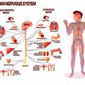
The human nervous system is a system of nerves that begins in the brain and is part of every area of your body from your fingertips to your eyes, from the skin on your toes to your bones, tendons and ligaments. The nervous system is designed to help humans perceive the world around them and to keep them safe.
A simple example of the human nervous system is when your finger or hand gets too close to a heat source and you instinctively pull back. Your brain and nerve endings feel the heat and signal your muscles to retract your hand.
Teaching the Human Nervous System
Teaching the human nervous system is a big job. It's a complicated system that can be difficult to teach. Teacher Planet is here to help. Beginning with the lesson plans you can teach the nervous system to your class room in an easy and enjoyable way. Add worksheets, clip art and activities to really help your young students absorb the science and learn the concepts. Finally, take advantage of the teaching resources to add depth and additional learning to your unit on the nervous system.
Coloring Pages
Copyright © 2001 - 2024 TeacherPlanet.com ®. All rights reserved. Privacy Statement and Disclaimer Notice

Sign up for our free weekly newsletter and receive
top education news, lesson ideas, teaching tips, and more!
No thanks, I don't need to stay current on what works in education!
- Create new account
- Reset your password
Register and get FREE resources and activities
Ready to unlock all our resources?
The human brain and nervous system
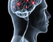
The human brain is like a powerful computer that stores our memory and controls how we think and react. It has evolved over time and features some incredibly intricate parts that scientists still struggle to understand.
The brain is the centre of the human nervous system , controlling our thoughts, movements, memories and decisions.
Our nervous system is the message centre of our body . Messages of the nervous system travel throughout the body to help the body function and stay safe. The nervous system is an amazing and complex network of cells that relay messages from all parts of our body to our brain.
The spinal cord is the main highway in which these messages travel. Messages can travel in such a short span of time so that it is almost immediate.
Top 10 facts
- Although the brain looks like a dull grey organ, it is actually mostly water. About 80% of our brain tissue is liquid . This is why it is important to drink lots of water – if you are dehydrated, your brain will not work as well.
- Our reactions to touch, danger and fear are so fast because the electrical signals in the nerve fibres travel around 280 kilometres per hour.
- Think about that famous image of a light bulb light up above someone’s head. The reality is not very different – our brain operates on the same electrical power as a 10v lamp.
- The brain contains billions of nerve cells that send and receive information around the body.
- The human brain is over three times as big as the brain of other mammals that are of similar body size.
- The human brain is protected by the skull (cranium), a 3D jigsaw of 29 bones (including the jaw) that form separately before birth and join as we grow to form a solid 'helmet' of bone.
- While your body rests at night, the brain is actually still active. Brain activity while sleeping is quite high: assessments say that the brain re-organises thoughts, processes the day you’ve just had and prepares itself for a new day.
- Dreams lasts between 2-3 seconds at the most. We mostly do not remember our dreams, just the ones that we have close to when we wake up.
- The brain of an adult human weighs around 3 pounds (1.5 kg). Although it makes up just 2% of the body's weight, it uses around 20% of its energy.
- Each side of the brain interacts largely with just one half of the body, but for reasons that are not yet fully understood the interaction is with opposite sides (the right side of the brain interacts with the left side of the body, and vice versa).

Boost Your Child's Learning Today!
- Start your child on a tailored learning programme
- Get weekly English & maths resources sent direct to your inbox
- Keep your child's learning on track
Did you know?
- The central nervous system is responsible for so many things we almost take for granted: tasting, smelling, seeing, hearing, thinking, dreaming, breathing, heart beating, moving, running, sleeping, laughing, singing, remembering, feeling pain or pleasure, painting, writing... You couldn't do any of these things without your central nervous system.
- Your brain is wrapped in three layers of tissue and floats in a special shock-proof fluid to stop it from getting bumped on the inside of your skull as your body moves around.
- Your brain is more powerful, more complex and cleverer than any computer ever built . It is constantly dealing with hundreds of messages from the world around you, and from your body, and telling your body what to do.
- Doctors and scientists have found that different parts of the brain are in charge of different things . The cerebellum controls and coordinates movements of the muscles, like walking or swinging the arms. This means that the movement is smooth and controlled and you don't fall over when you turn around. The outside layer of the cerebrum has special areas, which receive messages about sight, touch, hearing and taste. Other areas control movement, speech, learning, intelligence and personality. The brain stem is in charge of keeping the automatic systems of your body working. You don't have to think about breathing, you just do it automatically, but you can decide if you want to hold your breath for a short time. You don't have to think about your heart beating because your brain keeps it going automatically.
- Your brain keeps on growing until you are about 20 years old. By then the brain has made lots of links which it no longer needs so it is able to shed any unwanted connections and still have billions of brain cells left to cope with whatever you may want to do. You can still make new connections even when you are 100 years old!
- The human brain is so complex that doctors and scientists still don't know what some parts of it do. They do know that if the brain is damaged, the damage often cannot be repaired. You can't join brain cells together like you can bones. Doctors and scientists know how some of the brain works and can sometimes fix it when things go wrong, but the brain is truly amazing and we don't know all its secrets yet.
Look through the gallery and see if you can spot the following:
- What brains look like inside our skulls
- A nerve cell
- Scans of the brain
- Some of the many functions of the brain
The nervous system is made up of the brain, the spinal cord, and a large network of nerves that covers all parts of the body. Together, the nervous system helps different parts of our body communicate and allows our brain to control what is going on. Without the nervous system our brain wouldn’t work. It wouldn't know anything that was going on in the outside world and wouldn't be able to control our body.
The brain and the spinal cord make up what is called the central nervous system. The rest of the nerves together are called the peripheral nervous system.
Nerves are a little like wires that carry communication signals or impulses around the body. Inside each nerve is a bundle of nerve fibres. Some nerves are really long, like the ones that go all the way from your feet to your spinal cord. Nerve cells are called neurons . When a neuron is stimulated – by heat, cold, touch, sound vibrations or some other message – it begins to generate a tiny electrical pulse. This electricity and chemical change travels the full length of the neuron. But when it gets to the end of finger-like points at the end of the neuron, it needs help getting across to the next extended finger. That's where chemicals come in; the electrical pulse in the cells triggers the release of chemicals that carry the pulse to the next cell.
There are two main types of nerves: motor nerves and sensory nerves.
- Motor nerves – Motor nerves allow the brain to control our muscles. The brain sends signals over the motor nerves to tell our muscles to expand or contract so we can move.
- Sensory nerves – The second type of nerves are called sensory nerves. These nerves carry signals to the brain to tell it about what is going on in the outside world. They come from our skin (touch), nose (smell), eyes (sight), tongue (taste), nose (smell), and ears (hear).
Our bodies are incredibly clever. Sometimes we need to move so fast that our brains don't have time to think. So our body just bypasses the brain. This happens when we touch something hot. Our hand actually moves before the brain tells it to. The brain eventually finds out what is going on, but our body has done the clever thing and moved first.
The biggest part of the brain is the cerebrum . The cerebrum makes up 85% of the brain's weight. The cerebrum is the thinking part of the brain and it controls your voluntary muscles – the ones that move when you want them to. So you can't dance or kick a ball without your cerebrum!
When you're thinking hard, you're using your cerebrum. You need it to solve maths problems, read a map, and draw a picture. Your memory lives in the cerebrum – both short-term memory (what you ate for dinner last night) and long-term memory (the name of the place you went on holiday two years ago). The cerebrum also helps you reason , like when you know you need to put a bookmark in your book to keep your place.
The cerebrum has two halves, with one on either side of the head. Scientists think that the right half helps you think about abstract things like music, colours, and shapes. The left half is said to be more analytical , helping you with maths, logic, and speech. Scientists also know for sure that the right half of the cerebrum controls the actions of the left side of your body, and the left half controls the right side.
The brain is suspended in a special fluid , and floating in liquid acts as both a cushion to physical impact and a barrier to infections. As the brain is made up of mostly water, keeping it hydrated is crucial for it to work efficiently… so drink plenty of water!
Words to know:
Abstract - not relating to actual objects but expressing something that can only be understood by thinking Analytical - able to separate things into their parts in order to study or examine them, draw conclusions, or solve problems Automatic - done without conscious thought Complex - made up of many interrelated parts Evolved - to develop from an earlier biological form Interact - to be or become involved in communication, social activity, or work with somebody else or one another Intricate - containing many details or small parts that are combined in a particularly complex or skilful way Mammals - a class of warm-blooded vertebrate animals Network - a pattern or system that looks like a series of branching or interconnecting lines Organ - a complete and independent part of a plant or animal that has a specific function Relay - the passing on of something, especially a message or information received, to somebody else, or the process of being passed on Voluntary - resulting from somebody's own choice or decision
Related Videos
Just for fun...
- Explore the brain and how it works and test your frontal lobe, parietal lobe, temporal lobe and occipital lobe with some games
- A hands-on experiment to help you test how fast your brain sends messages to your body
- Help your brain remember things better with these fun exercises
- A word matching game
- Brain teasers and brain games for kids
- Try the Fastball Reaction Time game to see how fast your brain sends messages to your muscles
- Watch National Geographic Kids videos of brain games
Children's books about the brain
Find out more
- The Brain Explorer offers an tour around the different parts of the human brain
- Try some brain training activities
- A quiz about the brain and the nervous system
- More facts and information about the brain and the nervous system
- The funny bone (not actually a bone at all!) explained
- Understand more about neuroscience on the Brain Diaries website and play games and watch videos about how our brains grow and learn
- Find out more about how the brain works and how information is passed along the nervous system
- Loads of links to explain neuroscience to kids
- Watch a video that explains how our eyes and brain work together to make sense of the world
- Discover how your brain cells talk to each other and what different brains are made of
- Find out how Magnetic Resonance Imaging (MRI) is used to study the brain , with a children's guide
See for yourself
- Take a 3D tour of the brain
- See which parts of the brain work when different tasks are carried out
Give your child a headstart
- FREE articles & expert information
- FREE resources & activities
- FREE homework help

- school Campus Bookshelves
- menu_book Bookshelves
- perm_media Learning Objects
- login Login
- how_to_reg Request Instructor Account
- hub Instructor Commons
- Download Page (PDF)
- Download Full Book (PDF)
- Periodic Table
- Physics Constants
- Scientific Calculator
- Reference & Cite
- Tools expand_more
- Readability
selected template will load here
This action is not available.
15: Module 13- The Nervous System
- Last updated
- Save as PDF
- Page ID 34525
- 15.1: Introduction to the Nervous System
- 15.2: The Embryologic Perspective
- 15.3: The Central Nervous System
- 15.4: Circulation and the Central Nervous System
- 15.5: The Peripheral Nervous System
- 15.6: Video- The Unfixed Brain
- 15.7: Video- The Unfixed Spinal Cord
- 15.8: Case Study- The Brain
- 15.9: Additional Links
- 15.10: Glossary- The Nervous System
- 15.11: Practice Test- Anatomy of the Nervous System

- school Campus Bookshelves
- menu_book Bookshelves
- perm_media Learning Objects
- login Login
- how_to_reg Request Instructor Account
- hub Instructor Commons
- Download Page (PDF)
- Download Full Book (PDF)
- Periodic Table
- Physics Constants
- Scientific Calculator
- Reference & Cite
- Tools expand_more
- Readability
selected template will load here
This action is not available.
10: Overview of the Nervous System
- Last updated
- Save as PDF
- Page ID 7180

Nervous System Study Resources {Part 1}
Sharing is caring!

*contains affiliate links
Welcome to our study with Apologia’s Human Anatomy and Physiology. This week, we’ve been studying the nervous system.
I have always been fascinated by the nervous system. The nervous system is the command center of your body. It controls and coordinates your movements, processes, and thoughts. The nervous system is made up of the brain, the spinal cord, and the nerves that reach across the entire body. We’ve been having fun learning about each part with Apologia’s Human Anatomy and Physiology curriculum.
If you look at the body as a city. This city has lots of roads and highways. Last week, we learned about the circulatory system. Those roads carried blood cells, nutrients, and wastes through the body. The highways of the nervous system carry information.
The main nervous system highway is called the central nervous system. The central nervous system is made up of the brain and the spinal cord. The smaller roads of the nervous system are called the peripheral nervous system . The peripheral nervous system is made up of nerves that extend from the central nervous system to the outer edges of the body.
The Central Nervous System
As mentioned before, the central nervous system is made up of the brain and the spinal cord. The brain is made up of 3 main parts: the cerebrum , the cerebellum, and the brainstem . Each has a separate, important function. To help remember where those parts are, my daughter created a clay brain with each part a different color. (S he used polymer clay for this project. It’s so easy to work with and comes in lots of colors)

The cerebrum is where your thoughts and decisions happen. You think about what you want for dinner in the cerebrum. You decide you want to eat pizza and your cerebrum sends a message to your arm to reach for the phone and dial for delivery.

The cerebellum (in red) controls muscle movements you don’t think of. When you stand still, you might not think there are muscles working, but the cerebellum makes sure your muscles are doing the work of making sure you stay standing up. The cerebellum, also, helps with equilibrium.

The brainstem (in yellow) connects the brain to the spinal cord. It controls basic body functions like breathing and the regulation of the heartbeat. Once again, the brain makes sure these things happen automatically. You don’t even have to think about it.
The second main part of the central nervous system is the spinal cord . The job of the spinal cord is to take messages from the brain to the peripheral nervous system and from the peripheral nervous system back to the brain. It does this via nerves originating in the spinal cord and branching out to the rest of the body.
The Peripheral Nervous System
The peripheral nervous system consists of those nerves that branch off of the spinal cord and spread to the rest of the body. These nerves are made up of cells called neurons. To help learn the various parts of the neuron, my daughter made this neuron model out of clay and yarn.

One end of the neuron has branches called dendrites . Dendrites gather information and bring it back to the cell body. The cell body sends the information along the axon to another nerve cell. Most axons are wrapped in fatty tissue called the myelin sheath . The myelin sheath speeds up the transmission of information along the axon, which can vary in length from a millimeter to a meter or more. The end of the axon is called the terminal end . Instead of the terminal end of the axon connecting directly to the next neuron, the two cells are connected indirectly by a synapse .
A synapse is a tiny gap between neurons. In order for information from one neuron to get to the next neuron, chemicals called neurotransmitters are released. Neurotransmitters leave the terminal of the axon and travel across the synapse to the dendrite of the next neuron. Not all neurotransmitters are accepted by the dendrite, just the ones carrying the necessary information at the time.
As you can see, one end of the neuron collects information and the other transmits information. This means that information can only travel one way along a nerve. Neurons that collect information from the body and take it to the brain are called sensory neurons . Neurons that carry information from the brain to the body are called motor neurons .
More Nervous System Information
Nervous System Study Resources {Part 2} will dive into learning more details about the brain, spinal cord, and reflexes.
In the meantime…
- The crew at MeetPenny.com , had a fun time this week studying the nervous system, too. They have great nervous system ideas and games.
- DIY Nervous System Models
- Nervous System Study Ideas
- Nervous System Notebooking Pages
- Human Brain Coloring Book
- Make a Neuron
- Brain Games
- Neuroscience is Cool
- Nervous System printables
- Brain Game printables
- Make a Brain Hemisphere Hat
- Brain Imaging
- Neuroscience Lessons for Kids: lower elementary
- The Brain in Space
- The Brain is a Computer
- Your Wires Are Really Crossed: Communication in the Nervous System
- Anatomy of a Neuron video from Khan Academy – jr high/high school level
- Understanding Neurons video from MakeMeAGenius.com – primary/grade school level

- Biology Article
- Nervous System
Human Nervous System
Living organisms adapt to their moves and positions in response to the environmental changes for their protection or to their advantage. When an entity reacts to the changes in its surroundings, it is referred to as stimulus while the reaction to the stimulus is referred to as a response. Common stimuli are sound, light, air, heat, smell, taste, water and gravity.
Think of burning your finger of fracturing your bone without any pain sensation. It may certainly sound like a superpower or an ideal situation, however, when it comes to the standpoint of survival, it can be disastrous.
The characteristic behaviour of living entities is to respond to stimuli with the intervention of the nervous system. It is an organ system ascribed to send signals from the spinal cord and the brain throughout the body and then back from all the body parts to the brain. The neuron acts as the mediator and is the basic signalling unit of the nervous system.
Pain is the body’s way of letting us know that something is not right. It can prevent further injuries or push us to seek medical attention. Moreover, all of this is possible because humans can respond and react to stimuli due to control and coordination among the various organs and organ systems.
Control and Coordination in simple multicellular organisms take place through only the Nervous system which coordinates activities of our body. It is the control system for all our actions, thinking, and behaviour.
Refer more: Control and Coordination
Let us have a detailed look at the nervous system notes to explore what is the nervous system, and the different functions of the nervous system with the help of diagrams. Table of Contents
What is the Nervous System?
Human nervous system diagram, central nervous system, peripheral nervous system, recommended video:.

The nervous system or the neural system is a complex network of neurons specialized to carry messages . The complexity of the nervous system increases as we move towards higher animals.
For instance, cnidarians such as jellyfish have relatively simple nerve nets spread throughout their body. Crabs have a more complicated nervous system in the form of 2 nerve centers called dorsal ganglion and ventral ganglion.
As we move further up the ladder, higher organisms such as vertebrates have a developed brain. Moreover, it is one of the most complicated structures in the animal kingdom, containing billions of neurons, all intricately connected.
In the human body, the neural system integrates the activities of organs based on the stimuli, which the neurons detect and transmit. They transmit messages in the form of electrical impulses and convey messages to and from the sense organs. Thus, the nervous coordination involves the participation of the sense organs, nerves, spinal cord, and brain.
Also Read: Creutzfeldt-Jakob Disease

Diagram of the Human Nervous System
One of the most complex organ system to ever evolve, the human nervous system consists of two parts, namely:
- Central Nervous System (consists of the brain and spinal cord)
- Peripheral Nervous System (includes all the nerves of the body)
Central Nervous System (CNS) is often called the central processing unit of the body. It consists of the brain and the spinal cord.
The brain is one of the important, largest and central organ of the human nervous system. It is the control unit of the nervous system, which helps us in discovering new things, remembering and understanding, making decisions, and a lot more. It is enclosed within the skull, which provides frontal, lateral and dorsal protection. The human brain is composed of three major parts:
Forebrain : The anterior part of the brain, consists of Cerebrum, Hypothalamus and Thalamus.
Midbrain : The smaller and central part of the brainstem, consists of Tectum and Tegmentum.
Hindbrain : The central region of the brain, composed of Cerebellum, Medulla and Pons.
Also read: Human Brain
Spinal Cord
The spinal cord is a cylindrical bundle of nerve fibers and associated tissues enclosed within the spine and connect all parts of the body to the brain. It begins in continuation with the medulla and extends downwards. It is enclosed in a bony cage called vertebral column and surrounded by membranes called meninges. The spinal cord is concerned with spinal reflex actions and the conduction of nerve impulses to and from the brain.
Peripheral Nervous System (PNS) is the lateral part of the nervous system that develops from the central nervous system which connects different parts of the body with the CNS. We carry out both voluntary and involuntary actions with the help of peripheral nerves.
Also refer: Peripheral Nervous System
PNS includes two types of nerve fibers:
- Afferent nerve fibers – These are responsible for transmitting messages from tissues and organs to the CNS.
- Efferent nerve-fibers – These are responsible for conveying messages from CNS to the corresponding peripheral organ.
Classification of the peripheral nervous system:
Somatic neural system (SNS): It is the neural system that controls the voluntary actions in the body by transmitting impulses from CNS to skeletal muscle cells. It consists of the somatic nerves.
Autonomic neural system (ANS): The autonomic neural system is involved in involuntary actions like regulation of physiological functions (digestion, respiration, salivation, etc.). It is a self-regulating system which conveys the impulses from the CNS to the smooth muscles and involuntary organs (heart, bladder and pupil). The autonomic neural system can be further divided into:
- Sympathetic nervous system
- Parasympathetic nervous system

A Neuron is a structured and functional unit of the nervous system and unlike other cells, neurons are irregular in shape and able to conduct electrochemical signals. The different parts of a neuron are discussed below.
- Dendrite stretches out from the cell body of a neuron, and it is the shortest fibre in the cell body.
- Axon is the longest thread on the cell body of a neuron and has an insulating and protective sheath of myelin around it.
- Cell body consists of cytoplasm and nucleus.
- Synapse is the microscopic gap between a pair of adjacent neurons over which nerve impulses pass, when moving from one neuron to the other.
Explore more: Placebo Effect
Nerves are thread-like structures that emerge from the brain and spinal cord. It is responsible for carrying messages to all the parts of the body. There are three types of nerves. Some of these neurons can fire signals at speeds of over 119 m/s or above 428 km/h.
- Sensory nerves send messages from all the senses to the brain.
- Motor nerves carry messages from the brain to all the muscles.
- Mixed nerves carry both sensory and motor nerves.
Also read: Nerves
Cranial nerves begin from the brain as these nerves carry impulses to start from the central nervous system. Certain cranial nerves belong to the group of mixed nerves while certain ones fall under sensory nerves. Spinal nerves originate from the spinal cord. All the spinal nerves carry impulses to and from the central nervous system and these are part of mixed nerves. The above nervous system diagram depicts the various nerves arising from various parts of the body.
Learn more in detail about the Human Nervous System with diagrams or any other related topics by referring to the nervous system notes provided at BYJU’S website. Download BYJU’S app for further reference.
Frequently Asked Questions
What are the two divisions of the nervous system.
The human nervous system controls all activities of the body in a quicker fashion. It can be divided into the central nervous system and peripheral nervous system. The central nervous system includes spinal cord and brain and the peripheral covers the nerves branching from spinal cord and brain.
What are nerves and neurons?
Nerves are thread-like structures that emerge from the spinal cord and brain. These nerves are actual projections of neurons. A neuron is a basic structural and functional unit of a nervous system that conducts electrochemical signals.
What are cranial nerves?
The nerves that extend throughout the body on both sides and emerges directly from the brain stem and brain are called cranial nerves. They carry information from the brain to other parts, primarily to the neck and head.

Put your understanding of this concept to test by answering a few MCQs. Click ‘Start Quiz’ to begin!
Select the correct answer and click on the “Finish” button Check your score and answers at the end of the quiz
Visit BYJU’S for all Biology related queries and study materials
Your result is as below
Request OTP on Voice Call
Leave a Comment Cancel reply
Your Mobile number and Email id will not be published. Required fields are marked *
Post My Comment
Hello Byju’s I need to chat with you to clarify a doubt. Your app is so good
Really Byjus is a very good application in the case of study in a systematic knowledge so Byjus achievement is great
Thank you for helping with this concept, it gave me better information than other websites.
THIS IS SERIOUSLY A NICE SITE FOR KNOWLEDGE.
Thanks a lot for helping me
Thanks a lot for helping
This app is very awesome and also help me in doing my homework ..
Actually I need more of these to pass my test thanks for sharing these notes I am really grateful to you about these Bye
Byju’s always help me a lot. .This platform is really great.
- Share Share
Register with BYJU'S & Download Free PDFs
Register with byju's & watch live videos.

Things you buy through our links may earn Vox Media a commission
- When Did Everyone Get So ‘Dysregulated’?
How managing our mental health became a matter of monitoring our nervous systems.

What is wrong with you?” is a rich and open question. When it comes from other people, it is “aggressive” or “rude,” but when you pose it to yourself, it is endlessly fascinating. What is wrong with you?
For better or for worse, a buffet of terminology has emerged that might help explain your terminal unease. Maybe you are struggling with boundaries or your social circle is overrun with toxic people. Maybe your mother or roommate or ex-boyfriend is a narcissist . Maybe you are traumatized, which is perhaps one reason you are also anxiously attached. The problem with these words, of course, is that their impact fades with use, leaving us to hunt for new language that can justify our suffering. By early 2024, we seem to have landed on our latest favorite diagnosis: Have you considered the possibility that you might be dysregulated?
The odds seemed high. In a podcast interview last summer, an author explained that while discovering nonmonogamy had been a revelation, actually enacting it had initially caused “utter panic and dysregulation.” In a blog post on how to conquer dating anxiety, the Gottman Institute advises you to “surround yourself with emotionally safe people,” which “helps to regulate one’s nervous system.” On anonymous forums, people worried whether their dysregulation was affecting their work performance — a concern their bosses seemed to share, which is perhaps why corporate-wellness blogs and workplace “thought leaders” have advised managers to pay attention to their employees’ dysregulation. (“Is Your Team Struggling?” warned one headline. “Their Dysregulation May Impact Your Performance.”) School administrators reported that kids were showing up at school more dysregulated than ever. Their teachers weren’t far behind. In its course description for a training about workplace conflict, a college of social work in Ohio crisply identified the source of the problem: “Surprise! It’s not your age cohort, or workload, or low pay — it’s dysregulation!”
Often, the term appeared attached to formal diagnoses, as a symptom of ADHD or BPD or PTSD or ASD or generalized anxiety, but just as frequently it floated freely, explaining why you were yelling at your husband because — by way of hypothetical example — you couldn’t find the garlic powder and also your computer wasn’t working. Sometimes, people seemed to refer to a process taking place within the nervous system. (“Being sleep deprived can really dysregulate our control over the autonomic system,” offered the neuroguru Andrew Huberman.) Other times, the thing being dysregulated was one’s behavior or emotions. You were experiencing an outsized reaction to something that didn’t seem to warrant it; your behavior did not match the situation; your emotions were too big and intense and long-lasting, which was making it difficult to function. It could explain why you were breaking out or eating too much or not eating at all , why you’re thrill-seeking or sleeping poorly or exhausted ; or why you’re depressed or experiencing gastrointestinal distress. On one health podcast from January, a clinician-slash-trainer suggested dysregulation might be why, despite consistent exercise, she had, for years, struggled to gain muscle in her legs. It was a constant threat. “You can’t trust someone who purposely wants to dysregulate you,” advised a TikTok video, which has been viewed more than 100,000 times.
Until last year, I had only thought about one person as dysregulating me: my daughter, who is a baby. The concept had saturated parenting discourse, appearing in books and blogs and pastel slides on Instagram. The idea is that children’s meltdowns are not malicious or even conscious but instead a natural consequence of their developing brains. These, according to modern parenting experts like Dr. Becky , are episodes of dysregulation; the child has not yet learned to control their response to stress, and it is your job, as a parent, to help by modeling a Zen-like state yourself: a relaxed body, a slow heartbeat, a quiet mind. “A dysregulated adult,” the psychiatrist Bruce D. Perry said, in his 2021 book co-written with Oprah, “cannot regulate a dysregulated child.” In her new book, The 5 Principles of Parenting: Your Essential Guide to Raising Good Humans , developmental psychologist and guru Aliza Pressman lays out countless parental self-regulation strategies, including breathing, meditating, running your hands under cold water, and “playing with a pet.” On my neighborhood parental Facebook group, I learned about a $27 webinar that would teach me “how regulation and connection shape cooperation.” The pressure to be Zen could be daunting. “The phrase ‘a dysregulated parent can’t regulate a child,’” one parenting Instagram account argued , “is making parents dysregulated.”
Parenting Instagram is where I first encountered the concept, and sometime last summer, late in pregnancy, the algorithm cracked me, and then the Baader-Meinhof phenomenon did its thing. I couldn’t help but begin to narrate my own experience in regulatory terms ( I am dysregulated , I thought, crying in the shower). I couldn’t decide if it made things better, but it was so flexible. In a sentence, “dysregulated,” could replace everything from “panicked” or “overstimulated” or “irrationally flipping out” or “being in acute crisis,” and transform those messy conditions into something isolatable and objective.
That, of course, is what therapyspeak has always done: It simplifies, it distances, it clarifies. But unlike “triggered” or “boundaries” or other classics of the genre, “dysregulation” felt dispassionately medical, gesturing at the base mechanics of one’s misery. Perhaps mental health is simply a matter of monitoring one’s inner circuitry — an idea that slotted neatly into the lives and vocabularies of those who have learned to track heartbeats and sleep cycles and ovulation cycles and blood glucose levels and blood oxygen. Most tantalizing was that the word seemed to suggest a solution: Maybe if you could just tweak some inner levers and recalibrate some dials, you could at last reveal your best and calmest self.
Feeling bad is timeless, and so is the notion that the reason for your agony is that something in your bodily system is out of whack. Kathryn Tabb , a philosopher of science at Bard focused on psychiatric history, traces this pattern of diagnosis at least to Hippocrates, who believed that one’s health depended on the proper balance of the four humors. In his view, all problems stemmed from the proportions of those humors: Maybe a person had too much or too little blood or phlegm or bile, which was either too strong or too weak or improperly mixed. Then scientists discovered germs, which undermined the humors theory, but its underlying framework would prove infinitely adaptable.
In the 1990s, the logic of the humors found its place in the human brain. At the top of the decade, George H.W. Bush signed a presidential proclamation announcing that it was now the Decade of the Brain. New neuroimaging technology was converting neural activity into pictures, which were appearing everywhere from Newsweek to murder trials . A flood of research was giving us access to our own mental processes, and it looked like maybe we were going to solve philosophy, and perhaps finally understand ourselves. Neuroscience, Tom Wolfe wrote in 1996, was “on the threshold of a unified theory that will have an impact as powerful as that of Darwinism a hundred years ago.” (He wasn’t happy about it.) Prozac had just hit the U.S. market — followed by Zoloft, and then Paxil — and this new class of relatively gentle psychiatric drugs fundamentally changed how millions of Americans thought about their brains. “That’s probably when the idea really caught on that our emotional states have a neurobiological basis,” says Sally Satel , a psychiatrist and the co-author of Brainwashed: The Seductive Appeal of Mindless Neuroscience .
By then, scientists already understood that psychic stress — whether from war or avalanches or being late to work — caused dramatic changes in the nervous and endocrine systems, which manifested in physiological symptoms. But in the ’90s, a generation of researchers began proposing accounts of how stress not only worked upon the body but also lingered in it, creating a disequilibrium. In 1994, Robert Sapolsky, then an associate professor of biological sciences and neuroscience at Stanford, published Why Zebras Don’t Get Ulcers , explaining, in enthusiastic detail, the different ways that mammalian stress responses — the rush of adrenaline, shallow breath, and fast heart rate that were usually fantastic in acute physical emergencies like getting chased by a wolf — could start to wear down our various bodily systems in the face of modern, chronic stress (a passive-aggressive boss, the MTA). Bessel van der Kolk , then a psychiatrist at Harvard, published the first iteration of what would go on to become his best-selling manifesto, The Body Keeps the Score , which argued that trauma gets stored in the nervous system and physiologically altered how your body processes stressful events. Biophysicist Peter Levine, who wrote a popular self-help book on overcoming trauma, put forth a related theory: It wasn’t the trauma itself that caused long-lasting symptoms, but a “frozen residue of energy that has not been resolved and discharged; this residue remains trapped in the nervous system where it can wreak havoc on our bodies and spirits.”
What emerged was a popular understanding that extreme or chronic stress presented a regulatory problem, specifically in the autonomic nervous system. Two of the system’s components are the sympathetic nervous system and the parasympathetic nervous system. The first responds to danger or excitement by signaling the adrenal glands to release hormones like adrenaline and cortisol (also known as our fight-or-flight response), while the second promotes relaxation and recovery and basic bodily functions (“rest and digest”). One goes up, the other goes down, and vice versa. “Nervous system dysregulation” is what happens when the two are out of sync, and you stay stuck, either overactivated, or not activated enough. It wasn’t just the stress researchers and the traumatologists who were thinking about regulation. The neuropsychologist Russell Barkley had begun to argue that ADHD was not, in fact, just about deficient attention, but also deficient emotional self-regulation. Marsha Linehan, a former psychologist at the University of Washington, developed dialectical behavior therapy, or DBT, specifically to treat emotional dysregulation, which she argued was at the heart of borderline personality disorder. “She really shifted the way we look at personality disorders,” said Lina Perl , a clinical psychologist in New York who was first introduced to the word in graduate school. BPD wasn’t a fatal flaw intrinsic to your defective personality, but a regulatory issue, likely stemming from unmet childhood needs.
The dozen or so therapists, clinicians, and historians I spoke to couldn’t precisely pinpoint how or when “dysregulation” crossed over from the clinic into popular imagination, but they had theories. One obvious explanation is simply that as therapy and psychiatric diagnoses grew more mainstream, “dysregulation” rode its coattails. Several pointed to evolving understandings of various conditions. “To me, a smoking gun is the obsession with ADHD,” says Tabb, citing the recent explosion of newly diagnosed adults. Justin Baeder, an educational consultant and former school principal popular on TikTok, told me in the last year or so, he’d seen dysregulation morph. Once, it had been used in “a narrow technical sense,” generally describing students with disabilities, he told me. Now, “I’m hearing it used much more broadly to mean any kind of feelings — anytime a child is upset, they’re dysregulated.” It is, he suggests, a way to “medically explain away any type of bad behavior” (well-intentioned, but in his view, not especially productive).
Wellness culture predated the pandemic, but after years of inward focus, it got turbocharged. People had a lot of time to think, mostly about their health, and read, mostly about their trauma , and consume content on the internet, mostly about how to feel less bad. Regulation was an answer that seemed to cross demographic lines. Holistic influencers in bikinis were regulating (“ three ways to soothe your nervous system ”), but so were hyper-rational science bros. On his podcast, Huberman, in his trademark black button-down, offered all kinds of regulatory guidance for the chronic maximizer crowd. Jordan Peterson, wellness influencer for aggrieved men, extolled the virtues of emotional regulation. The proposed solutions were generally obvious — get sunlight, go to bed, meditate, eat breakfast.
It is true that neuroimaging shows brain activity and that stress is brain activity, and in that sense, sure, it would show up on a scan. If you found yourself, one day, in a state of what you called “dysregulation,” and at that moment you had an fMRI, it would show arousal centers activated in the brain, which you could see represented as bright glowing splotches. And if you had repeated scans, in different mental states, you would be able to see changes in the pattern or intensity of that activation. What the glowing splotches would not tell you is exactly what any of it means. Is dysregulation real? Of course it’s “real,” in the sense that yes, your experiences are mirrored in your brain, and yes, you can, at any given time, see activity that is different from other possible activity. But what are you supposed to do with that? “The line between the normal and the pathological is something we construct,” says Tabb. There’s a lot we still don’t know about the way our nervous system works in relationship with the rest of our body. “It’s as if we were outside the restaurant pressing our faces against the restaurant window looking in without being able to access what’s going inside the restaurant at all,” the medical historian Edward Shorter told me.
In some ways, “dysregulation” is an updated version of another science-coded phrase we used to like: “chemical imbalance.” Picture the Zoloft commercial from the early aughts that featured line drawings of “nerve A” and “nerve B” on a plain-white background. “Depression may be related to an imbalance of natural chemicals between nerve cells in the brain,” chirped the voiceover, as dots floated erratically between the two illustrated nerves. “I think it’s such a tantalizing metaphor because it’s so straightforward,” says Nate Greenslit, an anthropologist of science. “If you can think of it as a soupy mixture of different chemicals, and if those chemicals are out of balance with each other, then you will suffer as a consequence.” The only problem with Zoloft’s early diagram is that it isn’t true. A major 2022 umbrella review showed what had been an open secret in the field: There is no substantial evidence that low serotonin causes depression. A lot of people do do well on SSRIs, but we don’t know why. “There is no ‘chemical imbalance,’” University of Virginia sociologist Joseph E. Davis told me, bluntly. “That was a misnomer right from the start.” More recent commercials make no claims about root causes. These days, they tend to feature generic women doing normal things looking ambiently bummed.
Dysregulation doesn’t yet carry the same baggage. Functionally, it’s similar, in that it locates your problem in your biology rather than your personality, which is comforting. But unlike being “chemically imbalanced,” which sounds like an intractable, if treatable, feature of a person, being “dysregulated” sounds like a reassuringly temporary state . “It strikes me as more dynamic, less static, than the language of chemical imbalance,” agrees Greenslit. “Chemical imbalance is also very, very individualistic: You as an individual have programmatic biology, and so the solution is at an individual level.” Dysregulation, on the other hand, “implies relationships.” You’d be chemically imbalanced anywhere, but you couldn’t be dysregulated in a vacuum. “It doesn’t say there’s something inherently wrong with you,” the founding director of Health Justice Commons and senior adjunct lecturer at the California Institute of Integral Studies Mordecai Cohen Ettinger, told me. “It’s saying your body has been harmed, or decentered, or stressed from a set of traumatic events, so you are dysregulated, and that is just a normal biological consequence of what you’ve experienced.” In this sense, it is the best possible therapyspeak for this moment, when the climate is in crisis and there are microplastics in the water as well as a widespread hopelessness and a grim election looming and a pervasive sense of tenuousness, interpersonally and economically, and it is tiresome to list these things, I know, but isn’t it also true?
The fundamental appeal of regulatory language is the idea that there is a calm stasis to return to, a “you” separate from your acute distress. “I think from what I’ve heard from my clients, it most often refers to the sensation that they don’t feel like themselves,” said Lisa Daronatsy Kiyindou, a health and fitness trainer in New York. “They’re trying to get back to feeling more like themselves.” There is a kind of longing in dysregulation. People were struggling not to change, but to recapture an idea of who they’d been before. You may be a wreck on a hair trigger now , but that isn’t at the core of who you are . I am dysregulating , I thought, and was struck by the perverse optimism of it. I was a mess, but there was hope.
- remove interruptions
- mental health
The Cut Shop
Most viewed stories.
- Rose Hanbury Says She Didn’t Have an Affair With Prince William
- Am I a Rose Hanbury Stan Now?
- Lukas Gage Calls Marriage to Chris Appleton ‘Unhinged’
- Is This Really the Worst Place You Can Put a Birkin?
- ‘How I Paid Off $36,000 in Credit Card Debt’
- The Lure of Divorce
Editor’s Picks

Most Popular
What is your email.
This email will be used to sign into all New York sites. By submitting your email, you agree to our Terms and Privacy Policy and to receive email correspondence from us.
Sign In To Continue Reading
Create your free account.
Password must be at least 8 characters and contain:
- Lower case letters (a-z)
- Upper case letters (A-Z)
- Numbers (0-9)
- Special Characters (!@#$%^&*)
As part of your account, you’ll receive occasional updates and offers from New York , which you can opt out of anytime.
- U.S. Department of Health & Human Services
- Virtual Tour
- Staff Directory
- En Español
You are here
News releases.
News Release
Tuesday, March 12, 2024
NIH opens long COVID trials to evaluate treatments for autonomic nervous system dysfunction
Part of NIH’s RECOVER Initiative, trials will test at least three treatments for symptoms such as fast heart rate, dizziness and fatigue.
Two phase 2 clinical trials to test the safety and effectiveness of three treatments for adults with autonomic nervous system dysfunction from long COVID have begun. The autonomic nervous system acts largely unconsciously and regulates bodily functions, such as heart rate, digestion and respiratory rate. Symptoms associated with autonomic nervous system dysfunction have been among those that patients with long COVID say are most burdensome. The trials are part of the National Institutes of Health’s Researching COVID to Enhance Recovery (RECOVER) Initiative, a nationwide research program to fully understand, diagnose and treat long COVID. Other RECOVER phase 2 clinical trials testing treatments to address viral persistence and neurological symptoms, including cognitive dysfunction (like brain fog), launched in July 2023.
“As a long COVID patient, I know firsthand how disruptive and frightening symptoms including rapid heart rate, dizziness and fatigue can be. Patient representatives across RECOVER have also shared that these symptoms are some of the most debilitating symptoms of long COVID,” said Heather Marti, co-chair of the RECOVER National Community Engagement Group. “These trials are giving me and others with long COVID hope that it will restore our health and get us back to the lives we so desire.”
The two trials, collectively known as RECOVER-AUTONOMIC, are testing three potential treatments in adults who, following COVID-19, now have postural orthostatic tachycardia syndrome (POTS). An autonomic nervous system disorder, POTS is characterized by unexpected fast heart rate, dizziness, fatigue or a combination of these symptoms when a person stands up from sitting or lying down.
“The trials were developed with input from people living with long COVID, caregivers, community representatives, clinicians and scientists all with unique expertise in the field,” said Gary H. Gibbons, M.D., director of the National Heart, Lung, and Blood Institute at the NIH and co-chair of RECOVER. “We are grateful for their collective involvement which significantly shaped the trials and the choice of interventions.”
The trials will initially examine three potential treatments:
- Gamunex-C, a form of intravenous immunoglobulin (IVIG), contains antibodies to help the body protect itself against infection from various diseases and is given by intravenous infusion.
- Ivabradine, an oral medication that reduces heart rate.
- Coordinator-guided, non-drug care, which includes a series of activities managed through weekly phone calls with a care coordinator, such as wearing a compression belt and eating a high-salt diet, which are recommended for patients with POTS to counteract excessive loss of fluids.
“Patients who develop POTS after having COVID-19 are often severely limited by their symptoms, and there are no proven effective treatments,” said Christopher Granger, M.D., Duke University Medical Center, who is co-leading RECOVER-AUTONOMIC. “These interventions were selected because they have shown potential benefit in treating symptoms for POTS. The theory we’re testing is that they might also help individuals with long COVID.”
Participants will first be randomly assigned to receive either IVIG, ivabradine or a placebo. Participants will then be randomly assigned a second time to receive either coordinator-guided, non-drug care or what is considered the usual non-drug care for POTS following COVID-19, such as diet and lifestyle recommendations. RECOVER-AUTONOMIC is an adaptive clinical trial, meaning if additional potential interventions emerge, they can quickly be added and studied in the trial.
Researchers plan to enroll 380 total participants at 50 sites across the United States. Teams at the trial sites will recruit participants from their health systems and surrounding communities. The current list of sites for the trials can be found on ClinicalTrials.gov (search: NCT06305793, NCT06305806 and NCT06305780) and additional sites will be added to this list as they begin enrolling participants.
Diversity among the trial participants is a high priority for RECOVER. To support diverse and inclusive representation, study sites are chosen based on geographic location, their connections to communities, and their track records for enrolling diverse research participants.
With the launch of the RECOVER-AUTONOMIC trials, RECOVER is currently testing seven treatments across four clinical trials and continues to enroll participants. Those interested in learning more about RECOVER clinical trials should visit trials.recovercovid.org .
About RECOVER : The National Institutes of Health Researching COVID to Enhance Recovery (NIH RECOVER) Initiative brings together clinicians, scientists, caregivers, patients, and community members to understand, diagnose, and treat long COVID. RECOVER has created one of the largest and most diverse groups of long COVID study participants in the world. In addition, RECOVER clinical trials are testing potential interventions across five symptom focus areas. For more information, please visit recovercovid.org .
HHS Long COVID Coordination: This work is a part of the National Research Action Plan , a broader government-wide effort in response to the Presidential Memorandum directing the Secretary for the Department of Health and Human Services to mount a full and effective response to long COVID. Led by Assistant Secretary for Health Admiral Rachel Levine, the Plan and its companion Services and Supports for Longer-term Impacts of COVID-19 report lay the groundwork to advance progress in the prevention, diagnosis, treatment, and provision of services for individuals experiencing long COVID.
About the National Institutes of Health (NIH): NIH, the nation's medical research agency, includes 27 Institutes and Centers and is a component of the U.S. Department of Health and Human Services. NIH is the primary federal agency conducting and supporting basic, clinical, and translational medical research, and is investigating the causes, treatments, and cures for both common and rare diseases. For more information about NIH and its programs, visit www.nih.gov .
NIH…Turning Discovery Into Health ®
Connect with Us
- More Social Media from NIH
Advertisement

Abstract 2823: Development of an in vitro blood-brain barrier model of CCR7/CCL19 induced invasion of the central nervous system
- Split-Screen
- Article contents
- Figures & tables
- Supplementary Data
- Peer Review
- Get Permissions
- Cite Icon Cite
- Search Site
- Version of Record March 22 2024
Alondra Rodriguez , Cesar I. Cardona , Colin A. Bill , Charlotte M. Vines; Abstract 2823: Development of an in vitro blood-brain barrier model of CCR7/CCL19 induced invasion of the central nervous system. Cancer Res 15 March 2024; 84 (6_Supplement): 2823. https://doi.org/10.1158/1538-7445.AM2024-2823
Download citation file:
- Ris (Zotero)
- Reference Manager
Pediatric T-cell acute lymphoblastic leukemia (T-ALL), an aggressive hematological malignancy, is characterized by abnormal thymocyte proliferation, frequently with mutations of the Notch-1 gene and has an overall 10-year survival rate of ~27%. A marker of poor prognosis is the presence of T-ALL within the central nervous system (CNS), which is treated with intrathecal chemotherapy and cranial irradiation; two toxic treatments with high morbidities. Gene-expression profiling of Notch-1-induced adhesion regulators showed that C-C chemokine receptor 7 (CCR7) was highly expressed and was associated with leukemic T-cell invasion of the CNS. Normal functions of CCR7 include lymph node homing and immune cell trafficking in response to CCR7 ligands, CC-chemokine ligand 19 (CCL19) or CCL21. In several studies CCL19 has been linked to T-cell invasion of the CNS across the blood-brain barrier (BBB). We hypothesize that antagonizing CCR7 binding to CCL19 will block migration of T-ALL into the CNS and thereby alleviate the need for toxic therapies. To test our hypothesis, we are using receptor internalization assays, calcium mobilization assays, transwell migration assays and an in vitro model of an intact human BBB. We have used flow cytometry-based CCR7 internalization assays ± CCR7 antagonist and found that we can completely block CCL19-induced receptor internalization in human CCR7-expressing CCRF-CEM, and HuT-78 T-ALL cells, and chemotaxis via β1 integrins across a fibronectin-coated transwell membrane. Our BBB employs HBEC-5i, human cerebral microvascular endothelial cells, in transwell chambers coated with gelatin and fibronectin for the adhesion of endothelial cells. HBEC-5i cells are a suitable cell line to make a 3D model of the BBB, as they form an endothelial monolayer with tight junctions (TJ), which is crucial to maintaining the integrity and inhibiting permeability of the BBB. We are establishing a transwell migration assay to assess the migration of CCRF-CEM T-ALL cells across the HBEC-5i cell monolayer. The integrity of the HBEC-5i cell monolayer is assessed by trans-epithelial electrical resistance (TEER), which measures the electrical resistance across the HBEC-5i monolayer, a measure of TJ integrity. Using TEER measurements, we observe that HBEC-5i form a monolayer that reaches an intact monolayer functioning with TJs over 72-96 hours. We are using this model system to investigate the migration of CCR7-expressing CCRF-CEM cells through the BBB ± CCL19. Subsequently, we will add the CCL19 antagonist (CCL197-77) to determine if inhibiting CCR7 is sufficient to eliminate migration of the CCRF-CEM cells through the BBB. If successful, this model will allow us to test the contribution of CCR7 signaling to integrins, the contribution of other immune cell types and the overall molecular mechanisms that mediate invasion of the CNS in a 3-D model of the BBB.
Citation Format: Alondra Rodriguez, Cesar I. Cardona, Colin A. Bill, Charlotte M. Vines. Development of an in vitro blood-brain barrier model of CCR7/CCL19 induced invasion of the central nervous system [abstract]. In: Proceedings of the American Association for Cancer Research Annual Meeting 2024; Part 1 (Regular Abstracts); 2024 Apr 5-10; San Diego, CA. Philadelphia (PA): AACR; Cancer Res 2024;84(6_Suppl):Abstract nr 2823.
Citing articles via
Email alerts.
- Online First
- Collections
- Online ISSN 1538-7445
- Print ISSN 0008-5472
AACR Journals
- Blood Cancer Discovery
- Cancer Discovery
- Cancer Epidemiology, Biomarkers & Prevention
- Cancer Immunology Research
- Cancer Prevention Research
- Cancer Research
- Cancer Research Communications
- Clinical Cancer Research
- Molecular Cancer Research
- Molecular Cancer Therapeutics
- Info for Advertisers
- Information for Institutions/Librarians
- Privacy Policy
- Copyright © 2023 by the American Association for Cancer Research.
This Feature Is Available To Subscribers Only
Sign In or Create an Account
IMAGES
VIDEO
COMMENTS
T/F: The myelin sheath and nodes of Ranvier work together to enhance the speed of conduction of nerve impulses along axons. True. T/F: The sodium-potassium pump moves potassium ions from inside to outside the neuron's cell membrane. False. Potassium passively diffuses out of the cell. T/F: During depolarization, sodium ions move into the neuron.
The nervous system outside the brain and spinal cord. Enteric plexuses. Extensive networks of neurons located in the walls of organs of the gastrointestinal tract. Sensory receptors. Detect changes in the environment and stimulate neurons to send nerve impulses to the brain. Somatic Nervous System (SNS) Division of the peripheral nervous system ...
multipolar neuron. Neurons that carry impulses from peripheral receptors to the central nervous system are classified as neurons. They are typically neurons, though some are bipolar. sensory; unipolar. Neurons found within the central nervous system are classified as ___. Structurally, these are ___ neurons.
The nervous system is a network of neurons whose main feature is to generate, modulate and transmit information between all the different parts of the human body. This property enables many important functions of the nervous system, such as regulation of vital body functions ( heartbeat, breathing, digestion), sensation and body movements.
Nervous tissue, present in both the CNS and PNS, contains two basic types of cells: neurons and glial cells. A glial cell is one of a variety of cells that provide a framework of tissue that supports the neurons and their activities. The neuron is the more functionally important of the two, in terms of the communicative function of the nervous system.
Nervous System. The human nervous system is a system of nerves that begins in the brain and is part of every area of your body from your fingertips to your eyes, from the skin on your toes to your bones, tendons and ligaments. The nervous system is designed to help humans perceive the world around them and to keep them safe.
The nervous system also includes mixed nerves, which have separate sensory nerve fibers and motor nerve fibers. Nerves also can be classified as afferent and efferent. Afferent nerves feed input signals to the central nervous system for processing. ... Improved homework resources designed to support a variety of curriculum subjects and ...
The nervous system produces a response on the basis of the stimuli perceived by sensory structures. An obvious response would be the movement of muscles, such as withdrawing a hand from a hot stove, but there are broader uses of the term. The nervous system can cause the contraction of all three types of muscle tissue.
Figure 16.6.6 16.6. 6: In the autonomic nervous system, a preganglionic neuron (originating in the CNS) synapses to a neuron in a ganglion that, in turn, synapses on a target organ. Activation of the sympathetic nervous system causes release of norepinephrine on the target organ.
Animals constantly use their senses to gather information about their surroundings. Nerves in the body pick up this information. In most animals the nerves send the information to the brain or a similar organ. The brain makes sense of the information. It then sends a message back through the nerves to tell the body how to react. This network ...
The nervous system, illustrated in Figure 11.2.2, is the human organ system that coordinates all of the body's voluntary and involuntary actions by transmitting electrical signals to and from different parts of the body. Specifically, the nervous system extracts information from the internal and external environments using sensory receptors.
The nervous system has two main parts: Central nervous system (CNS) : Your brain and spinal cord make up your CNS. Your brain reads signals from your nerves to regulate how you think, move and feel. Peripheral nervous system (PNS): Your PNS is made up of a network of nerves. The nerves branch out from your spinal cord.
The brain is the centre of the human nervous system, controlling our thoughts, movements, memories and decisions. Our nervous system is the message centre of our body. Messages of the nervous system travel throughout the body to help the body function and stay safe. The nervous system is an amazing and complex network of cells that relay ...
15.10: Glossary- The Nervous System 15.11: Practice Test- Anatomy of the Nervous System 15: Module 13- The Nervous System is shared under a not declared license and was authored, remixed, and/or curated by LibreTexts.
two types of cells in the nervous system. Dendrites. The part of the neuron that carries impulses to the neuron cell body. Axon. The part of the neuron that carries impulses away from the neuron cell body. sensory neurons. The type of neuron that carry impulses to the brain and spinal cord. motor neurons.
The LibreTexts libraries are Powered by NICE CXone Expert and are supported by the Department of Education Open Textbook Pilot Project, the UC Davis Office of the Provost, the UC Davis Library, the California State University Affordable Learning Solutions Program, and Merlot. We also acknowledge previous National Science Foundation support under grant numbers 1246120, 1525057, and 1413739.
The nervous system is the command center of your body. It controls and coordinates your movements, processes, and thoughts. The nervous system is made up of the brain, the spinal cord, and the nerves that reach across the entire body. We've been having fun learning about each part with Apologia's Human Anatomy and Physiology curriculum.
Nervous systems are of two general types, diffuse and centralized. In the diffuse type of system, found in lower invertebrates, there is no brain, and neurons are distributed throughout the organism in a netlike pattern.In the centralized systems of higher invertebrates and vertebrates, a portion of the nervous system has a dominant role in coordinating information and directing responses.
The central nervous system is made up of the brain and spinal cord. It gathers information from all over the body and coordinates activity. We explore the types of cells involved, the regions of ...
Hank begins a series of videos on organ systems with a look at the nervous system and all of the things that it is responsible for in the body. References:ht...
Anatomy & Physiology Chapter 13 Homework. Get a hint. False. Actually, the PNS includes all neural structures outside of the brain and spinal cord. Click the card to flip 👆. The peripheral nervous system (PNS) includes the brain and spinal cord. True. False. Click the card to flip 👆.
In the human body, the neural system integrates the activities of organs based on the stimuli, which the neurons detect and transmit. They transmit messages in the form of electrical impulses and convey messages to and from the sense organs. Thus, the nervous coordination involves the participation of the sense organs, nerves, spinal cord, and ...
Two of the system's components are the sympathetic nervous system and the parasympathetic nervous system. The first responds to danger or excitement by signaling the adrenal glands to release hormones like adrenaline and cortisol (also known as our fight-or-flight response), while the second promotes relaxation and recovery and basic bodily ...
The autonomic nervous system acts largely unconsciously and regulates bodily functions, such as heart rate, digestion and respiratory rate. Symptoms associated with autonomic nervous system dysfunction have been among those that patients with long COVID say are most burdensome. The trials are part of the National Institutes of Health's ...
Abstract. Background: Central nervous system (CNS) metastasis from breast cancer can be challenging to treat due to the limited blood brain barrier penetration of many systemic therapies. PARP inhibitors (PARPi) block single-strand DNA break repairs and have activity in cancers with homologous recombination repair deficiency; they are FDA approved for patients with metastatic HER2 negative ...
Abstract. Background: BRAF alterations are present in a variety of primary central nervous system (CNS) tumors from 3% in glioblastomas to 60% in pleomorphic xanthoastrocytomas. BRAF V600E mutation is the most common BRAF alteration found in tumors and has been successfully targeted by combining BRAF/MEK inhibitors in different tumor types, including gliomas. We aimed to describe the clinical ...
Abstract. Pediatric T-cell acute lymphoblastic leukemia (T-ALL), an aggressive hematological malignancy, is characterized by abnormal thymocyte proliferation, frequently with mutations of the Notch-1 gene and has an overall 10-year survival rate of ~27%. A marker of poor prognosis is the presence of T-ALL within the central nervous system (CNS), which is treated with intrathecal chemotherapy ...
Study with Quizlet and memorize flashcards containing terms like 1. The word root ortho- means straight, upright, or vertical. 2. The word root -ergic means exhibiting or stimulating activity. 3. The word root pre- means before or in front of. 4. The word root splanchn- means viscera., 1. The vessels serving abdominal organs are collectively called splanchnic circulation. 2. Neurons which have ...