Left Brain vs. Right Brain: Hemisphere Function
Eagle Gamma
Freelance Writer
Eagle Gamma writes about science & technology for the BBC, the US Department of Energy, and popular magazines.
Learn about our Editorial Process
Saul Mcleod, PhD
Editor-in-Chief for Simply Psychology
BSc (Hons) Psychology, MRes, PhD, University of Manchester
Saul Mcleod, PhD., is a qualified psychology teacher with over 18 years of experience in further and higher education. He has been published in peer-reviewed journals, including the Journal of Clinical Psychology.

Hemispheric lateralization is the idea that both brain hemispheres are functionally different and that specific mental processes and behaviors are mainly controlled by one hemisphere rather than the other.

Left hemisphere function
The left hemisphere controls the right-hand side of the body and receives information from the right visual field, controlling speech, language, and recognition of words, letters, and numbers.
Right hemisphere function
The right hemisphere controls the left-hand side of the body and receives information from the left visual field, controlling creativity, context, and recognition of faces, places, and objects.
According to the left-brain, right-brain dominance theory, the left side of the brain is considered to be adept at tasks that are considered logical, rational, and calculating.
By contrast, the right side of the brain is best at artistic, creative, and spontaneous tasks (Corballis, 2014; Joseph, 1988).
Brain Lateralization
Brain lateralization has become a somewhat controversial topic. While evidence supports some mental capacity occurring predominantly on one side of the brain or the other, science has overturned several earlier notions relating to this topic.
Psychologists now consider functions like language, spatial processing, and certain broader tasks to have lateralization. Language uses several brain modules, many of which are situated on the left side of the brain (Taylor, 1990).
In fact, language represents one of the main areas of interest for brain lateralization and the function for which this neurological division was first found. Even the language neurons, however, may also be split among both halves of the brain (Riès et al., 2016), or located on the right side, which is more common in left-handed people (Beaumont, 2008).
The left side of the brain often contains language-processing regions such as Broca’s Area , which produces understandable sentences, as well as Wernicke’s area , which understands speech (Griggs, 2010).
Injuries to these areas result in speech pathologies, such as an inability to speak or usually listen (Broca, 1865; Pinel & Barnes, 2017). Other language functions, like associating emotions with phrases, occur on the right side of the brain (Kandel et al., 2012).
As such, language can be seen as bridging together both halves of the brain, but with some specific functionality located on one side or the other (Riès et al., 2016). Scientists have studied language, memory, and other topics through various methods, such as the “Wada test.”
This involves disabling one side of the brain chemically, then observing how the other side operates. Logical thinking, like language, often resides primarily on the left side of the brain (Dehaene, 1999).
Again, this applies more often to right-handed people, with the reverse holding true for many left-handed people. The right brain, by contrast, has more active involvement than the left in visual or spatial processing.
As such, this works while drawing, navigating around a room, or in other comparable situations. People who have right brain injuries may become clumsy or artistically inept (McGilchrist, 2019). The right brain also becomes active while recognizing faces.
As with other functionalities, there is some degree of symmetry. The left brain can also do facial recognition, but it is more perfunctory than the right brain’s work. The right brain deals with other social perception, too, like body posture (Lane & Nadel, 2002).
Another feature of the right brain is to focus one’s attention. When a person thinks about one topic, this lights up regions on the right side of the brain. Numerous other mental activities operate differently on each side of the brain.
For example, the left brain is more associated with positive emotions, while the right brain is more associated with negative emotions (Lane & Nadel, 2002). People with depression often suffer from a disproportionate ratio of right-to-left brain activity (Atchley et al., 2003; Hecht, 2010).
Comparing the left versus right sides of the brain, the left brain processes new information into an understanding of events (making it the “interpreter”), while the right brain accounts for social behaviors.
For example, the left brain may assess which actions would lead to eating food, while the right brain may nix some of these actions on the basis of social norms (one doesn’t just run through a crowd at a party to grab food).
The left brain can be seen as an analyst, breaking apart concepts into smaller, manageable chunks. By contrast, the right brain can be seen as a synthesist, developing a more cohesive view (McGilchrist, 2019).
Interestingly, the lateralization of the brain has deeper roots in the peripheral nervous systems (Craig 2005). Nerves throughout the body feed into and out of the brain.
The left brain largely receives connections from the parasympathetic system, while the right brain largely receives connections from the sympathetic system (Conesa 1995).
Together, the two halves of the brain work with the rest of the nervous system to maintain a homeostatic balance.
The two sides of the brain are connected together by several components called “commissural nerve tracts”, largely the corpus callosum.
This segment bridges the left and right brains, sharing information. In rare cases, people are born without the corpus callosum or have it surgically removed to reduce epileptic seizures.
These cases have revealed interesting information about the two halves of the brain. In people without a corpus callosum, the brain can reorganize to perform functions normally occurring on one side instead of the other.
This may even result in the person developing the use of brain regions on both sides for the same task, allowing, for example, a person to read two texts simultaneously, one on each side of the brain.
The brain can also reorganize itself under other conditions (Gómez-Robles et al., 2013). Often this involves using either a nearby region or a mirror opposite region to replace lost function. This shows how the brain structure has largely symmetrical functionality on the left versus right halves.
There are, however, some slight differences. The body is connected to the brain, so senses as well as control usually take place on the opposite side. The left brain senses and controls the right hand, the right foot, the right half of the visual field, the right ear, and so forth.
Scientists discovered this by electrically probing the brain and observing the body’s responses, which produced a map of the body’s associations in the brain.
The motor cortex produces motions for the opposite side. In right-handed people, the left motor cortex is usually larger than the right motor cortex. Left-handed people, by contrast, often have right-brain dominance.
In addition, some people are ambidextrous (able to use both sides effectively) or have mixed dominance (using the left side for some activities but the right side for others).
The brain evolved to have some asymmetry (Vallortigara & Rogers, 2005), which occurs at multiple levels, from the basic cell arrangements differing on each side to the right hemisphere sitting slightly forward of the left hemisphere (called Yakovlevian torque).
Numerous specific brain regions, like the parietal operculum or the central sulcus, have left-right asymmetry. People who have less brain asymmetry may suffer from less effective thought processes, even schizophrenia or mood disorders (Sun et al., 2015; Ribolsi et al., 2014).
Numerous other disorders also have bases in left-right brain problems (Royer et al., 2015). In one contentious theory, called bicameralism, the left-right human brain evolved only over the last three thousand years or so from having two different identities within it.
One of these minds would speak, issuing commands, while the other mind would listen, obeying. Split-brain patients often act as though they have two minds, which some neuroscientists argue may be the case (called “dual consciousness”).
For example, one side of the body may work to prevent the other side of the body from acting. This would put a more literal spin on the phrase “being of two minds.”
Roger W. Sperry, a twentieth-century neuroscientist, made numerous contributions to the understanding of the twin halves of the brain.
Sperry (1967) conducted investigations on split-brain patients, people whose left and right brains lack the normal connections between them. These people sometimes exhibit brain-side dominance, but they also display a range of distinctive behaviors from only one side or the other.
Sperry also studied animal subjects, rewiring their nervous systems to send signals to the opposite side of the body. This showed how some mental features have hard wiring on one side of the brain while other mental features can adapt to function correctly on either side of the brain.
Sperry’s work revealed that the left side of the brain contains critical modules for producing sentences but that the right side of the brain retains some language capacities, such as understanding the social context of speech. The psychology of left brain versus right brain dominance indicates that humans have brains with overlapping yet distinct halves.
Critical Evaluation
How lateralized are brain functions? Not nearly as much as people often think. While one’s brain lateralization can affect personality, this only has a small part in the overall development of an individual.
People generally use both sides of the brain equally. There are, however, numerous specific brain regions on either the left or right side, which can have powerful effects.
For example, a person who had part of the right prefrontal lobe removed became incapable of valuing long-term rewards over short-term considerations, while people with regions of the left brain removed exhibit different symptoms (Lane & Nadel, 2002).
The two sides of the brain have somewhat different contributions in many ways: how one thinks, how one perceives other people and the environment, how one feels (both consciously and unconsciously), how mentally healthy one is, and countless other facets of personality and behavior.
Left-handed people have right brain dominance for body control, which may also result in the more artistic personality for which such people are known. However, as can be seen by the fact that there are numerous right-handed artists as well as left-handed rational thinkers, brain lateralization only goes so far.
The notion of left-brain versus right-brain dominance has some basis, but it represents a false dichotomy. The complexity of the brain involves features on both sides working together, often communicating with each other through the center (Beaumont, 2008).
Many mental functions require both sides of the brain to work in unison, undermining the claim that either side outdoes the other. As a whole, the brain remains poorly understood, with scientists continuing to investigate (Halpern, 2005).
What we do know about left-brain versus right-brain dominance is that it seems to have specific patterns, such as language or logic often occurring in the left brain or emotion and social cognition often occurring in the right brain.
However, these sides can be reversed in individuals or more balanced between both sides. Also, all of these functionalities have at least some equivalent on the opposite side of the brain.
The brain has plasticity, and in cases such as injury, it will recruit other regions which can easily be located on the opposite side (Pulsifer, 2004).
However, each brain is unique. Some have different lateralization than others, and the location of functions can even develop during the course of one’s life.
What does the right side of the brain control?
The right side of the brain primarily controls spatial abilities, face recognition, visual imagery, music awareness, and artistic skills. It’s also linked to creativity, imagination, and intuition.
However, the concept of each brain hemisphere controlling distinct functions is an oversimplification; both hemispheres work together for most tasks.
What does the left side of the brain control?
The left side of the brain mainly controls logic-related tasks, such as science and mathematics, language processing, like grammar and vocabulary, and fact-based thinking. It’s also involved in analytical abilities and sequential processing.
Nevertheless, the notion of each brain hemisphere controlling distinct tasks is a simplification; in reality, both hemispheres collaborate for most activities.
Atchley, R. A., Ilardi, S. S., & Enloe, A. (2003). Hemispheric asymmetry in the processing of emotional content in word meanings: The effect of current and past depression. Brain and Language, 84 (1), 105–119.
Beaumont, G. J. (2008). Introduction to Neuropsychology (2nd ed.). The Guilford Press.
Broca, P. (1865). Sur le siège de la faculté du langage articulé. Bulletins de La Société d’anthropologie de Paris, 6 (1), 377–393.
Conesa, J. (1995). Electrodermal Palmar Asymmetry and Nostril Dominance. Perceptual and Motor Skills, 80 (1), 211–216.
Corballis, M. C. (2014). Left brain, right brain: facts and fantasies . PLoS Biol, 12 (1), e1001767.
Craig, A. D. B. (2005). Forebrain emotional asymmetry: a neuroanatomical basis? Trends in Cognitive Sciences, 9 (12), 566–571.
Dehaene, S. (1999). Sources of Mathematical Thinking: Behavioral and Brain-Imaging Evidence. Science, 284 (5416), 970–974.
Drew, W. (2020). Psychology . John Wiley & Sons.
Gómez-Robles, A., Hopkins, W. D., & Sherwood, C. C. (2013). Increased morphological asymmetry, evolvability and plasticity in human brain evolution. Proceedings of the Royal Society B: Biological Sciences, 280 (1761), 20130575.
Griggs, R. A. (2010). Psychology: A Concise Introduction (Third ed.). Worth Publishers.
Halpern, M. E. (2005). Lateralization of the Vertebrate Brain: Taking the Side of Model Systems. Journal of Neuroscience, 25 (45), 10351–10357.
Harenski, C. L., & Hamann, S. (2006). Neural correlates of regulating negative emotions related to moral violations. NeuroImage, 30 (1), 313–324.
Hecht, D. (2010). Depression and the hyperactive right-hemisphere. Neuroscience Research, 68 (2), 77–87.
Hines, T. (1987). Left Brain/Right Brain Mythology and Implications for Management and Training. The Academy of Management Review, 12 (4), 600.
Joseph, R. (1988). The right cerebral hemisphere: Emotion, music, visual‐spatial skills, body‐image, dreams, and awareness . Journal of Clinical Psychology, 44 (5), 630-673.
Kandel, E. R., Schwartz, J. H., Jessell, T. M., Siegelbaum, S. A., & Hudspeth, A. J. (2012). Principles of Neural Science, Fifth Edition (Principles of Neural Science (Kandel)) (5th ed.). McGraw-Hill Education / Medical.
Lane, R. D., & Nadel, L. (2002). Cognitive Neuroscience of Emotion (Series in Affective Science). Oxford University Press.
McGilchrist, I. (2019). The Master and His Emissary: The Divided Brain and the Making of the Western World (Second Edition, New Expanded ed.). Yale University Press.
Pinel, J. P. J., & Barnes, S. (2017). Biopsychology (10th Edition) (10th ed.). Pearson.
Pulsifer, M. B., Brandt, J., Salorio, C. F., Vining, E. P. G., Carson, B. S., & Freeman, J. M. (2004). The Cognitive Outcome of Hemispherectomy in 71 Children. Epilepsia, 45 (3), 243–254.
Ribolsi, M., Daskalakis, Z. J., Siracusano, A., & Koch, G. (2014). Abnormal Asymmetry of Brain Connectivity in Schizophrenia. Frontiers in Human Neuroscience, 8 , 1010.
Riès, S. K., Dronkers, N. F., & Knight, R. T. (2016). Choosing words: left hemisphere, right hemisphere, or both? Perspective on the lateralization of word retrieval. Annals of the New York Academy of Sciences, 13 69(1), 111–131.
Royer, C., Delcroix, N., Leroux, E., Alary, M., Razafimandimby, A., Brazo, P., Delamillieure, P., & Dollfus, S. (2015). Functional and structural brain asymmetries in patients with schizophrenia and bipolar disorders. Schizophrenia Research, 161 (2–3), 210–214.
Sun, Y., Chen, Y., Collinson, S. L., Bezerianos, A., & Sim, K. (2015). Reduced Hemispheric Asymmetry of Brain Anatomical Networks Is Linked to Schizophrenia: A Connectome Study. Cerebral Cortex , bhv255.
Taylor, I. (1990). Psycholinguistics: Learning and Using Language (1st ed.). Pearson.
Toga, A. W., & Thompson, P. M. (2003). Mapping brain asymmetry. Nature Reviews Neuroscience, 4 (1), 37–48.
Vallortigara, G., & Rogers, L. (2005). Survival with an asymmetrical brain: advantages and disadvantages of cerebral lateralization. Behavioral and Brain Sciences, 28 (4), 575–589.
Further Reading
- Gainotti, G. (2014). Why are the right and left hemisphere conceptual representations different?. Behavioral neurology, 2014.
- Macdonald, K., Germine, L., Anderson, A., Christodoulou, J., & McGrath, L. M. (2017). Dispelling the myth: Training in education or neuroscience decreases but does not eliminate beliefs in neuromyths. Frontiers in Psychology, 8, 1314.
- Corballis, M. C. (2014). Left brain, right brain: facts and fantasies. PLoS Biol, 12(1), e1001767.
- Nielsen, J. A., Zielinski, B. A., Ferguson, M. A., Lainhart, J. E., & Anderson, J. S. (2013). An evaluation of the left-brain vs. right-brain hypothesis with resting state functional connectivity magnetic resonance imaging. PloS one, 8(8), e71275.

What brain regions control our language? And how do we know this?

Senior Research Fellow and Head of the Epilepsy Neuroinformatics Laboratory, Florey Institute of Neuroscience and Mental Health
Disclosure statement
David Abbott receives fellowship funding from the Australian National Imaging Facility. He has received grants from the National Health and Medical Research Council (Australia), the Australian Research Council, and the National Institutes of Health (USA). David works at the Florey Institute of Neuroscience and Mental Health and has honorary affiliations with The University of Melbourne. The Florey acknowledges support from the Victorian Government and in particular the funding from the Operational Infrastructure Support Grant.
View all partners
The brain is key to our existence, but there’s a long way to go before neuroscience can truly capture its staggering capacity. For now, though, our Brain Control series explores what we do know about the brain’s command of six central functions: language, mood, memory, vision, personality and motor skills – and what happens when things go wrong.
When you read something, you first need to detect the words and then to interpret them by determining context and meaning. This complex process involves many brain regions.
Detecting text usually involves the optic nerve and other nerve bundles delivering signals from the eyes to the visual cortex at the back of the brain. If you are reading in Braille, you use the sensory cortex towards the top of the brain. If you listen to someone else reading, then you use the auditory cortex not far from your ears.
A system of regions towards the back and middle of your brain help you interpret the text. These include the angular gyrus in the parietal lobe, Wernicke’s area (comprising mainly the top rear portion of the temporal lobe), insular cortex , basal ganglia and cerebellum .
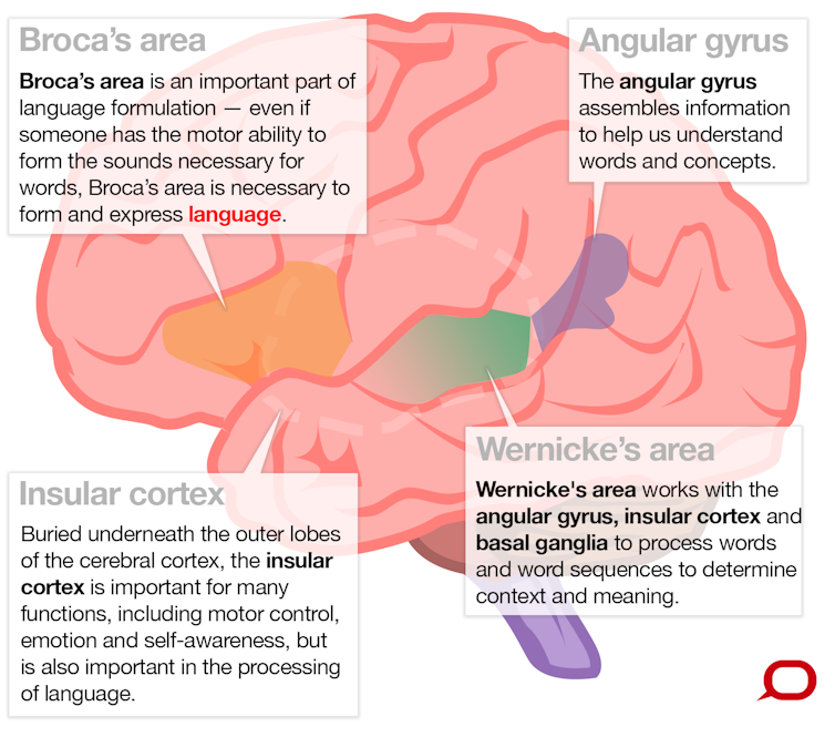
These regions work together as a network to process words and word sequences to determine context and meaning. This enables our receptive language abilities, which means the ability to understand language. Complementary to this is expressive language, which is the ability to produce language.
To speak sensibly, you must think of words to convey an idea or message, formulate them into a sentence according to grammatical rules and then use your lungs, vocal cords and mouth to create sounds. Regions in your frontal, temporal and parietal lobes formulate what you want to say and the motor cortex , in your frontal lobe, enables you to speak the words.
Most of this language-related brain activity is likely occurring in the left side of your brain. But some people use an even mix of both sides and, rarely, some have right dominance for language. There is an evolutionary view that specialisation of certain functions to one side or the other may be an advantage, as many animals, especially vertebrates, exhibit brain function with prominence on one side.
Why the left side is favoured for language isn’t known. But we do know that injury or conditions such as epilepsy, if it affects the left side of the brain early in a child’s development, can increase the chances language will develop on the right side. The chance of the person being left-handed is also increased. This makes sense, because the left side of the body is controlled by the motor cortex on the right side of the brain.

Selective problems
In 1861, French neurologist Pierre Paul Broca described a patient unable to speak who had no motor impairments to account for the inability. A postmortem examination showed a lesion in a large area towards the lower middle of his left frontal lobe particularly important in language formulation. This is now known as Broca’s area .
The clinical symptom of being unable to speak despite having the motor skills is known as expressive aphasia, or Broca’s aphasia.
In 1867, Carl Wernicke observed an opposite phenomenon. A patient was able to speak but not understand language. This is known as receptive aphasia, or Wernicke’s aphasia. The damaged region, as you might correctly guess, is the Wernicke’s area mentioned above.
Scientists have also observed injured patients with other selective problems , such as an inability to understand most words except nouns; or words with unusual spelling, such as those with silent consonants, like reign.
These difficulties are thought to arise from damage to selective areas or connections between regions in the brain’s language network. However, precise localisation can often be difficult given the complexity of individuals’ symptoms and the uncontrolled nature of their brain injury.
We also know the brain’s language regions work together as a co-ordinated network , with some parts involved in multiple functions and a level of redundancy in some processing pathways. So it’s not simply a matter of one brain region doing one thing in isolation.
How do we know all this?
Before advanced medical imaging, most of our knowledge came from observing unfortunate patients with injuries to particular brain parts. One could relate the approximate region of damage to their specific symptoms. Broca’s and Wernicke’s observations are well-known examples.
Other knowledge was inferred from brain-stimulation studies. Weak electrical stimulation of the brain while a patient is awake is sometimes performed in patients undergoing surgery to remove a lesion such as a tumour. The stimulation causes that part of the brain to stop working for a few seconds, which can enable the surgeon to identify areas of critically important function to avoid damaging during surgery.
In the mid-20th century, this helped neurosurgeons discover more about the localisation of language function in the brain . It was clearly demonstrated that while most people have language originating on the left side of their brain, some could have language originating on the right.
Towards the later part of the 20th century, if a surgeon needed to find out which side of your brain was responsible for language – so he didn’t do any damage – he would put to sleep one side of your brain with an anaesthetic. The doctor would then ask you a series of questions, determining your language side from your ability or inability to answer them. This invasive test (which is less often used today due to the availability of functional brain imaging) is known as the Wada test , named after Juhn Wada, who first described it just after the second world war.
Brain imaging
Today, we can get a much better view of brain function by using imaging techniques, especially magnetic resonance imaging (MRI), a safe procedure that uses magnetic fields to take pictures of your brain.
Using MRI to measure brain function is called functional MRI (fMRI), which detects signals from magnetic properties of blood in vessels supplying oxygen to brain cells. The fMRI signal changes depending on whether the blood is carrying oxygen , which means it slightly reduces the magnetic field, or has delivered up its oxygen, which slightly increases the magnetic field.
A few seconds after brain neurons become active in a brain region, there is an increase in freshly oxygenated blood flow to that brain part, much more than required to satisfy the oxygen demand of the neurons. This is what we see when we say a brain region is activated during certain functions.
Brain-imaging methods have revealed that much more of our brain is involved in language processing than previously thought. We now know that numerous regions in every major lobe (frontal, parietal, occipital and temporal lobes; and the cerebellum, an area at the bottom of the brain) are involved in our ability to produce and comprehend language.
Functional MRI is also becoming a useful clinical tool. In some centres it has replaced the Wada test to determine where language is in the brain .
Scientists are also using fMRI to build up a finer picture of how the brain processes language by designing experiments that compare which areas are active during various tasks. For instance, researchers have observed differences in brain language regions of dyslexic children compared to those without dyslexia.
Researchers compared fMRI images of groups of children with and without dyslexia while they performed language-related tasks. They found that dyslexic children had, on average, less activity in Broca’s area mainly on the left during this task. They also had less activity in or near Wernicke’s area on the left and right, and a portion of the front of the temporal lobe on the right.
Could this type of brain imaging provide a diagnostic signature of dyslexia? This is a work-in-progress, but we hope further study will one day lead to a robust, objective and early brain-imaging test for dyslexia and other disorders.
Want to know how the brain controls your mood? Read today’s accompanying piece here .
- Brain regions
- Language processing
- Brain Control series
Lecturer / Senior Lecturer - Marketing
Communications and Engagement Officer, Corporate Finance Property and Sustainability
Assistant Editor - 1 year cadetship
Executive Dean, Faculty of Health
Lecturer/Senior Lecturer, Earth System Science (School of Science)
How the brain controls our speech
Speaking requires both sides of the brain. Each hemisphere takes over a part of the complex task of forming sounds, modulating the voice and monitoring what has been said. However, the distribution of tasks is different than has been thought up to now, as an interdisciplinary team of neuroscientists and phoneticians at Goethe University Frankfurt and the Leibniz-Centre General Linguistics Berlin has discovered: it is not just the right hemisphere that analyses how we speak -- the left hemisphere also plays a role.
Until now, it has been assumed that the spoken word arises in left side of the brain and is analysed by the right side. According to accepted doctrine, this means that when we learn to speak English and for example practice the sound equivalent to "th," the left side of the brain controls the motor function of the articulators like the tongue, while the right side analyses whether the produced sound actually sounds as we intended.
The division of labour actually follows different principles, as Dr Christian Kell from the Department of Neurology at Goethe University explains: "While the left side of the brain controls temporal aspects such as the transition between speech sounds, the right hemisphere is responsible for the control of the sound spectrum. When you say 'mother', for example, the left hemisphere primarily controls the dynamic transitions between "th" and the vowels, while the right hemisphere primarily controls the sounds themselves." His team, together with the phonetician Dr Susanne Fuchs, was able to demonstrate this division of labour in temporal and spectral control of speech for the first time in studies in which speakers were required to talk while their brain activities were recorded using functional magnetic resonance imaging.
A possible explanation for this division of labour between the two sides of the brain is that the left hemisphere generally analyses fast processes such as the transition between speech sounds better than the right hemisphere. The right hemisphere could be better at controlling the slower processes required for analysing the sound spectrum. A previous study on hand motor function that was published in the scientific publication "elife" demonstrates that this is in fact the case. Kell and his team wanted to learn why the right hand was preferentially used for the control of fast actions and the left hand preferred for slow actions. For example, when cutting bread, the right hand is used to slice with the knife while the left hand holds the bread.
In the experiment, scientists had right-handed test persons tap with both hands to the rhythm of a metronome. In one version they were supposed to tap with each beat, and in another only with every fourth beat. As it turned out, the right hand was more precise during the quick tapping sequence and the left hemisphere, which controls the right side of the body, exhibited increased activity. Conversely, tapping with the left hand corresponded better with the slower rhythm and resulted in the right hemisphere exhibiting increased activity.
Taken together, the two studies create a convincing picture of how complex behaviour -- hand motor functions and speech -- are controlled by both cerebral hemispheres. The left side of the brain has a preference for the control of fast processes while the right side tends to control the slower processes in parallel.
- Language Acquisition
- Neuroscience
- Brain-Computer Interfaces
- Intelligence
- Brain Injury
- Child Development
- Educational Psychology
- Procrastination
- Left-handed
- Neocortex (brain)
- Human brain
- Psychedelic drug
Story Source:
Materials provided by Goethe University Frankfurt . Note: Content may be edited for style and length.
Journal Reference :
- Mareike Floegel, Susanne Fuchs, Christian A. Kell. Differential contributions of the two cerebral hemispheres to temporal and spectral speech feedback control . Nature Communications , 2020; 11 (1) DOI: 10.1038/s41467-020-16743-2
Cite This Page :
Explore More
- Controlling Shape-Shifting Soft Robots
- Brain Flexibility for a Complex World
- ONe Nova to Rule Them All
- AI Systems Are Skilled at Manipulating Humans
- Planet Glows With Molten Lava
- A Fragment of Human Brain, Mapped
- Symbiosis Solves Long-Standing Marine Mystery
- Surprising Common Ideas in Environmental ...
- 2D All-Organic Perovskites: 2D Electronics
- Generative AI That Imitates Human Motion
Trending Topics
Strange & offbeat.
Masks Strongly Recommended but Not Required in Maryland, Starting Immediately
Due to the downward trend in respiratory viruses in Maryland, masking is no longer required but remains strongly recommended in Johns Hopkins Medicine clinical locations in Maryland. Read more .
- Vaccines
- Masking Guidelines
- Visitor Guidelines

Brain Anatomy and How the Brain Works
What is the brain.
The brain is a complex organ that controls thought, memory, emotion, touch, motor skills, vision, breathing, temperature, hunger and every process that regulates our body. Together, the brain and spinal cord that extends from it make up the central nervous system, or CNS.
What is the brain made of?
Weighing about 3 pounds in the average adult, the brain is about 60% fat. The remaining 40% is a combination of water, protein, carbohydrates and salts. The brain itself is a not a muscle. It contains blood vessels and nerves, including neurons and glial cells.
What is the gray matter and white matter?
Gray and white matter are two different regions of the central nervous system. In the brain, gray matter refers to the darker, outer portion, while white matter describes the lighter, inner section underneath. In the spinal cord, this order is reversed: The white matter is on the outside, and the gray matter sits within.

Gray matter is primarily composed of neuron somas (the round central cell bodies), and white matter is mostly made of axons (the long stems that connects neurons together) wrapped in myelin (a protective coating). The different composition of neuron parts is why the two appear as separate shades on certain scans.

Each region serves a different role. Gray matter is primarily responsible for processing and interpreting information, while white matter transmits that information to other parts of the nervous system.
How does the brain work?
The brain sends and receives chemical and electrical signals throughout the body. Different signals control different processes, and your brain interprets each. Some make you feel tired, for example, while others make you feel pain.
Some messages are kept within the brain, while others are relayed through the spine and across the body’s vast network of nerves to distant extremities. To do this, the central nervous system relies on billions of neurons (nerve cells).
Main Parts of the Brain and Their Functions
At a high level, the brain can be divided into the cerebrum, brainstem and cerebellum.

The cerebrum (front of brain) comprises gray matter (the cerebral cortex) and white matter at its center. The largest part of the brain, the cerebrum initiates and coordinates movement and regulates temperature. Other areas of the cerebrum enable speech, judgment, thinking and reasoning, problem-solving, emotions and learning. Other functions relate to vision, hearing, touch and other senses.
Cerebral Cortex
Cortex is Latin for “bark,” and describes the outer gray matter covering of the cerebrum. The cortex has a large surface area due to its folds, and comprises about half of the brain’s weight.
The cerebral cortex is divided into two halves, or hemispheres. It is covered with ridges (gyri) and folds (sulci). The two halves join at a large, deep sulcus (the interhemispheric fissure, AKA the medial longitudinal fissure) that runs from the front of the head to the back. The right hemisphere controls the left side of the body, and the left half controls the right side of the body. The two halves communicate with one another through a large, C-shaped structure of white matter and nerve pathways called the corpus callosum. The corpus callosum is in the center of the cerebrum.
The brainstem (middle of brain) connects the cerebrum with the spinal cord. The brainstem includes the midbrain, the pons and the medulla.
- Midbrain. The midbrain (or mesencephalon) is a very complex structure with a range of different neuron clusters (nuclei and colliculi), neural pathways and other structures. These features facilitate various functions, from hearing and movement to calculating responses and environmental changes. The midbrain also contains the substantia nigra, an area affected by Parkinson’s disease that is rich in dopamine neurons and part of the basal ganglia, which enables movement and coordination.
- Pons. The pons is the origin for four of the 12 cranial nerves, which enable a range of activities such as tear production, chewing, blinking, focusing vision, balance, hearing and facial expression. Named for the Latin word for “bridge,” the pons is the connection between the midbrain and the medulla.
- Medulla. At the bottom of the brainstem, the medulla is where the brain meets the spinal cord. The medulla is essential to survival. Functions of the medulla regulate many bodily activities, including heart rhythm, breathing, blood flow, and oxygen and carbon dioxide levels. The medulla produces reflexive activities such as sneezing, vomiting, coughing and swallowing.
The spinal cord extends from the bottom of the medulla and through a large opening in the bottom of the skull. Supported by the vertebrae, the spinal cord carries messages to and from the brain and the rest of the body.
The cerebellum (“little brain”) is a fist-sized portion of the brain located at the back of the head, below the temporal and occipital lobes and above the brainstem. Like the cerebral cortex, it has two hemispheres. The outer portion contains neurons, and the inner area communicates with the cerebral cortex. Its function is to coordinate voluntary muscle movements and to maintain posture, balance and equilibrium. New studies are exploring the cerebellum’s roles in thought, emotions and social behavior, as well as its possible involvement in addiction, autism and schizophrenia.
Brain Coverings: Meninges
Three layers of protective covering called meninges surround the brain and the spinal cord.
- The outermost layer, the dura mater , is thick and tough. It includes two layers: The periosteal layer of the dura mater lines the inner dome of the skull (cranium) and the meningeal layer is below that. Spaces between the layers allow for the passage of veins and arteries that supply blood flow to the brain.
- The arachnoid mater is a thin, weblike layer of connective tissue that does not contain nerves or blood vessels. Below the arachnoid mater is the cerebrospinal fluid, or CSF. This fluid cushions the entire central nervous system (brain and spinal cord) and continually circulates around these structures to remove impurities.
- The pia mater is a thin membrane that hugs the surface of the brain and follows its contours. The pia mater is rich with veins and arteries.

Lobes of the Brain and What They Control
Each brain hemisphere (parts of the cerebrum) has four sections, called lobes: frontal, parietal, temporal and occipital. Each lobe controls specific functions.

- Frontal lobe. The largest lobe of the brain, located in the front of the head, the frontal lobe is involved in personality characteristics, decision-making and movement. Recognition of smell usually involves parts of the frontal lobe. The frontal lobe contains Broca’s area, which is associated with speech ability.
- Parietal lobe. The middle part of the brain, the parietal lobe helps a person identify objects and understand spatial relationships (where one’s body is compared with objects around the person). The parietal lobe is also involved in interpreting pain and touch in the body. The parietal lobe houses Wernicke’s area, which helps the brain understand spoken language.
- Occipital lobe. The occipital lobe is the back part of the brain that is involved with vision.
- Temporal lobe. The sides of the brain, temporal lobes are involved in short-term memory, speech, musical rhythm and some degree of smell recognition.
Deeper Structures Within the Brain
Pituitary gland.
Sometimes called the “master gland,” the pituitary gland is a pea-sized structure found deep in the brain behind the bridge of the nose. The pituitary gland governs the function of other glands in the body, regulating the flow of hormones from the thyroid, adrenals, ovaries and testicles. It receives chemical signals from the hypothalamus through its stalk and blood supply.
Hypothalamus
The hypothalamus is located above the pituitary gland and sends it chemical messages that control its function. It regulates body temperature, synchronizes sleep patterns, controls hunger and thirst and also plays a role in some aspects of memory and emotion.
Small, almond-shaped structures, an amygdala is located under each half (hemisphere) of the brain. Included in the limbic system, the amygdalae regulate emotion and memory and are associated with the brain’s reward system, stress, and the “fight or flight” response when someone perceives a threat.
Hippocampus
A curved seahorse-shaped organ on the underside of each temporal lobe, the hippocampus is part of a larger structure called the hippocampal formation. It supports memory, learning, navigation and perception of space. It receives information from the cerebral cortex and may play a role in Alzheimer’s disease.
Pineal Gland
The pineal gland is located deep in the brain and attached by a stalk to the top of the third ventricle. The pineal gland responds to light and dark and secretes melatonin, which regulates circadian rhythms and the sleep-wake cycle.
Ventricles and Cerebrospinal Fluid
Deep in the brain are four open areas with passageways between them. They also open into the central spinal canal and the area beneath arachnoid layer of the meninges.
The ventricles manufacture cerebrospinal fluid , or CSF, a watery fluid that circulates in and around the ventricles and the spinal cord, and between the meninges. CSF surrounds and cushions the spinal cord and brain, washes out waste and impurities, and delivers nutrients.

Blood Supply to the Brain
Two sets of blood vessels supply blood and oxygen to the brain: the vertebral arteries and the carotid arteries.
The external carotid arteries extend up the sides of your neck, and are where you can feel your pulse when you touch the area with your fingertips. The internal carotid arteries branch into the skull and circulate blood to the front part of the brain.
The vertebral arteries follow the spinal column into the skull, where they join together at the brainstem and form the basilar artery , which supplies blood to the rear portions of the brain.
The circle of Willis , a loop of blood vessels near the bottom of the brain that connects major arteries, circulates blood from the front of the brain to the back and helps the arterial systems communicate with one another.

Cranial Nerves
Inside the cranium (the dome of the skull), there are 12 nerves, called cranial nerves:
- Cranial nerve 1: The first is the olfactory nerve, which allows for your sense of smell.
- Cranial nerve 2: The optic nerve governs eyesight.
- Cranial nerve 3: The oculomotor nerve controls pupil response and other motions of the eye, and branches out from the area in the brainstem where the midbrain meets the pons.
- Cranial nerve 4: The trochlear nerve controls muscles in the eye. It emerges from the back of the midbrain part of the brainstem.
- Cranial nerve 5: The trigeminal nerve is the largest and most complex of the cranial nerves, with both sensory and motor function. It originates from the pons and conveys sensation from the scalp, teeth, jaw, sinuses, parts of the mouth and face to the brain, allows the function of chewing muscles, and much more.
- Cranial nerve 6: The abducens nerve innervates some of the muscles in the eye.
- Cranial nerve 7: The facial nerve supports face movement, taste, glandular and other functions.
- Cranial nerve 8: The vestibulocochlear nerve facilitates balance and hearing.
- Cranial nerve 9: The glossopharyngeal nerve allows taste, ear and throat movement, and has many more functions.
- Cranial nerve 10: The vagus nerve allows sensation around the ear and the digestive system and controls motor activity in the heart, throat and digestive system.
- Cranial nerve 11: The accessory nerve innervates specific muscles in the head, neck and shoulder.
- Cranial nerve 12: The hypoglossal nerve supplies motor activity to the tongue.
The first two nerves originate in the cerebrum, and the remaining 10 cranial nerves emerge from the brainstem, which has three parts: the midbrain, the pons and the medulla.
Find a Treatment Center
- Neurology and Neurosurgery
Find Additional Treatment Centers at:
- Howard County Medical Center
- Sibley Memorial Hospital
- Suburban Hospital
Request an Appointment

Facioscapulohumeral Muscular Dystrophy in Children

Moyamoya Disease
Motor Stereotypies
Related Topics
- U.S. Department of Health & Human Services
- Virtual Tour
- Staff Directory
- En Español
You are here
Nih research matters.
February 13, 2024
How the brain produces speech
At a glance.
- Researchers identified how neurons in the human brain encode various elements of speech.
- The findings might be used to help develop treatments for speech and language disorders.

Speech and language depend on our ability to produce a wide variety of sounds in a specific order. How the neurons in the human brain work together to plan and produce speech remains poorly understood.
To begin to address this question, an NIH-funded team of researchers, led by Drs. Ziv Williams and Sydney Cash at Massachusetts General Hospital, recorded neuron activity during natural speech in five native English speakers. The experiments were done while participants were having electrodes implanted for deep brain stimulation. The researchers recorded neurons in a prefrontal brain region known to be involved in word planning and sentence construction. They used high-density arrays of tiny electrodes that could record signals from many individual neurons at once. Their results appeared in Nature on January 31, 2024.
The scientists found that the activity of almost half the neurons depended on the particular sounds, or phonemes, in the word about to be said. Some neurons, for instance, became more active ahead of speaking the sounds for “p” or “b”, which involve stopping airflow at the lips. Others did so ahead of speaking “k” or “g” sounds, which are formed by the tongue against the soft palate. Moreover, certain neurons seemed to reflect the specific combination of phonemes in the upcoming word. The team found that they could predict the phonemes that made up the word about to be spoken based on the activity of these neurons.
For about a quarter of the neurons, activity further reflected specific syllables, or ordered sequences of phonemes that may be all or part of a word. The team could predict the syllables in the upcoming word using the activity from these neurons. These neurons did not respond to the phonemes in the syllable by themselves. Nor did they respond to the phonemes out of order or split across different syllables.
A minority of neurons responded to the presence of prefixes or suffixes. These are examples of morphemes, or groups of sounds that carry specific meanings. The presence of morphemes in the upcoming word could be predicted from these neurons’ activities.
The team also found that different sets of neurons activated in a specific order. The morpheme neurons activated first, around 400 milliseconds (ms) before the utterance. Phoneme neurons activated next, around 200 ms before the utterance. Syllable neurons activated last, around 70 ms before utterance. Most neurons responded to the same feature (phoneme, syllable, or morpheme) both before and during the utterance. But the activity patterns during the utterance differed from those before it. Finally, the team found that neurons that responded to speech sounds during speaking differed from those that responded to those same speech sounds during listening.
In an accompanying paper in the same issue of Nature , another research team used the same technique to examine how neurons in another area of the brain respond while listening to speech. They similarly found that single neurons encoded different speech sound cues.
The findings suggest how various elements of speech are encoded in the brain, and how the brain combines these elements to form spoken words. This information might aid in developing brain-machine interfaces that can synthesize speech. Such devices could help a range of patients with conditions that impair speech.
“Disruptions in the speech and language networks are observed in a wide variety of neurological disorders—including stroke, traumatic brain injury, tumors, neurodegenerative disorders, neurodevelopmental disorders, and more,” says co-author Dr. Arjun Khanna. “Our hope is that a better understanding of the basic neural circuitry that enables speech and language will pave the way for the development of treatments for these disorders.”
—by Brian Doctrow, Ph.D.
Related Links
- Scientists Translate Brain Activity into Music
- Brain Decoder Turns a Person’s Brain Activity into Words
- Understanding How the Brain Tracks Social Status and Competition
- Study Reveals Brain Networks Critical for Conversation
- Device Allows Paralyzed Man to Communicate with Words
- How the Human Brain Tracks Location
- Memories Involve Replay of Neural Firing Patterns
- Scientists Create Speech Using Brain Signals
- How The Brain Keeps Track of Time
- Brain Basics: Know Your Brain
References: Single-neuronal elements of speech production in humans. Khanna AR, Muñoz W, Kim YJ, Kfir Y, Paulk AC, Jamali M, Cai J, Mustroph ML, Caprara I, Hardstone R, Mejdell M, Meszéna D, Zuckerman A, Schweitzer J, Cash S, Williams ZM. Nature . 2024 Jan 31. doi: 10.1038/s41586-023-06982-w. Online ahead of print. PMID: 38297120.
Funding: NIH’s National Institute of Neurological Disorders and Stroke (NINDS), National Institute of Mental Health (NIMH), and National Institute on Deafness and other Communication Disorders (NIDCD); Canadian Institutes of Health Research; Foundations of Human Behavior Initiative; Tiny Blue Dot Foundation; American Association of University Women.
Connect with Us
- More Social Media from NIH
NOTICE MyAANS, password-protected resources, and purchases are currently experiencing issues and are unavailable. We are working to get this fixed as soon as possible.
The site navigation utilizes arrow, enter, escape, and space bar key commands. Left and right arrows move across top level links and expand / close menus in sub levels. Up and Down arrows will open main level menus and toggle through sub tier links. Enter and space open menus and escape closes them as well. Tab will move on to the next part of the site rather than go through menu items.
Anatomy of the Brain
The brain serves many important functions. It gives meaning to things that happen in the world surrounding us. Through the five senses of sight, smell, hearing, touch and taste, the brain receives messages, often many at the same time.
The brain controls thoughts, memory and speech, arm and leg movements and the function of many organs within the body. It also determines how people respond to stressful situations (i.e. writing of an exam, loss of a job, birth of a child, illness, etc.) by regulating heart and breathing rates. The brain is an organized structure, divided into many components that serve specific and important functions.
The weight of the brain changes from birth through adulthood. At birth, the average brain weighs about one pound, and grows to about two pounds during childhood. The average weight of an adult female brain is about 2.7 pounds, while the brain of an adult male weighs about three pounds.
The Nervous System
The nervous system is commonly divided into the central nervous system and the peripheral nervous system. The central nervous system is made up of the brain, its cranial nerves and the spinal cord. The peripheral nervous system is composed of the spinal nerves that branch from the spinal cord and the autonomous nervous system (divided into the sympathetic and parasympathetic nervous system).
The Cell Structure of the Brain
The brain is made up of two types of cells: neurons and glial cells, also known as neuroglia or glia. The neuron is responsible for sending and receiving nerve impulses or signals. Glial cells are non-neuronal cells that provide support and nutrition, maintain homeostasis, form myelin and facilitate signal transmission in the nervous system. In the human brain, glial cells outnumber neurons by about 50 to one. Glial cells are the most common cells found in primary brain tumors.
When a person is diagnosed with a brain tumor, a biopsy may be done, in which tissue is removed from the tumor for identification purposes by a pathologist. Pathologists identify the type of cells that are present in this brain tissue, and brain tumors are named based on this association. The type of brain tumor and cells involved impact patient prognosis and treatment.
The Meninges
The brain is housed inside the bony covering called the cranium. The cranium protects the brain from injury. Together, the cranium and bones that protect the face are called the skull. Between the skull and brain is the meninges, which consist of three layers of tissue that cover and protect the brain and spinal cord. From the outermost layer inward they are: the dura mater, arachnoid and pia mater.
Dura Mater: In the brain, the dura mater is made up of two layers of whitish, nonelastic film or membrane. The outer layer is called the periosteum. An inner layer, the dura, lines the inside of the entire skull and creates little folds or compartments in which parts of the brain are protected and secured. The two special folds of the dura in the brain are called the falx and the tentorium. The falx separates the right and left half of the brain and the tentorium separates the upper and lower parts of the brain.
Arachnoid: The second layer of the meninges is the arachnoid. This membrane is thin and delicate and covers the entire brain. There is a space between the dura and the arachnoid membranes that is called the subdural space. The arachnoid is made up of delicate, elastic tissue and blood vessels of varying sizes.
Pia Mater: The layer of meninges closest to the surface of the brain is called the pia mater. The pia mater has many blood vessels that reach deep into the surface of the brain. The pia, which covers the entire surface of the brain, follows the folds of the brain. The major arteries supplying the brain provide the pia with its blood vessels. The space that separates the arachnoid and the pia is called the subarachnoid space. It is within this area that cerebrospinal fluid flows.
Cerebrospinal Fluid
Cerebrospinal fluid (CSF) is found within the brain and surrounds the brain and the spinal cord. It is a clear, watery substance that helps to cushion the brain and spinal cord from injury. This fluid circulates through channels around the spinal cord and brain, constantly being absorbed and replenished. It is within hollow channels in the brain, called ventricles, that the fluid is produced. A specialized structure within each ventricle, called the choroid plexus, is responsible for the majority of CSF production. The brain normally maintains a balance between the amount of CSF that is absorbed and the amount that is produced. However, disruptions in this system may occur.
The Ventricular System
The ventricular system is divided into four cavities called ventricles, which are connected by a series of holes, called foramen, and tubes.
Two ventricles enclosed in the cerebral hemispheres are called the lateral ventricles (first and second). They each communicate with the third ventricle through a separate opening called the Foramen of Munro. The third ventricle is in the center of the brain, and its walls are made up of the thalamus and hypothalamus.
The third ventricle connects with the fourth ventricle through a long tube called the Aqueduct of Sylvius.
CSF flowing through the fourth ventricle flows around the brain and spinal cord by passing through another series of openings.
Brain Components and Functions
The brainstem is the lower extension of the brain, located in front of the cerebellum and connected to the spinal cord. It consists of three structures: the midbrain, pons and medulla oblongata. It serves as a relay station, passing messages back and forth between various parts of the body and the cerebral cortex. Many simple or primitive functions that are essential for survival are located here.
The midbrain is an important center for ocular motion while the pons is involved with coordinating eye and facial movements, facial sensation, hearing and balance.
The medulla oblongata controls breathing, blood pressure, heart rhythms and swallowing. Messages from the cortex to the spinal cord and nerves that branch from the spinal cord are sent through the pons and the brainstem. Destruction of these regions of the brain will cause "brain death." Without these key functions, humans cannot survive.
The reticular activating system is found in the midbrain, pons, medulla and part of the thalamus. It controls levels of wakefulness, enables people to pay attention to their environments and is involved in sleep patterns.
Originating in the brainstem are 10 of the 12 cranial nerves that control hearing, eye movement, facial sensations, taste, swallowing and movements of the face, neck, shoulder and tongue muscles. The cranial nerves for smell and vision originate in the cerebrum. Four pairs of cranial nerves originate from the pons: nerves five through eight.
The cerebellum is located at the back of the brain beneath the occipital lobes. It is separated from the cerebrum by the tentorium (fold of dura). The cerebellum fine tunes motor activity or movement, e.g. the fine movements of fingers as they perform surgery or paint a picture. It helps one maintain posture, sense of balance or equilibrium, by controlling the tone of muscles and the position of limbs. The cerebellum is important in one's ability to perform rapid and repetitive actions such as playing a video game. In the cerebellum, right-sided abnormalities produce symptoms on the same side of the body.
The cerebrum, which forms the major portion of the brain, is divided into two major parts: the right and left cerebral hemispheres. The cerebrum is a term often used to describe the entire brain. A fissure or groove that separates the two hemispheres is called the great longitudinal fissure. The two sides of the brain are joined at the bottom by the corpus callosum. The corpus callosum connects the two halves of the brain and delivers messages from one half of the brain to the other. The surface of the cerebrum contains billions of neurons and glia that together form the cerebral cortex.
The cerebral cortex appears grayish brown in color and is called the "gray matter." The surface of the brain appears wrinkled. The cerebral cortex has sulci (small grooves), fissures (larger grooves) and bulges between the grooves called gyri. Scientists have specific names for the bulges and grooves on the surface of the brain. Decades of scientific research have revealed the specific functions of the various regions of the brain. Beneath the cerebral cortex or surface of the brain, connecting fibers between neurons form a white-colored area called the "white matter."
The cerebral hemispheres have several distinct fissures. By locating these landmarks on the surface of the brain, it can effectively be divided into pairs of "lobes." Lobes are simply broad regions of the brain. The cerebrum or brain can be divided into pairs of frontal, temporal, parietal and occipital lobes. Each hemisphere has a frontal, temporal, parietal and occipital lobe. Each lobe may be divided, once again, into areas that serve very specific functions. The lobes of the brain do not function alone: they function through very complex relationships with one another.
Messages within the brain are delivered in many ways. The signals are transported along routes called pathways. Any destruction of brain tissue by a tumor can disrupt the communication between different parts of the brain. The result will be a loss of function such as speech, the ability to read or the ability to follow simple spoken commands. Messages can travel from one bulge on the brain to another (gyri to gyri), from one lobe to another, from one side of the brain to the other, from one lobe of the brain to structures that are found deep in the brain, e.g. thalamus, or from the deep structures of the brain to another region in the central nervous system.
Research has determined that touching one side of the brain sends electrical signals to the other side of the body. Touching the motor region on the right side of the brain would cause the opposite side or the left side of the body to move. Stimulating the left primary motor cortex would cause the right side of the body to move. The messages for movement and sensation cross to the other side of the brain and cause the opposite limb to move or feel a sensation. The right side of the brain controls the left side of the body and vice versa. So if a brain tumor occurs on the right side of the brain that controls the movement of the arm, the left arm may be weak or paralyzed.
Cranial Nerves
There are 12 pairs of nerves that originate from the brain itself. These nerves are responsible for very specific activities and are named and numbered as follows:
- Olfactory: Smell
- O ptic: Visual fields and ability to see
- Oculomotor: Eye movements; eyelid opening
- Trochlear: Eye movements
- Trigeminal: Facial sensation
- Abducens: Eye movements
- Facial: Eyelid closing; facial expression; taste sensation
- Auditory/vestibular: Hearing; sense of balance
- Glossopharyngeal: Taste sensation; swallowing
- Vagus: Swallowing; taste sensation
- Accessory : Control of neck and shoulder muscles
- Hypoglossal: Tongue movement
Hypothalamus
The hypothalamus is a small structure that contains nerve connections that send messages to the pituitary gland. The hypothalamus handles information that comes from the autonomic nervous system. It plays a role in controlling functions such as eating, sexual behavior and sleeping; and regulates body temperature, emotions, secretion of hormones and movement. The pituitary gland develops from an extension of the hypothalamus downwards and from a second component extending upward from the roof of the mouth.
Frontal Lobes
The frontal lobes are the largest of the four lobes responsible for many different functions. These include motor skills such as voluntary movement, speech, intellectual and behavioral functions. The areas that produce movement in parts of the body are found in the primary motor cortex or precentral gyrus. The prefrontal cortex plays an important part in memory, intelligence, concentration, temper and personality.
The premotor cortex is a region found beside the primary motor cortex. It guides eye and head movements and a person’s sense of orientation. Broca's area, important in language production, is found in the frontal lobe, usually on the left side.
Occipital Lobes
These lobes are located at the back of the brain and enable humans to receive and process visual information. They influence how humans process colors and shapes. The occipital lobe on the right interprets visual signals from the left visual space, while the left occipital lobe performs the same function for the right visual space.
Parietal Lobes
These lobes interpret simultaneously, signals received from other areas of the brain such as vision, hearing, motor, sensory and memory. A person’s memory, and the new sensory information received, give meaning to objects.
Temporal Lobes
These lobes are located on each side of the brain at about ear level, and can be divided into two parts. One part is on the bottom (ventral) of each hemisphere, and the other part is on the side (lateral) of each hemisphere. An area on the right side is involved in visual memory and helps humans recognize objects and peoples' faces. An area on the left side is involved in verbal memory and helps humans remember and understand language. The rear of the temporal lobe enables humans to interpret other people’s emotions and reactions.
Limbic System
This system is involved in emotions. Included in this system are the hypothalamus, part of the thalamus, amygdala (active in producing aggressive behavior) and hippocampus (plays a role in the ability to remember new information).
Pineal Gland
This gland is an outgrowth from the posterior or back portion of the third ventricle. In some mammals, it controls the response to darkness and light. In humans, it has some role in sexual maturation, although the exact function of the pineal gland in humans is unclear.
Pituitary Gland
The pituitary is a small gland attached to the base of the brain (behind the nose) in an area called the pituitary fossa or sella turcica. The pituitary is often called the "master gland" because it controls the secretion of hormones. The pituitary is responsible for controlling and coordinating the following:
- Growth and development
- The function of various body organs (i.e. kidneys, breasts and uterus)
- The function of other glands (i.e. thyroid, gonads, and adrenal glands)
Posterior Fossa
This is a cavity in the back part of the skull which contains the cerebellum, brainstem and cranial nerves 5-12.
The thalamus serves as a relay station for almost all information that comes and goes to the cortex. It plays a role in pain sensation, attention and alertness. It consists of four parts: the hypothalamus, the epythalamus, the ventral thalamus and the dorsal thalamus. The basal ganglia are clusters of nerve cells surrounding the thalamus.
Language and Speech Functions
In general, the left hemisphere or side of the brain is responsible for language and speech. Because of this, it has been called the "dominant" hemisphere. The right hemisphere plays a large part in interpreting visual information and spatial processing. In about one-third of individuals who are left-handed, speech function may be located on the right side of the brain. Left-handed individuals may need specialized testing to determine if their speech center is on the left or right side prior to any surgery in that area.
Many neuroscientists believe that the left hemisphere and perhaps other portions of the brain are important in language. Aphasia is simply a disturbance of language. Certain parts of the brain are responsible for specific functions in language production. There are many types of aphasias, each depending upon the brain area that is affected, and the role that area plays in language production.
There is an area in the frontal lobe of the left hemisphere called Broca’s area. It is next to the region that controls the movement of facial muscles, tongue, jaw and throat. If this area is destroyed, a person will have difficulty producing the sounds of speech, because of the inability to move the tongue or facial muscles to form words. A person with Broca's aphasia can still read and understand spoken language, but has difficulty speaking and writing.
There is a region in the left temporal lobe called Wernicke's area. Damage to this area causes Wernicke's aphasia. An individual can make speech sounds, but they are meaningless (receptive aphasia) because they do not make any sense.
The AANS does not endorse any treatments, procedures, products or physicians referenced in these patient fact sheets. This information is provided as an educational service and is not intended to serve as medical advice. Anyone seeking specific neurosurgical advice or assistance should consult his or her neurosurgeon, or locate one in your area through the AANS’ “Find a Board-certified Neurosurgeon” online tool.
Find a Board-certified Neurosurgeon Near You
Support nref.
Make a difference in one minute. Text "Neurosurgery" to 41444 and follow the prompts to donate easily from your mobile device.

Support the NREF When You Shop
Register with iGive.com or AmazonSmile and designate the NREF as your charity.
Scientists unlock secret of how the brain encodes speech
- Feinberg School of Medicine
People like the late Stephen Hawking can think about what they want to say, but are unable to speak because their muscles are paralyzed. In order to communicate, they can use devices that sense a person’s eye or cheek movements to spell out words one letter at a time. However, this process is slow and unnatural.
Scientists want to help these completely paralyzed, or “locked-in,” individuals communicate more intuitively by developing a brain machine interface to decode the commands the brain is sending to the tongue, palate, lips and larynx (articulators.)
In other words, the person would simply try to say words and the brain machine interface (BMI) would translate into speech.
New research from Northwestern Medicine and Weinberg College of Arts and Sciences has moved science closer to creating speech brain machine interfaces by unlocking new information about how the brain encodes speech.
Scientists have discovered the brain controls speech production in a similar manner to how it controls the production of arm and hand movements. To do this, researchers recorded signals from two parts of the brain and decoded what these signals represented. Scientists found the brain represents both the goals of what we are trying to say (speech sounds like “pa” and “ba”) and the individual movements that we use to achieve those goals (how we move our lips, palate, tongue and larynx). The different representations occur in two different parts of the brain.
“This can help us build better speech decoders for BMIs, which will move us closer to our goal of helping people that are locked-in speak again,” said lead author Dr. Marc Slutzky, associate professor of neurology and of physiology at Northwestern University Feinberg School of Medicine and a Northwestern Medicine neurologist.
The study was published Sept. 26 in the Journal of Neuroscience.
The discovery could also potentially help people with other speech disorders, such as apraxia of speech, which is seen in children as well as after stroke in adults. In speech apraxia, an individual has difficulty translating speech messages from the brain into spoken language.
How words are translated from your brain into speech
Speech is composed of individual sounds, called phonemes, that are produced by coordinated movements of the lips, tongue, palate and larynx. However, scientists didn’t know exactly how these movements, called articulatory gestures, are planned by the brain. In particular, it was not fully understood how the cerebral cortex controls speech production, and no evidence of gesture representation in the brain had been shown.
“We hypothesized speech motor areas of the brain would have a similar organization to arm motor areas of the brain,” Slutzky said. “The precentral cortex would represent movements (gestures) of the lips, tongue, palate and larynx, and the higher level cortical areas would represent the phonemes to a greater extent.”
That’s exactly what they found. “We studied two parts of the brain that help to produce speech,” Slutzky said. “The precentral cortex represented gestures to a greater extent than phonemes. The inferior frontal cortex, which is a higher level speech area, represented both phonemes and gestures.”
Chatting up patients in brain surgery to decode their brain signals
Northwestern scientists recorded brain signals from the cortical surface using electrodes placed in patients undergoing brain surgery to remove brain tumors. The patients had to be awake during their surgery, so researchers asked them to read words from a screen.
After the surgery, scientists marked the times when the patients produced phonemes and gestures. Then they used the recorded brain signals from each cortical area to decode which phonemes and gestures had been produced, and measured the decoding accuracy. The brain signals in the precentral cortex were more accurate at decoding gestures than phonemes, while those in the inferior frontal cortex were equally good at decoding both phonemes and gestures. This information helped support linguistic models of speech production. It will also help guide engineers in designing brain machine interfaces to decode speech from these brain areas.
The next step for the research is to develop an algorithm for brain machine interfaces that would not only decode gestures but also combine those decoded gestures to form words.
This was an interdisciplinary, cross-campus investigation; authors included a neurosurgeon, a neurologist, a computer scientist, a linguist, and biomedical engineers. In addition to Slutzky, Northwestern authors are Emily M. Mugler, Matthew C. Tate (neurological surgery), Jessica W. Templer (neurology) and Matthew A. Goldrick (linguistics).
The paper is titled “Differential Representation of Articulatory Gestures and Phonemes in Precentral and Inferior Frontal Gyri.”
This work was supported in part by the Doris Duke Charitable Foundation, Northwestern Memorial Foundation Dixon Translational Research Award (including partial funding from National Center for Advancing Translational Sciences, UL1TR000150 and UL1TR001422), NIH grants F32DC015708and R01NS094748and National Science Foundation1321015.
Editor’s Picks

This algorithm makes robots perform better
‘the night watchman’ named next one book selection, six northwestern faculty elected to american academy of arts and sciences, related stories.

The childhood of Hans Christian Andersen explored in musical at Northwestern University
Northwestern academy supports evanston students, trethewey named to the academy of american poets.
- Type 2 Diabetes
- Heart Disease
- Digestive Health
- Multiple Sclerosis
- Diet & Nutrition
- Supplements
- Health Insurance
- Public Health
- Patient Rights
- Caregivers & Loved Ones
- End of Life Concerns
- Health News
- Thyroid Test Analyzer
- Doctor Discussion Guides
- Hemoglobin A1c Test Analyzer
- Lipid Test Analyzer
- Complete Blood Count (CBC) Analyzer
- What to Buy
- Editorial Process
- Meet Our Medical Expert Board
Left-Sided Stroke Signs, Long-Term Effects, and Treatment
What happens if the dominant side of your brain is affected by a stroke?
- Long-Term Effects
A left-sided stroke is a stroke that damages the left side of the brain . This type of stroke typically causes language and speech problems, as well as physical symptoms that affect the right side of the body. A stroke can be ischemic (caused by an obstruction) or hemorrhagic (caused by bleeding in the brain).
A stroke is an emergency that requires immediate medical attention. Treatment may include blood thinners, fluid management, and/or medication or surgery to remove a blood clot or relieve pressure in the skull. Recovery may include physical therapy, occupational therapy, and speech therapy.
This article discusses the types of left-sided strokes, as well as signs, effects, treatment, and prevention.
Verywell / Daniel Fishel
Types of Left-Sided Strokes
Most of the time, strokes are caused by insufficient blood supply to a region of the brain. Ischemia is a lack of blood supply due to blockage or narrowing of a blood vessel.
Sometimes, a stroke can occur due to a hemorrhage (bleeding) often caused by a leaking blood vessel.
An ischemic stroke causes damage to a region of the brain that is supplied by the blood vessel that is obstructed (blocked).
This type of stroke can occur due to atherosclerotic disease within the blood vessels of the brain (hardening and narrowing due to a buildup of cholesterol plaques). Changes in blood vessels leading to narrowing and ischemic stroke may also be caused by high blood pressure, diabetes, and smoking.
Ischemic strokes may also occur due to a blood clot traveling to the brain from the heart or the carotid artery.
Hemorrhagic
A hemorrhagic stroke can occur due to a ruptured brain aneurysm (a bulging area in the wall of an artery), a damaged blood vessel that leaks, or damage that occurred during an ischemic stroke .
A hemorrhagic stroke can cause tissue death (infarction) in the area of the brain that is supplied by the bleeding blood vessel. Additionally, the accumulation of blood can cause further damage in nearby areas.
Brain damage from a hemorrhagic stroke can lead to seizures (uncontrolled electrical disturbances in the brain) due to irritation from the bleeding. In some people, seizures can persist even after the blood is completely reabsorbed. This most commonly occurs when the blood affects an area of the brain that controls motor function.
Left-sided strokes occur about as frequently as right-sided strokes. According to the Centers for Disease Control and Prevention (CDC), every year, more than 795,000 people in the United States have a stroke.
Signs of a Left-Sided Stroke
There are several signs of a left-sided stroke . They include:
- Weakness in the face, arm, and/or leg on the right side of the body
- Decreased sensation on the right side of the body
- Effortful or slurred speech
- Speaking fluently but with incorrect or nonword content
- Difficulty understanding language
- Changes in visual perception
- Severe and sudden head pain
- Sudden dizziness or loss of balance
- Left-sided sensory and motor symptoms if the stroke involves areas known as the cerebellum and brain stem
Get immediate medical attention if you or someone else experiences any of these symptoms. A stroke is a medical emergency that can worsen quickly, causing disability or death. The long-term effects can be minimized if treatment is started promptly.
Left-sided strokes vs. right-sided strokes
A left-sided stroke affects the right side of a person's body, while a right-sided stroke affects the left side. Left-sided strokes also cause problems with speech, while right-sided strokes cause problems with seeing or responding to things that occur on the left side of the body.
Long-Term Effects of a Left-Sided Stroke
The lasting effects of a left-sided stroke range from mild to severe, depending on the size of the stroke and the timing of treatment.
Paralysis and Weakness
Hemiplegia and hemiparesis can occur on the right side of the body after a left-sided stroke:
- Hemiplegia is complete paralysis, and it can affect the right side of the face, arm, and/or leg after a left-sided stroke.
- Hemiparesis is partial weakness with some residual strength.
Immediately after a left-sided stroke, right-body hemiplegia or hemiparesis will be apparent. After months or longer, the weak areas of the body can become gradually stronger and more coordinated. Individuals with moderate-to-severe weakness are more likely to experience spasticity, with stiffness and tightness of the muscles.
Decreased Sensation on the Right Side
Diminished sensation on the right side of the body can occur after a stroke involving the left sensory cortex or the left internal capsule. Diminished sensation can involve the face, arm, and/or leg, and sometimes the torso.
Sensory impairment can indirectly affect your ability to control your body because you rely on sensory feedback to coordinate your movements.
In addition to decreased sensation, sometimes paresthesias can occur in the same areas that have diminished sensation. Paresthesias involve numbness, tingling, burning, or a sense of pins and needles. They can occur when the specific area of the body is touched or without a trigger.
Speech and Language Problems
A left-sided stroke can cause a range of problems with speech, language, and swallowing.
Left-sided strokes are known to cause aphasia, which is a language deficit. There are several types of aphasia, and they occur when one or more of the speech areas of the brain are damaged.
Wernicke’s aphasia, also called fluent aphasia, causes difficulty understanding language. A person who has this type of aphasia can speak fluently, but the words do not make sense. Wernicke’s aphasia can occur when there is damage to the language area near the left sensory cortex of the brain.
Broca’s aphasia is a type of language deficit in which a person may have full or mildly impaired language comprehension, with difficulty forming words and sentences. This type of aphasia occurs when there is damage to the language area near the left motor cortex of the brain.
Language Centers
Aphasia occurs when the language areas of the brain are damaged. Language is usually located on the left hemisphere of the brain. For nearly all people who are right-handed, the language functions are located on the left side of the brain. Some people who are left-handed have the language centers on the right side of the brain.
A stroke can cause difficulty swallowing, which is also known as dysphagia. This can happen if the stroke affects the part of the brain that coordinates your swallowing muscles.
Stoke may also cause problems with swallowing because of its effect on the muscles around your mouth, your ability to sit up straight, and how you use eating utensils.
Apraxia of Speech
Speech apraxia is difficulty with the brain's control of the motor movements of speech. Unlike dysphagia, which is not specific to damage on one side of the brain and affects speech and swallowing, speech apraxia is a type of language impairment.
Apraxia of speech can occur as a result of damage to the left insular cortex, an area deep in the left hemisphere of the brain.
Vision Problems
After a left-sided stroke involving the temporal lobe, parietal lobe, or occipital cortex at the back of the brain, a person can have vision defects on the right side. Homonymous hemianopia from a left-sided stroke is vision loss on the right visual field of both eyes. It can affect the upper or lower field of vision or both.
Cognitive Impairments
After a stroke affecting the left hemisphere of the brain, a person can develop difficulty thinking and making decisions. These cognitive deficits, often described as executive dysfunction, can occur due to damage of the left frontal lobe, left temporal lobe, or left parietal lobe. Generally, a larger stroke is expected to cause more severe cognitive impairments.
Treatment for Left-Sided Strokes
A stroke should be treated immediately after symptoms begin. Calling for emergency transport to the hospital is important to get rapid and proper medical attention on arrival.
Treatment for an Ischemic Stroke
The first line of treatment for an ischemic stroke is a medication called tissue plasminogen activator (tPA). It helps break up the blood clot that is causing the blockage in your brain. This medication is usually given within the first three hours after symptoms begin. Depending on the circumstances, some healthcare providers may give tPA up to four-and-a-half hours after symptoms start.
If you can't have tPA, you may be given a blood thinner to help stop the blood clot from growing and prevent new ones from forming.
Some people require surgery to remove the clot in the brain, called a thrombectomy. During this procedure, the surgeon places a long, flexible tube into an artery in your thigh and maneuvers it into the blocked artery. A surgical device is then used to remove the blockage.
Treatment for a Hemorrhagic Stroke
Treatment for a hemorrhagic stroke typically includes blood pressure medication to help reduce the strain on the blood vessels in your brain. You may also receive vitamin K, which can help stop bleeding.
Medical procedures used to treat a hemorrhagic stroke include surgery to clamp the aneurysm and stop the bleeding. A similar procedure called coil embolization involves threading a long, flexible tube through an artery in your upper thigh and into the brain. A tiny coil that helps blood clot is then inserted into the aneurysm.
You may also need surgery to drain excess fluid or pooled blood. Sometimes, a part of the skull is temporarily removed in order to relieve pressure. You may also need a blood transfusion to replace blood lost during the stroke or during surgery.
Rehabilitation and Recovery
After your condition is stabilized, treatment focuses on recovery and rehabilitation . Patients who qualify for and complete a course of intensive/acute rehabilitation may have better outcomes and lower mortality (rates of death) than those who do not get this care.
After a stroke, reach out for support to get the help you need to recover. It can take time and hard work, but it is important to be patient and maintain connections. Friends and family can help greatly by learning the effects of a left-sided stroke and modifying expectations, especially with communication.
All neurological rehabilitation takes time but can substantially help a person improve their ability to communicate with others and function independently.
- Physical Therapy: A physical therapist can help you improve your motor control. Your physical therapist will have you do exercises that help strengthen your muscles and improve your balance. This work can help you regain your ability to stand and walk.
- Occupational Therapy: An occupational therapist will work with you on the skills you need to manage your daily life. This may include eating, dressing, bathing, and cooking. Working with an occupational therapist can help you regain your independence.
- Speech Therapy: A speech therapist will work with you if you are having trouble speaking or comprehending speech. A speech therapist can also help you with reading and writing. Speech therapy can also help you manage cognitive difficulties and swallowing problems.
The time it takes to recover from a left-sided stroke depends on how severe the stroke was and the amount of damage done. It can take months or longer to improve after a stroke. A person may recover almost completely or have substantial permanent handicaps after a stroke.
Prevention
After you have had a stroke, you are at higher risk of having another one. It is important that you follow your healthcare provider's guidance for preventing another stroke.
Stroke prevention is a comprehensive strategy that involves reducing the risk of cerebrovascular disease. Prevention involves medication, diet, and lifestyle approaches.
Stroke prevention includes:
- Smoking cessation
- Cholesterol and triglyceride control
- Blood pressure control
- Blood sugar control
- Management of heart disease, including abnormal heart rhythms such as atrial fibrillation
A left-sided stroke affects the left side of the brain and the right side of the body. This type of stroke can also cause cognitive and language problems, which can include difficulty with comprehension and/or speech.
A stroke is a medical emergency, and immediate treatment can help prevent disability or death. Recovery after a left-sided stroke involves physical rehabilitation, as well as speech and language therapy.
Johns Hopkins Medicine. Stroke .
Centers for Disease Control and Prevention. Stroke facts .
American Association of Neurological Surgeons. Stroke .
National Institute of Health. Dysphagia .
Conterno M, Kümmerer D, Dressing A, Glauche V, Urbach H, Weiller C, Rijntjes M. Speech apraxia and oral apraxia: association or dissociation? A multivariate lesion-symptom mapping study in acute stroke patients . Exp Brain Res. 2021. doi:10.1007/s00221-021-06224-3
Meier EL, Kelly CR, Goldberg EB, Hillis AE. Executive control deficits and lesion correlates in acute left hemisphere stroke survivors with and without aphasia . Brain Imaging Behav . 2021:1–10. doi:10.1007/s11682-021-00580-y
National Lung, Heart, and Blood Institute. Stroke treatment .
American Stroke Association. Rehab therapy after a stroke .
By Heidi Moawad, MD Dr. Moawad is a neurologist and expert in brain health. She regularly writes and edits health content for medical books and publications.
Thank you for visiting nature.com. You are using a browser version with limited support for CSS. To obtain the best experience, we recommend you use a more up to date browser (or turn off compatibility mode in Internet Explorer). In the meantime, to ensure continued support, we are displaying the site without styles and JavaScript.
- View all journals
- Explore content
- About the journal
- Publish with us
- Sign up for alerts
- 13 May 2024
Brain-reading device is best yet at decoding ‘internal speech’
- Miryam Naddaf
You can also search for this author in PubMed Google Scholar

Illustration showing the supramarginal gyrus (orange), a region of the brain involved in speech. Credit: My Box/Alamy
Scientists have developed brain implants that can decode internal speech — identifying words that two people spoke in their minds without moving their lips or making a sound.
Although the technology is at an early stage — it was shown to work with only a handful of words, and not phrases or sentences — it could have clinical applications in future.
Similar brain–computer interface (BCI) devices, which translate signals in the brain into text, have reached speeds of 62–78 words per minute for some people . But these technologies were trained to interpret speech that is at least partly vocalized or mimed.
The latest study — published in Nature Human Behaviour on 13 May 1 — is the first to decode words spoken entirely internally, by recording signals from individual neurons in the brain in real time.
“It's probably the most advanced study so far on decoding imagined speech,” says Silvia Marchesotti, a neuroengineer at the University of Geneva, Switzerland.
“This technology would be particularly useful for people that have no means of movement any more,” says study co-author Sarah Wandelt, a neural engineer who was at the California Institute of Technology in Pasadena at the time the research was done. “For instance, we can think about a condition like locked-in syndrome.”
Mind-reading tech
The researchers implanted arrays of tiny electrodes in the brains of two people with spinal-cord injuries. They placed the devices in the supramarginal gyrus (SMG), a region of the brain that had not been previously explored in speech-decoding BCIs.
Figuring out the best places in the brain to implant BCIs is one of the key challenges for decoding internal speech, says Marchesotti. The authors decided to measure the activity of neurons in the SMG on the basis of previous studies showing that this part of the brain is active in subvocal speech and in tasks such as deciding whether words rhyme.
Two weeks after the participants were implanted with microelectrode arrays in their left SMG, the researchers began collecting data. They trained the BCI on six words (battlefield, cowboy, python, spoon, swimming and telephone) and two meaningless pseudowords (nifzig and bindip). “The point here was to see if meaning was necessary for representation,” says Wandelt.
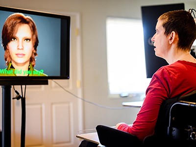
The rise of brain-reading technology: what you need to know
Over three days, the team asked each participant to imagine speaking the words shown on a screen and repeated this process several times for each word. The BCI then combined measurements of the participants’ brain activity with a computer model to predict their internal speech in real time.
For the first participant, the BCI captured distinct neural signals for all of the words and was able to identify them with 79% accuracy. But the decoding accuracy was only 23% for the second participant, who showed preferential representation for ‘spoon’ and ‘swimming’ and had fewer neurons that were uniquely active for each word. “It's possible that different sub-areas in the supramarginal gyrus are more, or less, involved in the process,” says Wandelt.
Christian Herff, a computational neuroscientist at Maastricht University in the Netherlands, thinks these results might highlight the different ways in which people process internal speech. “Previous studies showed that there are different abilities in performing the imagined task and also different BCI control abilities,” adds Marchesotti.
The authors also found that 82–85% of neurons that were active during internal speech were also active when the participants vocalized the words. But some neurons were active only during internal speech, or responded differently to specific words in the different tasks.
Although the study represents significant progress in decoding internal speech, clinical applications are still a long way off, and many questions remain unanswered.
“The problem with internal speech is we don't know what’s happening and how is it processed,” says Herff. For example, researchers have not been able to determine whether the brain represents internal speech phonetically (by sound) or semantically (by meaning). “What I think we need are larger vocabularies” for the experiments, says Herff.
Marchesotti also wonders whether the technology can be generalized to people who have lost the ability to speak, given that the two study participants are able to talk and have intact brain speech areas. “This is one of the things that I think in the future can be addressed,” she says.
The next step for the team will be to test whether the BCI can distinguish between the letters of the alphabet. “We could maybe have an internal speech speller, which would then really help patients to spell words,” says Wandelt.
doi: https://doi.org/10.1038/d41586-024-01424-7
Wandelt, S. K. et al. Nature Hum. Behav . https://doi.org/10.1038/s41562-024-01867-y (2024).
Article Google Scholar
Download references
Reprints and permissions
Related Articles

- Medical research
- Neuroscience

Human embryos embrace asymmetry to form the body
News 13 MAY 24

Hacking the immune system could slow ageing — here’s how
News Feature 07 MAY 24

‘Orangutan, heal thyself’: First wild animal seen using medicinal plant
News 02 MAY 24

How does ChatGPT ‘think’? Psychology and neuroscience crack open AI large language models
News Feature 14 MAY 24

Cubic millimetre of brain mapped in spectacular detail
News 09 MAY 24

Retuning of hippocampal representations during sleep
Article 08 MAY 24

Found: the dial in the brain that controls the immune system
News 01 MAY 24
Junior and Senior Staff Scientists in microfluidics & optics
Seeking staff scientists with expertise in microfluidics or optics to support development of new technology to combat antimicrobial resistance
Boston, Massachusetts (US)
Harvard Medical School Systems Biology Department
Postdoctoral Fellow in Biophysics
Seeking ambitious biophysicists to fight antimicrobial resistance with new technology at the interface of microfluidics and optics
Recruitment of Principal Investigators by the School of Life Sciences, Peking University
The School of Life Sciences at Peking University is actively seeking talents, with an emphasis on bioinformatics/computational biology/AI/RNA biology.
Beijing (CN)
School of Life Sciences, Peking University
2024 Recruitment notice Shenzhen Institute of Synthetic Biology: Shenzhen, China
The wide-ranging expertise drawing from technical, engineering or science professions...
Shenzhen,China
Shenzhen Institute of Synthetic Biology
Head of Operational Excellence
In this key position, you’ll be responsible for ensuring efficiency and quality in journal workflows through continuous improvement and innovation.
United States (US) - Remote
American Physical Society
Sign up for the Nature Briefing newsletter — what matters in science, free to your inbox daily.
Quick links
- Explore articles by subject
- Guide to authors
- Editorial policies
- Português Br
- Journalist Pass
5 things to know about stroke
Mayo Clinic Staff
Share this:

A stroke can happen at any time and to anyone. You might be talking to your loved one and notice they're suddenly slurring their words. Or, while grocery shopping, you realize you can't move your hand to pick up a jar from the shelf. You can go from feeling as usual to feeling sick within a matter of seconds to minutes.
Here are five key things to know about stroke:
1. strokes affect the oxygen and nutrients supplied to your brain..
Strokes occur when nutrients and oxygen are not delivered to the brain through blood vessels, leading to the death of brain cells. This lack of delivery can be caused by a clot in a blood vessel obstructing the blood flow to the brain, known as an ischemic stroke, or when a blood vessel ruptures and prevents blood flow to the brain, known as a hemorrhagic stroke.
Sometimes, the obstruction to the blood flow and the resulting symptoms are caused by a temporary clot and are transient, resulting in a mini-stroke or transient ischemic attack, or TIA.
2. Strokes can happen to anyone.
Strokes can happen to anyone regardless of age, gender or race. Certain risk factors can put you at a higher risk of stroke .
Risk factors are divided into two categories:
- Controllable — the ones you can control or improve
- Uncontrollable — those that are not within your control
Common controllable risk factors include:
- Atrial fibrillation, which increases stroke risk by five times
- Excessive alcohol intake — an average of more than one drink per day for women or more than two drinks a day for men
- High blood pressure
- High cholesterol
- Illicit drug use
- Obstructive sleep apnea
- Physical inactivity
- Sickle cell anemia
- Smoking or vaping
Uncontrollable risk factors include:
- Increasing age
3. Be prepared to spot the signs of a stroke.
Learn to recognize the signs of stroke quickly.
The American Stroke Association lists these symptoms to help you know when to seek medical care:
- F = Face drooping: Ask the person to smile and see if the smile is uneven.
- A = Arm weakness: Ask the person to raise both arms and see if one arm drifts down.
- S = Speech difficulty: Ask the person to speak and see if the speech is slurred.
- T = Time to call 911: Stroke is an emergency. Call 911 at once. Note the time when any of the symptoms first appear.
Other stroke symptoms to watch for include:
- Numbness of the face, arm or leg, especially on one side of the body
- Sudden confusion, trouble speaking or difficulty understanding speech
- Sudden onset severe headache with no known cause
- Sudden vision issues, such as trouble seeing in one or both eyes
- Trouble walking, loss of balance, dizziness or coordination
If you or someone you are with have any stroke-like symptoms, seek immediate medical care.
4. A stroke is a medical emergency.
Every second counts when someone is experiencing a stroke. Once a stroke starts, the brain loses around 1.9 million neurons each minute. For every hour without treatment, the brain loses as many neurons as it typically does in nearly 3.6 years of regular aging.
While waiting for paramedics, do these things if possible:
- If the person is conscious, lay them down on their side with their head slightly raised and supported to prevent falls.
- Loosen any restrictive clothing that could cause breathing difficulties.
- If weakness is obvious in any limb, support it and avoid pulling on it when moving the person.
- If the person is unconscious, check their breathing and pulse, and put them on their side.
- If they do not have a pulse or are not breathing, start CPR straight away.
- Do not give them anything to eat or drink if you feel they may have trouble swallowing.
5. Women have an increased risk of stroke.
According to the American Stroke Association, stroke is the third most common cause of death in women. Over 90,000 women die from a stroke in the U.S. each year. Every 1 in 5 women will have a stroke , and about 55,000 more women than men have a stroke each year, with Black women having the highest prevalence of stroke.
The risk of stroke increases in women who have atrial fibrillation, migraines with aura, smoke, take birth control pills, use hormonal replacement therapy, are pregnant or have preeclampsia.
Talk to your healthcare team about your stroke risk and ways to lower your risk by addressing controllable factors.
Prashant Natteru, M.B.B.S., M.D. , is a neurologist in La Crosse , Wisconsin.
This article first published on the Mayo Clinic Health System blog .
- In case you missed it: This week’s Top 5 stories on social media Mayo Clinic Q and A: Can honey help my cough?
Related Articles

Watch CBS News
Randy Travis sings again, courtesy of AI
How the parasite that RFK Jr. said he contracted infects the brain

A little-known parasitic infection in the brain received a jolt of attention Wednesday when presidential candidate Robert F. Kennedy Jr. said he once suffered from it.
The condition, known as neurocysticercosis, is a brain infection linked to larvae from pork tapeworms. It's rare: It hospitalizes roughly 1,000 to 2,000 people every year in the U.S.
Neurocysticercosis causes seizures, headaches, blindness, blurred vision, dizziness, psychosis or memory loss. In some cases, it may even be fatal.
The infection typically follows a sequence of events: People eat raw or undercooked pork that carries a tapeworm. They then shed tapeworm eggs in their stool and contaminate food or surfaces by not washing their hands. As a result, they or those around them who eat that food or touch those surfaces can accidentally swallow tapeworm eggs.
Once they are swallowed, the eggs hatch into larvae, which can move from the intestine to the brain. The larvae form fluid-filled pockets, or cysts, resembling tiny, clear balloons about a centimeter in diameter.
The eggs are “real sticky,” said Dr. Clinton White, an infectious disease professor at the University of Texas Medical Branch at Galveston. “They get on people’s hands and under their fingernails. Hand-to-mouth I think is an important route of transmission.”
Overall, the condition is poorly understood by health care providers, according to the Centers for Disease Control and Prevention, which considers neurocysticercosis to be a “neglected parasitic infection.”
“It’s an important disease and gets ignored a lot,” White said.
On Wednesday, The New York Times reported that Kennedy, who is running for president as an independent, once experienced a parasitic brain infection.
Kennedy’s campaign press secretary, Stefanie Spear, said in a statement responding to the Times article: “Mr. Kennedy traveled extensively in Africa, South America, and Asia in his work as an environmental advocate, and in one of those locations contracted a parasite. The issue was resolved more than 10 years ago, and he is in robust physical and mental health.”
According to the Times, Kennedy said in a 2012 deposition that, two years earlier, doctors had detected an abnormal spot on his brain amid symptoms of memory loss and mental fogginess. One doctor concluded it was caused by a worm that got into his brain and then died, Kennedy told the Times.
NBC News was not able to immediately verify the details of the Times report.
The Times also reported that Kennedy said he did not know what type of parasite it was. However, in an interview on the web radio show “ Pushing the Limit s” on Wednesday, Kennedy told host Brian Shapiro that the infection was neurocysticercosis and that the parasite "comes from eating undercooked pork."
Among tapeworms, pork tapeworms are most commonly associated with brain infections.
In March, doctors in Florida published a case study about a 52-year-old man who developed severe migraines from neurocysticercosis. The man had eaten undercooked bacon for most of his life , so doctors presumed he had acquired a pork tapeworm, shed the tapeworm’s eggs and then ingested them, leading to his brain infection.
Larval cysts from neurocysticercosis often live in the brain for around five to 10 years. When they start to die, the body’s immune system attacks them, and the inflammation can lead to epileptic seizures or life-threatening brain swelling. Some cases of neurocysticercosis are asymptomatic.
In countries where pork tapeworms are endemic, neurocysticercosis causes around 30% of all epilepsy cases , according to the World Health Organization.
“It’s a major neurologic infection worldwide,” White said.
“Most U.S. cases are imported,” he added. “They’re in people that acquire the infection in endemic areas — particularly in villages, say, in Mexico or Central or South America.”
The infection is also endemic in sub-Saharan Africa and several parts of Asia.
White said neurocysticercosis is starting to be diagnosed more in the U.S., most likely because doctors are becoming more aware of it.
“It’s probably the parasitic disease that causes the most problems in this country,” he said.
While symptoms may resolve on their own without any treatment, some patients may require seizure medications or a combination of steroids and anti-parasitic drugs. In severe cases, patients may need surgery to remove the cysts.
Aria Bendix is the breaking health reporter for NBC News Digital.
- Election 2024
- Entertainment
- Newsletters
- Photography
- Personal Finance
- AP Investigations
- AP Buyline Personal Finance
- AP Buyline Shopping
- Press Releases
- Israel-Hamas War
- Russia-Ukraine War
- Global elections
- Asia Pacific
- Latin America
- Middle East
- Election Results
- Delegate Tracker
- AP & Elections
- Auto Racing
- 2024 Paris Olympic Games
- Movie reviews
- Book reviews
- Personal finance
- Financial Markets
- Business Highlights
- Financial wellness
- Artificial Intelligence
- Social Media
Illness took away her voice. AI created a replica she carries in her phone
A team of Rhode Island doctors show that artificial intelligence voice-cloning technology that has triggered widespread fears of misuse, can be tremendously helpful to some people. (AP Video: Rodrique Ngowi) The Associated Press and OpenAI have a licensing and technology agreement that allows OpenAI access to part of AP’s text archives. AP is solely responsible for all content.
Alexis Bogan, whose speech was impaired by a brain tumor, uses an AI powered smartphone app to create a audible drink order at a Starbucks drive-thru on Monday, April 29, 2024, in Lincoln, R.I. The app converts her typed entries into a verbal message created using her original voice. (AP Photo/Steven Senne)
- Copy Link copied
Alexis Bogan types a response to a reporter’s question with an app which approximates her lost voice, Thursday, March 11, 2024, at Rhode Island Hospital in Providence, R.I. Doctors treating Bogan, whose speech was impaired by a brain tumor, used a voice-cloning tool from OpenAI to recreate her previous voice. (AP Photo/Josh Reynolds)
Alexis Bogan, whose speech was impaired by a brain tumor, uses mobile phone with an app that features a voice-cloning tool to order a drink at a Starbucks drive-thru Monday, April 29, 2024, in Lincoln, R.I. Doctors treating Bogan are recreating her original voice using a voice-cloning tool from OpenAI. (AP Photo/Steven Senne)
Dr. Rohaid Ali plays a video from a high school project made by his patient Alexis Bogan on Thursday, March 11, 2024, at Rhode Island Hospital in Providence, R.I. Doctors treating Bogan, whose speech was impaired by a brain tumor, used the recorded sample of her speech and a voice-cloning tool from OpenAI to recreate her previous voice. Neurosurgeon Dr. Konstantina Svokos, right, looks on. (AP Photo/Josh Reynolds)
Alexis Bogan, center, and her mother Pamela Bogan, right, react to hearing a recreation of her lost voice from a prompt typed by Dr. Fatima Mirza, left, on Thursday, March 11, 2024, at Rhode Island Hospital in Providence, R.I. Doctors treating Bogan, who’s speech was impaired by a brain tumor, used a voice-cloning tool from OpenAI to recreate her previous voice. (AP Photo/Josh Reynolds)
PROVIDENCE, R.I. (AP) — The voice Alexis “Lexi” Bogan had before last summer was exuberant.
She loved to belt out Taylor Swift and Zach Bryan ballads in the car. She laughed all the time — even while corralling misbehaving preschoolers or debating politics with friends over a backyard fire pit. In high school, she was a soprano in the chorus.
Then that voice was gone.
Doctors in August removed a life-threatening tumor lodged near the back of her brain. When the breathing tube came out a month later, Bogan had trouble swallowing and strained to say “hi” to her parents. Months of rehabilitation aided her recovery, but her speech is still impaired. Friends, strangers and her own family members struggle to understand what she is trying to tell them.
In April, the 21-year-old got her old voice back. Not the real one, but a voice clone generated by artificial intelligence that she can summon from a phone app. Trained on a 15-second time capsule of her teenage voice — sourced from a cooking demonstration video she recorded for a high school project — her synthetic but remarkably real-sounding AI voice can now say almost anything she wants.
She types a few words or sentences into her phone and the app instantly reads it aloud.
“Hi, can I please get a grande iced brown sugar oat milk shaken espresso,” said Bogan’s AI voice as she held the phone out her car’s window at a Starbucks drive-thru.
Experts have warned that rapidly improving AI voice-cloning technology can amplify phone scams, disrupt democratic elections and violate the dignity of people — living or dead — who never consented to having their voice recreated to say things they never spoke.
It’s been used to produce deepfake robocalls to New Hampshire voters mimicking President Joe Biden. In Maryland, authorities recently charged a high school athletic director with using AI to generate a fake audio clip of the school’s principal making racist remarks.
But Bogan and a team of doctors at Rhode Island’s Lifespan hospital group believe they’ve found a use that justifies the risks. Bogan is one of the first people — the only one with her condition — who have been able to recreate a lost voice with OpenAI’s new Voice Engine. Some other AI providers, such as the startup ElevenLabs, have tested similar technology for people with speech impediments and loss — including a lawyer who now uses her voice clone in the courtroom.
“We’re hoping Lexi’s a trailblazer as the technology develops,” said Dr. Rohaid Ali, a neurosurgery resident at Brown University’s medical school and Rhode Island Hospital. Millions of people with debilitating strokes, throat cancer or neurogenerative diseases could benefit, he said.
“We should be conscious of the risks, but we can’t forget about the patient and the social good,” said Dr. Fatima Mirza, another resident working on the pilot. “We’re able to help give Lexi back her true voice and she’s able to speak in terms that are the most true to herself.”
Mirza and Ali, who are married, caught the attention of ChatGPT-maker OpenAI because of their previous research project at Lifespan using the AI chatbot to simplify medical consent forms for patients. The San Francisco company reached out while on the hunt earlier this year for promising medical applications for its new AI voice generator.
Bogan was still slowly recovering from surgery. The illness started last summer with headaches, blurry vision and a droopy face, alarming doctors at Hasbro Children’s Hospital in Providence. They discovered a vascular tumor the size of a golf ball pressing on her brain stem and entangled in blood vessels and cranial nerves.
“It was a battle to get control of the bleeding and get the tumor out,” said pediatric neurosurgeon Dr. Konstantina Svokos.
The tumor’s location and severity coupled with the complexity of the 10-hour surgery damaged Bogan’s control of her tongue muscles and vocal cords, impeding her ability to eat and talk, Svokos said.
“It’s almost like a part of my identity was taken when I lost my voice,” Bogan said.
The feeding tube came out this year. Speech therapy continues, enabling her to speak intelligibly in a quiet room but with no sign she will recover the full lucidity of her natural voice.
“At some point, I was starting to forget what I sounded like,” Bogan said. “I’ve been getting so used to how I sound now.”
Whenever the phone rang at the family’s home in the Providence suburb of North Smithfield, she would push it over to her mother to take her calls. She felt she was burdening her friends whenever they went to a noisy restaurant. Her dad, who has hearing loss, struggled to understand her.
Back at the hospital, doctors were looking for a pilot patient to experiment with OpenAI’s technology.
“The first person that came to Dr. Svokos’ mind was Lexi,” Ali said. “We reached out to Lexi to see if she would be interested, not knowing what her response would be. She was game to try it out and see how it would work.”
Bogan had to go back a few years to find a suitable recording of her voice to “train” the AI system on how she spoke. It was a video in which she explained how to make a pasta salad.
Her doctors intentionally fed the AI system just a 15-second clip. Cooking sounds make other parts of the video imperfect. It was also all that OpenAI needed — an improvement over previous technology requiring much lengthier samples.
They also knew that getting something useful out of 15 seconds could be vital for any future patients who have no trace of their voice on the internet. A brief voicemail left for a relative might have to suffice.
When they tested it for the first time, everyone was stunned by the quality of the voice clone. Occasional glitches — a mispronounced word, a missing intonation — were mostly imperceptible. In April, doctors equipped Bogan with a custom-built phone app that only she can use.
“I get so emotional every time I hear her voice,” said her mother, Pamela Bogan, tears in her eyes.
“I think it’s awesome that I can have that sound again,” added Lexi Bogan, saying it helped “boost my confidence to somewhat where it was before all this happened.”
She now uses the app about 40 times a day and sends feedback she hopes will help future patients. One of her first experiments was to speak to the kids at the preschool where she works as a teaching assistant. She typed in “ha ha ha ha” expecting a robotic response. To her surprise, it sounded like her old laugh.
She’s used it at Target and Marshall’s to ask where to find items. It’s helped her reconnect with her dad. And it’s made it easier for her to order fast food.
Bogan’s doctors have started cloning the voices of other willing Rhode Island patients and hope to bring the technology to hospitals around the world. OpenAI said it is treading cautiously in expanding the use of Voice Engine, which is not yet publicly available.
A number of smaller AI startups already sell voice-cloning services to entertainment studios or make them more widely available. Most voice-generation vendors say they prohibit impersonation or abuse, but they vary in how they enforce their terms of use.
“We want to make sure that everyone whose voice is used in the service is consenting on an ongoing basis,” said Jeff Harris, OpenAI’s lead on the product. “We want to make sure that it’s not used in political contexts. So we’ve taken an approach of being very limited in who we’re giving the technology to.”
Harris said OpenAI’s next step involves developing a secure “voice authentication” tool so that users can replicate only their own voice. That might be “limiting for a patient like Lexi, who had sudden loss of her speech capabilities,” he said. “So we do think that we’ll need to have high-trust relationships, especially with medical providers, to give a little bit more unfettered access to the technology.”
Bogan has impressed her doctors with her focus on thinking about how the technology could help others with similar or more severe speech impediments.
“Part of what she has done throughout this entire process is think about ways to tweak and change this,” Mirza said. “She’s been a great inspiration for us.”
While for now she must fiddle with her phone to get the voice engine to talk, Bogan imagines an AI voice engine that improves upon older remedies for speech recovery — such as the robotic-sounding electrolarynx or a voice prosthesis — in melding with the human body or translating words in real time.
She’s less sure about what will happen as she grows older and her AI voice continues to sound like she did as a teenager. Maybe the technology could “age” her AI voice, she said.
For now, “even though I don’t have my voice fully back, I have something that helps me find my voice again,” she said.
The Associated Press and OpenAI have a licensing and technology agreement that allows OpenAI access to part of AP’s text archives.

R.F.K. Jr. Says Doctors Found a Dead Worm in His Brain
The presidential candidate has faced previously undisclosed health issues, including a parasite that he said ate part of his brain.
Robert F. Kennedy Jr. has emphasized his vitality and relative youth compared with the leading Democratic and Republican candidates. Credit... Eduardo Munoz/Reuters
Supported by
- Share full article

By Susanne Craig
- May 8, 2024
In 2010, Robert F. Kennedy Jr. was experiencing memory loss and mental fogginess so severe that a friend grew concerned he might have a brain tumor. Mr. Kennedy said he consulted several of the country’s top neurologists, many of whom had either treated or spoken to his uncle, Senator Edward M. Kennedy, before his death the previous year of brain cancer.
Several doctors noticed a dark spot on the younger Mr. Kennedy’s brain scans and concluded that he had a tumor, he said in a 2012 deposition reviewed by The New York Times. Mr. Kennedy was immediately scheduled for a procedure at Duke University Medical Center by the same surgeon who had operated on his uncle , he said.
While packing for the trip, he said, he received a call from a doctor at NewYork-Presbyterian Hospital who had a different opinion: Mr. Kennedy, he believed, had a dead parasite in his head.
The doctor believed that the abnormality seen on his scans “was caused by a worm that got into my brain and ate a portion of it and then died,” Mr. Kennedy said in the deposition.
Now an independent presidential candidate, the 70-year-old Mr. Kennedy has portrayed his athleticism and relative youth as an advantage over the two oldest people to ever seek the White House: President Biden, 81, and former President Donald J. Trump, 77. Mr. Kennedy has secured a place on the ballots in Utah, Michigan, Hawaii and, his campaign says, California and Delaware. His intensive efforts to gain access in more states could put him in a position to tip the election.
He has gone to lengths to appear hale, skiing with a professional snowboarder and with an Olympic gold medalist who called him a “ripper” as they raced down the mountain. A camera crew was at his side while he lifted weights, shirtless, at an outdoor gym in Venice Beach.
Still, over the years, he has faced serious health issues, some previously undisclosed, including the apparent parasite.
For decades, Mr. Kennedy suffered from atrial fibrillation, a common heartbeat abnormality that increases the risk of stroke or heart failure. He has been hospitalized at least four times for episodes, although in an interview with The Times this winter, he said he had not had an incident in more than a decade and believed the condition had disappeared.
About the same time he learned of the parasite, he said, he was also diagnosed with mercury poisoning, most likely from ingesting too much fish containing the dangerous heavy metal, which can cause serious neurological issues.
“I have cognitive problems, clearly,” he said in the 2012 deposition. “I have short-term memory loss, and I have longer-term memory loss that affects me.”
In the interview with The Times, he said he had recovered from the memory loss and fogginess and had no aftereffects from the parasite, which he said had not required treatment. Asked last week if any of Mr. Kennedy’s health issues could compromise his fitness for the presidency, Stefanie Spear, a spokeswoman for the Kennedy campaign, told The Times, “That is a hilarious suggestion, given the competition.”
The campaign declined to provide his medical records to The Times. Neither President Biden nor Mr. Trump has released medical records in this election cycle. However, the White House put out a six-page health summary for President Biden in February. Mr. Trump released a three-paragraph statement from his doctor in November.
On Wednesday afternoon, hours after this article was published, Mr. Kennedy posted a comment on his X profile. “I offer to eat 5 more brain worms and still beat President Trump and President Biden in a debate,” the post read. “I feel confident in the result even with a six-worm handicap.”
Doctors who have treated parasitic infections and mercury poisoning said both conditions can sometimes permanently damage brain function, but patients also can have temporary symptoms and mount a full recovery.
Some of Mr. Kennedy’s health issues were revealed in the 2012 deposition, which he gave during divorce proceedings from his second wife, Mary Richardson Kennedy. At the time, Mr. Kennedy was arguing that his earning power had been diminished by his cognitive struggles.
Mr. Kennedy provided more details, including about the apparent parasite, in the phone interview with The Times, conducted when he was on the cusp of getting on his first state ballot. His campaign declined to answer follow-up questions.
In the days after the 2010 call from NewYork-Presbyterian, Mr. Kennedy said in the interview, he underwent a battery of tests. Scans over many weeks showed no change in the spot on his brain, he said.
Doctors ultimately concluded that the cyst they saw on scans contained the remains of a parasite. Mr. Kennedy said that he did not know the type of parasite or where he might have contracted it, though he suspected it might have been during a trip through South Asia.
Several infectious disease experts and neurosurgeons said in separate interviews with The Times that, based on what Mr. Kennedy described, they believed it was likely a pork tapeworm larva. The doctors have not treated Mr. Kennedy and were speaking generally.
Dr. Clinton White, a professor of infectious diseases at the University of Texas Medical Branch in Galveston, said microscopic tapeworm eggs are sticky and easily transferred from one person to another. Once hatched, the larvae can travel in the bloodstream, he said, “and end up in all kinds of tissues.”
Though it is impossible to know, he added that it is unlikely that a parasite would eat a part of the brain, as Mr. Kennedy described. Rather, Dr. White said, it survives on nutrients from the body. Unlike tapeworm larvae in the intestines, those in the brain remain relatively small, about a third of an inch.
Some tapeworm larvae can live in a human brain for years without causing problems. Others can wreak havoc, often when they start to die, which causes inflammation. The most common symptoms are seizures, headaches and dizziness.
There are roughly 2,000 hospitalizations for the condition, known as neurocysticercosis, each year in the United States, according to the journal Emerging Infectious Diseases .
Scott Gardner, curator of the Manter Laboratory for Parasitology at the University of Nebraska-Lincoln, said that once any worm is in a brain, cells calcify around it. “And you’re going to basically have almost like a tumor that’s there forever. It’s not going to go anywhere.”
Dr. Gardner said it was possible a worm would cause memory loss. However, severe memory loss is more often associated with another health scare Mr. Kennedy said he had at the time: mercury poisoning.
Mr. Kennedy said he was then subsisting on a diet heavy on predatory fish, notably tuna and perch, both known to have elevated mercury levels. In the interview with The Times, he said that he had experienced “severe brain fog” and had trouble retrieving words. Mr. Kennedy, an environmental lawyer who has railed against the dangers of mercury contamination in fish from coal-fired power plants , had his blood tested.
He said the tests showed his mercury levels were 10 times what the Environmental Protection Agency considers safe.
At the time, Mr. Kennedy also was a few years into his crusade against thimerosal, a mercury-containing preservative used in some vaccines. He is a longtime vaccine skeptic who has falsely linked childhood inoculations to a rise in autism, as well as to other medical conditions .
In the interview, Mr. Kennedy said he was certain his diet had caused the poisoning. “ I loved tuna fish sandwiches. I ate them all the time,” he said.
The Times described Mr. Kennedy’s symptoms to Elsie Sunderland, an environmental chemist at Harvard who has not spoken to Mr. Kennedy and responded generally about the condition.
She said the mercury levels that Mr. Kennedy described were high, but not surprising for someone consuming that quantity and type of seafood.
Mr. Kennedy said he made changes after these two health scares, including getting more sleep, traveling less and reducing his fish intake.
He also underwent chelation therapy, a treatment that binds to metals in the body so they can be expelled. It is generally given to people contaminated by metals, such as lead and zinc, in industrial accidents. Dr. Sunderland said that when mercury poisoning is clearly diet-related, she would simply recommend that the person stop eating fish. But another doctor who spoke to The Times said she would advise chelation therapy for the levels Mr. Kennedy said he had.
Mr. Kennedy’s heart issue began in college, he said, when it started beating out of sync.
In 2001 he was admitted to a hospital in Seattle while in town to give a speech, according to news reports. He was treated, and released the next day. He was hospitalized at least three additional times between September 2011 and early 2012, including once in Los Angeles, he said in the deposition. On that visit, he said, doctors used a defibrillator to shock his heart to reset the rhythm.
He said in the deposition that stress, caffeine and a lack of sleep triggered the condition. “It feels like there’s a bag of worms in my chest. I can feel immediately when it goes out,” he said.
He also said in the deposition and the interview that he had contracted hepatitis C through intravenous drug use in his youth. He said he had been treated and had no lingering effects from the infection.
Mr. Kennedy has spoken publicly about one other major health condition — spasmodic dysphonia, a neurological disorder that causes his vocal cords to squeeze too close together and explains his hoarse, sometimes strained voice.
He first noticed it when he was 42 years old, he said in the deposition. Mr. Kennedy for years made a significant amount of money giving speeches , and that business fell off as the condition worsened, he said.
He told an interviewer last year that he had recently undergone a procedure available in Japan to implant titanium between his vocal cords to keep them from involuntarily constricting.
Susanne Craig is an investigative reporter. She has written about the finances of Donald J. Trump and Robert F. Kennedy Jr. and has been a journalist for more than 30 years. More about Susanne Craig
Our Coverage of the 2024 Election
Presidential Race
Donald Trump leads President Biden in five crucial battleground states, a new set of polls shows , as young and nonwhite voters express discontent with the president over the economy and the war in Gaza.
With Democratic Senate candidates running well ahead of Biden , the new battleground polling shows a ticket-splitting pattern, Nate Cohn writes .
In an extended riff at a rally in New Jersey, Trump compared migrants to Hannibal Lecte r, the fictional serial killer and cannibal from “The Silence of the Lambs.”
Dodging the Question: Leading Republicans, including several of Trump’s potential running mates, have refused to say flatly that they will accept the outcome of the election .
West Virginia Senate Race: Gov. Jim Justice’s companies have long had a reputation for not paying their debts. But that may be catching up to them as Justice campaigns for a seat in the Senate.
Ohio Senate Race: Bernie Moreno, the Republican challenging Senator Sherrod Brown, tells a riches-to-rags-to-riches tale. But the reality isn’t so tidy .
Maryland Senate Race: The Democratic Senate primary between Angela Alsobrooks, the Prince George’s County executive, and Representative David Trone has grown tighter as they vie to take on Larry Hogan, the popular former two-term Republican governor.
Advertisement
We asked a parasite expert about RFK Jr.'s claim that a worm ate his brain. Here's what they said.
- Did a worm eat Robert F. Kennedy Jr.'s brain?
- The third-party presidential candidate said in a 2012 deposition that a doctor suggested a parasite hurt him.
- We spoke to an expert, who said a brain-eating tapeworm larvae would be impossible.
Brain-eating worms? Not likely.
A shocking report in The New York Times on Wednesday revealed that independent presidential candidate Robert F. Kennedy Jr. suggested in a 2012 deposition that doctors had found a dead worm in his brain.
In the court proceeding — part of his divorce from his second wife — Kennedy said that he had short-term and long-term memory loss, according to The Times.
Kennedy said he had visited doctors in 2010 who thought he had a brain tumor, but another doctor suggested that a dark spot on Kennedy's brain scan was "caused by a worm that got into my brain and ate a portion of it and then died," The Times reported.
Kennedy argued in the deposition that he couldn't make as much money due to his health, and also revealed that he had mercury poisoning around the same time.
Kennedy has portrayed himself as the younger, more healthy alternative to the other two men running for president, Joe Biden and Donald Trump.
His campaign's press secretary confirmed that Kennedy was infected with a parasite 10 years ago and said it was resolved. His campaign told Business Insider that Kennedy is in "robust physical and mental health" and said questioning his fitness is a "hilarious suggestion, given his competition."
Related stories
But could a parasitic worm even cause that kind of damage? One medical expert told Business Insider that Kennedy's version of events doesn't quite add up.
Dr. Janina Caira, a University of Connecticut professor and tapeworm specialist, told BI that Kennedy's parasite sounds more like the larvae of a pork tapeworm.
That would be rare, Caira said in an email. Humans can be infected with the adult worm by eating undercooked pork, but can only be infected with the larvae after eating food or drinking water contaminated by the feces of someone with an adult tapeworm infection.
"This typically happens in areas with poor sanitation," Caira said. "So, it is possible that he could have contracted the infection in South Asia if he came into contact with food or water contaminated with eggs of the tapeworm."
But there's no way the larvae could have consumed Kennedy's brain tissue.
"Absolutely not," Caira wrote.
She said the larvae don't have mouths or digestive systems. Instead, they absorb nutrients through the surface of their bodies. While Caira said it is possible that a worm could do some "mechanical damage" to nearby brain tissue, the larvae are very small, and a single one "would not cause much damage."
That lines up with what experts, who were skeptical of the details, told The New York Times.
However, Dr. Peter Hotez, a pediatrician and global health advocate who is a professor of pediatrics and molecular virology & microbiology at Baylor College of Medicine, wrote on X that "neuroparasitic diseases" and "parasitic worms have a huge impact on the human brain."
Hotez said the diseases are seen in poor populations, with a "surprising amount of illness" in southern states and Texas. He said his team at the National School of Tropical Medicine is working on low-cost vaccines to prevent the conditions.
Watch: McConnell freezes again, raising concern for the 81-year-old Senator's health
- Main content
Why your sleeping position is shortening your life
Back, front or side? The position you favour has the potential to trigger its own set of health issues, from acid reflux to brain health
How many of us monitor our sleep posture? We have a favourite position; we fall asleep and that’s generally as far as it goes. But sleep position can have profound implications not only for the quality of sleep, but also for long-term health. Indeed, in the worst-case scenarios, a bad sleep posture may be slowly killing you.
Despite the impact sleep posture can have on conditions such as dementia and heart disease, research is limited and tends to focus on aches and pains. But back pain is just one implication of an unsuitable sleep position.
Chartered physiotherapist, sleep expert and author of The Good Sleep Guide , Sammy Margo, explains: “Sleep positions can significantly affect your overall health, comfort, and the quality of your sleep . Each position has its pros and cons and understanding them can help you make adjustments for better sleep and health outcomes.”
Dr Kat Lederle is a sleep scientist and the author of Sleep Sense . She points out that lifestyle factors in the day are usually the cause of postural problems at night.
“What you do in the day generally triggers the pain and discomfort that is felt when you sleep in certain positions. One of the most common contributing factors to this is a sedentary lifestyle, so it is important to move regularly during the day.”
The health risks of your sleep position – and how to mitigate them
Side sleeping.

Side sleeping is the most common position but there are health implications for certain people depending on whether they lie on their left or right side.
It is advised that pregnant women and anyone who suffers from acid reflux or gastroesophageal reflux disease (GERD) or other gut problems sleep on their left side.
“This is because the stomach is lower than your oesophagus,” explains Margo.
People with heart conditions , on the other hand, are advised to try sleeping on their right side to alleviate pressure on the heart. Studies show that when people lie on their left side the position of their heart shifts due to gravity. This causes changes in the heart’s electrical activity. Tissues and structures between the lungs hold the heart in place when you sleep on your right side.
Sleep position may also have an impact on brain health . During sleep the brain’s glymphatic system “washes” waste toxins away from the brain. There is evidence that suggests this process works better when we sleep on our right side.
“That is potentially of interest to people at risk of dementia or Alzheimer’s or any kind of neurodegenerative disease,” says Lederle.
Postural problems can occur with side sleepers depending on body shape, as Margo explains. “Women with hourglass figures sleeping on a soft mattress will sink into a banana shape and that will cause a strain on the spine and hips. While men who side-sleep can tend to get more pain in their shoulders as they get older and their muscles weaken.”
Side sleeping can also cause wrinkles and breast sagging because the skin on the face can get pressed against bedding and gravity can pull breast tissue and stretch skin.
One 2022 study by Beijing Forestry University and Chenzhou Vocational Technical College looked at the relationship between sleeping position and sleep quality. It used flexible wearable sensors to monitor sleep position and turning frequency. It concluded that subjects without sleep disorders who prefer to sleep on their side will sleep better than those who like to sleep on their back and that a higher frequency of turning during sleep will reduce sleep quality.
Another study published in 2021 looked at relationships between sleep posture, back pain and quality of sleep. It reported that positions in which the spine was twisted can cause tissue microdamage and muscle spasms. The study compared common positions such as supine (back sleeping), provocative side lying (where the sleeper twists at the hip with one leg over the other), protected side lying (where the sleeper places a hand between the thighs and crosses the other arm over the chest), and prone (front sleeping).
It concluded that while it is not known if sleep posture is a risk factor for acute onset or recurrent back pain, participants with symptoms and stiffness in the morning spent more of the night in provocative (i.e. twisted at the hip) sleep postures.
To mitigate some of the problems associated with side sleeping Margo recommends using a thick pillow to align the head and neck with your spine and placing a pillow between your knees to support your hips and reduce strain on your lower back.
Back sleeping

One of the most common health problems associated with back sleeping is sleep apnoea, a condition whereby the soft tissue at the back of the throat relaxes and collapses the airway causing snoring and interrupted breathing.
Lederle explains: “This has implications for wider health and often goes hand in hand with obesity. It disrupts the continuity and quality of sleep. It can lead to tiredness, which can be a problem for people driving. There are also physical health implications. We know that poor quality sleep raises the risk of diabetes, heart disease and other comorbidities. Sleep apnoea opens the door to all these other conditions.”
One way to try to lessen the problem is to sleep in an elevated position.
However, for those who suffer from back and neck pain , back sleeping is often the best option.
Margo says: “The optimal position for spine alignment is lying on your back with a pillow under the knees to soften the back. This position preserves the natural contours of your spine. It can also minimise wrinkles.”
She also points out that for those who do not suffer from sleep apnoea, back sleeping can be a good position to train yourself into as you get older as back sleepers tend to have less back pain and back sleeping is also required for post-operative patients.
Front sleeping

While stomach sleeping may reduce snoring because it can help keep the airways more open than back sleeping, it is the position most likely to lead to increased neck and back pain.
“Twisting your neck to the side puts strain on your neck, and stomach sleeping can also arch your spine,” explains Margo.
“Direct pressure on the face can contribute to wrinkles over time,” she adds.
To help alleviate postural pain front sleepers are advised to use a thin pillow or no pillow at all to keep the neck in a more neutral position and to place a pillow under the pelvis to help keep the lower back supported.
How to change your sleep position
It is normal to move around at night, some people are more active than others and if you move, it is not always indicative of problematic sleep.
If you want to change your regular sleep position, gradually train yourself. For example, if you want to change from a back sleeper to a side sleeper lie on your favoured side for five minutes the first night and then roll onto your back. The following night increase to six minutes, then seven and so on. Start slowly and build up until you get used to the position.
Recommended
Why your pillow is ruining your sleep – and how to fix it
- Facebook Icon
- WhatsApp Icon
IMAGES
VIDEO
COMMENTS
Medically reviewed by Heidi Moawad, M.D. — By Jared C. Pistoia, ND on February 22, 2023. The left side of your brain controls voice and articulation. The Broca's area, in the frontal part of the ...
This involves disabling one side of the brain chemically, then observing how the other side operates. Logical thinking, like language, often resides primarily on the left side of the brain (Dehaene, 1999). Again, this applies more often to right-handed people, with the reverse holding true for many left-handed people.
The terms left-brained and right-brained refer to the two hemispheres of the brain's cerebrum. The left hemisphere generally focuses more on speech and language, while the right hemisphere manages creativity and spatial relation (e.g., the ability to focus on a rabbit in a forest without noticing each tree).
The brain's left half is primarily responsible for speech and abstract thinking. It also controls the right side of the body. ... a 2020 review suggests that the left side of the brain is the ...
Broca area, region of the brain that contains neurons involved in speech function. This area, located in the frontal part of the left hemisphere of the brain, was discovered in 1861 by French surgeon Paul Broca, who found that it serves a vital role in the generation of articulate speech. Learn how the right and left hemispheres of the brain ...
By. Yves Boubenec. In the human brain, the perception and production of speech requires the tightly coordinated activity of neurons across diverse regions of the cerebral cortex. Writing in Nature ...
Regions in your frontal, temporal and parietal lobes formulate what you want to say and the motor cortex, in your frontal lobe, enables you to speak the words. Most of this language-related brain ...
When a person experiences a stroke, brain tumor, or injury that affects the dominant side of the brain, the ability to use language is often disrupted. The language areas of the brain include several structures that are located in the frontal, temporal, and parietal lobes. A stroke or another injury to any of these specialized language regions ...
How the brain controls our speech Date: June 10, 2020 Source: Goethe University Frankfurt Summary: Speaking requires both sides of the brain. Each hemisphere takes over a part of the complex task ...
The frontal lobe is the part of the brain that controls speech, language, and motor skills. ... This is different for each person, but most people store language and speech on the left side of ...
Neuroscientists reveal how the brain controls our speech. Speaking requires both sides of the brain. Each hemisphere takes over a part of the complex task of forming sounds, modulating the voice ...
The parietal lobe houses Wernicke's area, which helps the brain understand spoken language. Occipital lobe. The occipital lobe is the back part of the brain that is involved with vision. Temporal lobe. The sides of the brain, temporal lobes are involved in short-term memory, speech, musical rhythm and some degree of smell recognition.
Their results appeared in Nature on January 31, 2024. The scientists found that the activity of almost half the neurons depended on the particular sounds, or phonemes, in the word about to be said. Some neurons, for instance, became more active ahead of speaking the sounds for "p" or "b", which involve stopping airflow at the lips.
Taken together, the two studies create a convincing picture of how complex behaviour - hand motor functions and speech - are controlled by both cerebral hemispheres. The left side of the brain has a preference for the control of fast processes while the right side tends to control the slower processes in parallel.
The brain controls thoughts, memory and speech, arm and leg movements and the function of many organs within the body. It also determines how people respond to stressful situations (i.e. writing of an exam, loss of a job, birth of a child, illness, etc.) by regulating heart and breathing rates. ... The right side of the brain controls the left ...
Left-side strokes are more likely to affect speech. Right-brain strokes can affect spatial recognition. The effects of a stroke depend on the area of the brain affected and the severity of the stroke.
Right hemisphere brain damage, or RHD, is damage to the right side of the brain. Our brains have two sides, or hemispheres. In most people, language skills are in the left side of the brain. The right side controls attention, memory, reasoning, and problem solving. RHD may lead to problems with these important thinking skills.
Right hemisphere damage (RHD; also known as "right hemisphere disorder" and "right hemisphere brain damage") is an acquired brain injury—usually secondary to stroke or TBI—that causes impairments in language and other cognitive domains that affect communication. Syntax, grammar, phonological processing, and word retrieval typically ...
Scientists have discovered the brain controls speech production in a similar manner to how it controls the production of arm and hand movements. To do this, researchers recorded signals from two parts of the brain and decoded what these signals represented. Scientists found the brain represents both the goals of what we are trying to say ...
Strokes in the left hemisphere of the brain can affect speech, memory, and movement on the right side of the body. Learn about signs and treatment options for left-sided strokes. ... Unlike dysphagia, which is not specific to damage on one side of the brain and affects speech and swallowing, speech apraxia is a type of language impairment.
Singing therapy for individuals who cannot talk, which focuses on activating healthy areas in the right side of the brain to compensate for affected language skills on the left side; Learning to compensate for speech and language difficulties by using non-verbal techniques or augmentative and alternative communication
Scientists have developed brain implants that can decode internal speech — identifying words that two people spoke in their minds without moving their lips or making a sound. Although the ...
Dysarthria is a motor speech disorder in which the muscles you use to produce speech are damaged, paralyzed or weakened. ... Traumatic brain injury: Some 10% to 65% of people with traumatic brain injury have dysarthria. ... Dysarthria-like symptoms are sometimes a side effect of certain medications, such as sedatives and antiseizure drugs.
Here are five key things to know about stroke: 1. Strokes affect the oxygen and nutrients supplied to your brain. Strokes occur when nutrients and oxygen are not delivered to the brain through blood vessels, leading to the death of brain cells. This lack of delivery can be caused by a clot in a blood vessel obstructing the blood flow to the ...
In 2013 country singer Randy Travis suffered a massive stroke, which paralyzed his right side and damaged an area of his brain that controls speech and language. He has not quite recovered the ...
The condition, known as neurocysticercosis, is a brain infection linked to larvae from pork tapeworms. It's rare: It hospitalizes roughly 1,000 to 2,000 people every year in the U.S ...
Alexis Bogan, center, and her mother Pamela Bogan, right, react to hearing a recreation of her lost voice from a prompt typed by Dr. Fatima Mirza, left, on Thursday, March 11, 2024, at Rhode Island Hospital in Providence, R.I. Doctors treating Bogan, who's speech was impaired by a brain tumor, used a voice-cloning tool from OpenAI to recreate ...
Mr. Trump released a three-paragraph statement from his doctor in November. On Wednesday afternoon, hours after this article was published, Mr. Kennedy posted a comment on his X profile. "I ...
Geoff Weiss and Paul Squire. May 8, 2024, 1:49 PM PDT. Robert F. Kennedy Jr.'s claim that a worm ate his brain is most likely not accurate, a parasite expert told BI. Mario Tama/Getty Images. Did ...
Studies show that when people lie on their left side the position of their heart shifts due to gravity. This causes changes in the heart's electrical activity. Tissues and structures between the ...