
- school Campus Bookshelves
- menu_book Bookshelves
- perm_media Learning Objects
- login Login
- how_to_reg Request Instructor Account
- hub Instructor Commons
- Download Page (PDF)
- Download Full Book (PDF)
- Periodic Table
- Physics Constants
- Scientific Calculator
- Reference & Cite
- Tools expand_more
- Readability
selected template will load here
This action is not available.


8.27: Assignment- Mitosis and Meiosis Worksheets
- Last updated
- Save as PDF
- Page ID 44406
Use the two documents linked below to complete an internet hands-on activity involving mitosis and meiosis. During these activities you will demonstrate your understanding of cell division by identifying and drawing various stages of these events as well as answering questions about each.
Download Worksheets
You may chose to print these sheets, write your answers out and then rescan the documents to create your single submission file. Or you may chose to type your answers into the Word document and submit that file. Do keep in mind that you must complete the drawing activities as well so in some fashion you have to get those images into your final file.
See this example for one way you may insert your images.
Basic Requirements (the assignment will not be accepted or assessed unless the follow criteria have been met):
- Assignment has been proofread and does not contain any major spelling or grammatical errors
- Assignment includes appropriate references
- Assignment includes answers to all questions and required images
Contributors and Attributions
- Authored by : Shelli Carter. Provided by : Columbia Basin College. Located at : https://www.columbiabasin.edu/ . License : CC BY: Attribution
- Mitosis--Internet Lesson. Provided by : Biologycorner. Located at : https://www.biologycorner.com/worksheets/mitosis.html . License : CC BY-NC: Attribution-NonCommercial
- Meiosis--Internet Lesson. Provided by : Biologycorner. Located at : https://www.biologycorner.com//worksheets/meiosis_internet.html . License : CC BY-NC: Attribution-NonCommercial

The Biology Corner
Biology Teaching Resources

Meiosis Worksheet
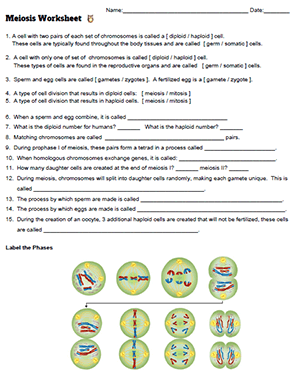
This worksheet is intended to reinforce concepts related to meiosis and sexual reproduction. Students compare terms such as diploid and haploid, mitosis and meiosis, and germ cells and somatic cells.
Meiosis can be a difficult concept to understand because it is a reduction division that results in unique gametes due to crossing-over that occurs in prophase I of meiosis.
It may take several different learning activities for students to really get the concept of independent assortment, which results in unique combinations of chromosomes in the gametes produced from meiosis.
These notes and Google slides can serve as an introduction to the topic which also includes an amazing video on meiosis from the Amoeba Sisters.
Other activities may include hands-on modeling of meiosis and crossing-over with popbeads and/or playdoh . I find this to be the most effective way for students to visualize how each gamete can receive a different combination of chromosome (independent assortment).
Grade Level: 8-12 Time Required: 10-15 minutes
HS-LS3-2 Make and defend a claim based on evidence that inheritable genetic variations may result from: (1) new genetic combinations through meiosis, (2) viable errors occurring during replication, and/or (3) mutations caused by environmental factors.
11.1 The Process of Meiosis
Learning objectives.
By the end of this section, you will be able to do the following:
- Describe the behavior of chromosomes during meiosis, and the differences between the first and second meiotic divisions
- Describe the cellular events that take place during meiosis
- Explain the differences between meiosis and mitosis
- Explain the mechanisms within the meiotic process that produce genetic variation among the haploid gametes
Sexual reproduction requires the union of two specialized cells, called gametes , each of which contains one set of chromosomes. When gametes unite, they form a zygote , or fertilized egg that contains two sets of chromosomes. (Note: Cells that contain one set of chromosomes are called haploid ; cells containing two sets of chromosomes are called diploid .) If the reproductive cycle is to continue for any sexually reproducing species, then the diploid cell must somehow reduce its number of chromosome sets to produce haploid gametes; otherwise, the number of chromosome sets will double with every future round of fertilization. Therefore, sexual reproduction requires a nuclear division that reduces the number of chromosome sets by half.
Most animals and plants and many unicellular organisms are diploid and therefore have two sets of chromosomes. In each somatic cell of the organism (all cells of a multicellular organism except the gametes or reproductive cells), the nucleus contains two copies of each chromosome, called homologous chromosomes . Homologous chromosomes are matched pairs containing the same genes in identical locations along their lengths. Diploid organisms inherit one copy of each homologous chromosome from each genetic contributor.
Meiosis is the nuclear division that forms haploid cells from diploid cells, and it employs many of the same cellular mechanisms as mitosis. However, as you have learned, mitosis produces daughter cells whose nuclei are genetically identical to the original parent nucleus. In mitosis, both the parent and the daughter nuclei are at the same “ploidy level”—diploid in the case of most multicellular animals. Plants use mitosis to grow as sporophytes, and to grow and produce eggs and sperm as gametophytes; so they use mitosis for both haploid and diploid cells (as well as for all other ploidies). In meiosis, the starting nucleus is always diploid and the daughter nuclei that result are haploid. To achieve this reduction in chromosome number, meiosis consists of one round of chromosome replication followed by two rounds of nuclear division. Because many events that occur during each of the division stages are analogous to the events of mitosis, the same stage names are assigned. However, because there are two rounds of division, the major process and the stages are designated with a “I” or a “II.” Thus, meiosis I is the first round of meiotic division and consists of prophase I, prometaphase I, and so on. Likewise, Meiosis II (during which the second round of meiotic division takes place) includes prophase II, prometaphase II, and so on.
Meiosis is preceded by an interphase consisting of G 1 , S, and G 2 phases, which are nearly identical to the phases preceding mitosis. The G 1 phase (the “first gap phase”) is focused on cell growth. During the S phase—the second phase of interphase—the cell copies or replicates the DNA of the chromosomes. Finally, in the G 2 phase (the “second gap phase”) the cell undergoes the final preparations for meiosis.
During DNA duplication in the S phase, each chromosome is replicated to produce two identical copies— sister chromatids that are held together at the centromere by cohesin proteins, which hold the chromatids together until anaphase II. (Note: these chromosome copies are called sister chromatids regardless of whether they are in a female gamete or a male gamete.)
Early in prophase I, before the chromosomes can be seen clearly with a microscope, the homologous chromosomes are attached at their tips to the nuclear envelope by proteins. As the nuclear envelope begins to break down, the proteins associated with homologous chromosomes bring the pair closer together. Recall that in mitosis, homologous chromosomes do not pair together. The synaptonemal complex , a lattice of proteins between the homologous chromosomes, first forms at specific locations and then spreads outward to cover the entire length of the chromosomes. The tight pairing of the homologous chromosomes is called synapsis . In synapsis , the genes on the chromatids of the homologous chromosomes are aligned precisely with each other. The synaptonemal complex supports the exchange of chromosomal segments between homologous nonsister chromatids—a process called crossing over . Crossing over can be observed visually after the exchange as chiasmata (singular = chiasma) ( Figure 11.3 ).
In humans, even though the X and Y sex chromosomes are not completely homologous (that is, most of their genes differ), there is a small region of homology that allows the X and Y chromosomes to pair up during prophase I. A partial synaptonemal complex develops only between the regions of homology.
Located at intervals along the synaptonemal complex are large protein assemblies called recombination nodules . These assemblies mark the points of later chiasmata and mediate the multistep process of crossover —or genetic recombination—between the nonsister chromatids. Near the recombination nodule, the double-stranded DNA of each chromatid is cleaved, the cut ends are modified, and a new connection is made between the nonsister chromatids. As prophase I progresses, the synaptonemal complex begins to break down and the chromosomes begin to condense. When the synaptonemal complex is gone, the homologous chromosomes remain attached to each other at the centromere and at chiasmata. The chiasmata remain until anaphase I. The number of chiasmata varies according to the species and the length of the chromosome. There must be at least one chiasma per chromosome for proper separation of homologous chromosomes during meiosis I, but there may be as many as 25. Following crossover, the synaptonemal complex breaks down and the cohesin connection between homologous pairs is removed. At the end of prophase I, the pairs are held together only at the chiasmata ( Figure 11.4 ). These pairs are called tetrads because a total of four sister chromatids of each pair of homologous chromosomes are now visible.
The crossover events are the first source of genetic variation in the nuclei produced by meiosis. A single crossover event between homologous nonsister chromatids leads to a reciprocal exchange of equivalent DNA between an egg-derived chromosome and a sperm-derived chromosome. When a recombinant sister chromatid is moved into a gamete cell it will carry a combination of maternal and paternal genes that did not exist before the crossover. Crossover events can occur almost anywhere along the length of the synapsed chromosomes. Different cells undergoing meiosis will therefore produce different recombinant chromatids, with varying combinations of maternal and parental genes. Multiple crossovers in an arm of the chromosome have the same effect, exchanging segments of DNA to produce genetically recombined chromosomes.
Prometaphase I
The key event in prometaphase I is the attachment of the spindle fiber microtubules to the kinetochore proteins at the centromeres. Kinetochore proteins are multiprotein complexes that bind the centromeres of a chromosome to the microtubules of the mitotic spindle. Microtubules grow from microtubule-organizing centers (MTOCs). In animal cells, MTOCs are centrosomes located at opposite poles of the cell. The microtubules from each pole move toward the middle of the cell and attach to one of the kinetochores of the two fused homologous chromosomes. Each member of the homologous pair attaches to a microtubule extending from opposite poles of the cell so that in the next phase, the microtubules can pull the homologous pair apart. A spindle fiber that has attached to a kinetochore is called a kinetochore microtubule . At the end of prometaphase I, each tetrad is attached to microtubules from both poles, with one homologous chromosome facing each pole. The homologous chromosomes are still held together at the chiasmata. In addition, the nuclear membrane has broken down entirely.
Metaphase I
During metaphase I, the homologous chromosomes are arranged at the metaphase plate —roughly in the midline of the cell, with the kinetochores facing opposite poles. Each homologous pair is oriented randomly at the equator. For example, if the two homologous members of chromosome 1 are labeled a and b , then the chromosomes could line up a-b or b-a. This is important in determining the genes carried by a gamete, as each will only receive one of the two homologous chromosomes. (Recall that homologous chromosomes are not identical. They contain different versions of the same genes, and after recombination during crossing over, each gamete will have a unique genetic makeup that has never existed before.)
The randomness in the alignment of recombined chromosomes at the metaphase plate, coupled with the crossing over events between nonsister chromatids, are responsible for much of the genetic variation in the offspring. To clarify this further, remember that the homologous chromosomes of a sexually reproducing organism are originally inherited as two separate sets, one from each parent. Using humans as an example, one set of 23 chromosomes is present in the egg cell, often called maternal chromosomes because the genetic contributor is often the mother. The other set of 23 chromosomes is contained in the sperm, and the genetic contributor is called a father who provides the paternal chromosomes. Every cell of the multicellular offspring has copies of the original two sets of homologous chromosomes. When the offspring human creates their own gametes through meiosis, the two sets of chromosomes will be rearranged. In prophase I of meiosis, the homologous chromosomes form the tetrads. In metaphase I, these pairs line up at the midway point between the two poles of the cell to form the metaphase plate. Because there is an equal chance that a microtubule fiber will encounter a maternally or paternally inherited chromosome, the arrangement of the tetrads at the metaphase plate is random. Thus, any maternally inherited chromosome may face either pole. Likewise, any paternally inherited chromosome may also face either pole. The orientation of each tetrad is independent of the orientation of the other 22 tetrads.
This event—the random (or independent ) assortment of homologous chromosomes at the metaphase plate—is the second mechanism that introduces variation into the gametes or spores. In each cell that undergoes meiosis, the arrangement of the tetrads is different. The number of variations is dependent on the number of chromosomes making up a set. There are two possibilities for orientation at the metaphase plate; the possible number of alignments therefore equals 2 n in a diploid cell, where n is the number of chromosomes per haploid set. Humans have 23 chromosome pairs, which results in over eight million (2 23 ) possible genetically-distinct gametes just from the random alignment of chromosomes at the metaphase plate. This number does not include the variability that was previously produced by crossing over between the nonsister chromatids. Given these two mechanisms, it is highly unlikely that any two haploid cells resulting from meiosis will have the same genetic composition ( Figure 11.5 ).
To summarize, meiosis I creates genetically diverse gametes in two ways. First, during prophase I, crossover events between the nonsister chromatids of each homologous pair of chromosomes generate recombinant chromatids with new combinations of maternal and paternal genes. Second, the random assortment of tetrads on the metaphase plate produces unique combinations of maternal and paternal chromosomes that will make their way into the gametes.
In anaphase I, the microtubules pull the linked chromosomes apart. The sister chromatids remain tightly bound together at the centromere. The chiasmata are broken in anaphase I as the microtubules attached to the fused kinetochores pull the homologous chromosomes apart ( Figure 11.6 ).
Telophase I and Cytokinesis
In telophase, the separated chromosomes arrive at opposite poles. The remainder of the typical telophase events may or may not occur, depending on the species. In some organisms, the chromosomes “decondense” and nuclear envelopes form around the separated sets of chromatids produced during telophase I. In other organisms, cytokinesis —the physical separation of the cytoplasmic components into two daughter cells—occurs without reformation of the nuclei. In nearly all species of animals and some fungi, cytokinesis separates the cell contents via a cleavage furrow (constriction of the actin ring that leads to cytoplasmic division). In plants, a cell plate is formed during cell cytokinesis by Golgi vesicles fusing at the metaphase plate. This cell plate will ultimately lead to the formation of cell walls that separate the two daughter cells.
Two haploid cells are the result of the first meiotic division of a diploid cell. The cells are haploid because at each pole, there is just one of each pair of the homologous chromosomes. Therefore, only one full set of the chromosomes is present. This is why the cells are considered haploid—there is only one chromosome set, even though each chromosome still consists of two sister chromatids. Recall that sister chromatids are merely duplicates of one of the two homologous chromosomes (except for changes that occurred during crossing over). In meiosis II, these two sister chromatids will separate, creating four haploid daughter cells.
Link to Learning
Review the process of meiosis, observing how chromosomes align and migrate, at Meiosis: An Interactive Animation .
In some species, cells enter a brief interphase, or interkinesis , before entering meiosis II. Interkinesis lacks an S phase, so chromosomes are not duplicated. The two cells produced in meiosis I go through the events of meiosis II in synchrony. During meiosis II, the sister chromatids within the two daughter cells separate, forming four new haploid gametes. The mechanics of meiosis II are similar to mitosis, except that each dividing cell has only one set of homologous chromosomes, each with two chromatids. Therefore, each cell has half the number of sister chromatids to separate out as a diploid cell undergoing mitosis. In terms of chromosomal content, cells at the start of meiosis II are similar to haploid cells in G 2 , preparing to undergo mitosis.
Prophase II
If the chromosomes decondensed in telophase I, they condense again. If nuclear envelopes were formed, they fragment into vesicles. The MTOCs that were duplicated during interkinesis move away from each other toward opposite poles, and new spindles are formed.
Prometaphase II
The nuclear envelopes are completely broken down, and the spindle is fully formed. Each sister chromatid forms an individual kinetochore that attaches to microtubules from opposite poles.
Metaphase II
The sister chromatids are maximally condensed and aligned at the equator of the cell.
Anaphase II
The sister chromatids are pulled apart by the kinetochore microtubules and move toward opposite poles. Nonkinetochore microtubules elongate the cell.
Telophase II and Cytokinesis
The chromosomes arrive at opposite poles and begin to decondense. Nuclear envelopes form around the chromosomes. If the parent cell was diploid, as is most commonly the case, then cytokinesis now separates the two cells into four unique haploid cells. The cells produced are genetically unique because of the random assortment of paternal and maternal homologs and because of the recombination of maternal and paternal segments of chromosomes (with their sets of genes) that occurs during crossover. The entire process of meiosis is outlined in Figure 11.7 .
Comparing Meiosis and Mitosis
Mitosis and meiosis are both forms of division of the nucleus in eukaryotic cells. They share some similarities, but also exhibit a number of distinct processes that lead to very different outcomes ( Figure 11.8 ). Mitosis is a single nuclear division that results in two nuclei that are usually partitioned into two new cells. The nuclei resulting from a mitotic division are genetically identical to the original nucleus. They have the same number of sets of chromosomes: one set in the case of haploid cells and two sets in the case of diploid cells. In contrast, meiosis consists of two nuclear divisions resulting in four nuclei that are usually partitioned into four new, genetically distinct cells. The four nuclei produced during meiosis are not genetically identical, and they contain one chromosome set only. This is half the number of chromosome sets of the original cell, which is diploid.
The main differences between mitosis and meiosis occur in meiosis I, which is a very different nuclear division than mitosis. In meiosis I, the homologous chromosome pairs physically meet and are bound together with the synaptonemal complex. Following this, the chromosomes develop chiasmata and undergo crossover between nonsister chromatids. In the end, the chromosomes line up along the metaphase plate as tetrads—with kinetochore fibers from opposite spindle poles attached to each kinetochore of a homolog to form a tetrad. All of these events occur only in meiosis I.
When the chiasmata resolve and the tetrad is broken up with the homologous chromosomes moving to one pole or another, the ploidy level—the number of sets of chromosomes in each future nucleus—has been reduced from two to one. For this reason, meiosis I is referred to as a reductional division . There is no such reduction in ploidy level during mitosis.
Meiosis II is analogous to a mitotic division. In this case, the duplicated chromosomes (only one set of them) line up on the metaphase plate with divided kinetochores attached to kinetochore fibers from opposite poles. During anaphase II, as in mitotic anaphase, the kinetochores divide and one sister chromatid—now referred to as a chromosome—is pulled to one pole while the other sister chromatid is pulled to the other pole. If it were not for the fact that there had been crossover, the two products of each individual meiosis II division would be identical (as in mitosis). Instead, they are different because there has always been at least one crossover per chromosome. Meiosis II is not a reduction division because although there are fewer copies of the genome in the resulting cells, there is still one set of chromosomes, as there was at the end of meiosis I.
Evolution Connection
The mystery of the evolution of meiosis.
Some characteristics of organisms are so widespread and fundamental that it is sometimes difficult to remember that they evolved like other simple traits. Meiosis is such an extraordinarily complex series of cellular events that biologists have had trouble testing hypotheses concerning how it may have evolved. Although meiosis is inextricably entwined with sexual reproduction and its advantages and disadvantages, it is important to separate the questions of the evolution of meiosis and the evolution of sex, because early meiosis may have been advantageous for different reasons than it is now. Thinking outside the box and imagining what the early benefits from meiosis might have been is one approach to uncovering how it may have evolved.
Meiosis and mitosis share obvious cellular processes, and it makes sense that meiosis evolved from mitosis. The difficulty lies in the clear differences between meiosis I and mitosis. Adam Wilkins and Robin Holliday 1 summarized the unique events that needed to occur for the evolution of meiosis from mitosis. These steps are homologous chromosome pairing and synapsis, crossover exchanges, sister chromatids remaining attached during anaphase, and suppression of DNA replication in interphase. They argue that the first step is the hardest and most important and that understanding how it evolved would make the evolutionary process clearer. They suggest genetic experiments that might shed light on the evolution of synapsis.
There are other approaches to understanding the evolution of meiosis in progress. Different forms of meiosis exist in single-celled protists. Some appear to be simpler or more “primitive” forms of meiosis. Comparing the meiotic divisions of different protists may shed light on the evolution of meiosis. Marilee Ramesh and colleagues 2 compared the genes involved in meiosis in protists to understand when and where meiosis might have evolved. Although research is still ongoing, recent scholarship into meiosis in protists suggests that some aspects of meiosis may have evolved later than others. This kind of genetic comparison can tell us what aspects of meiosis are the oldest and what cellular processes they may have borrowed from in earlier cells.
Click through the steps of this interactive animation to compare the meiotic process of cell division to that of mitosis at How Cells Divide .
- 1 Adam S. Wilkins and Robin Holliday, “The Evolution of Meiosis from Mitosis,” Genetics 181 (2009): 3–12.
- 2 Marilee A. Ramesh, Shehre-Banoo Malik and John M. Logsdon, Jr, “A Phylogenetic Inventory of Meiotic Genes: Evidence for Sex in Giardia and an Early Eukaryotic Origin of Meiosis,” Current Biology 15 (2005):185–91.
As an Amazon Associate we earn from qualifying purchases.
This book may not be used in the training of large language models or otherwise be ingested into large language models or generative AI offerings without OpenStax's permission.
Want to cite, share, or modify this book? This book uses the Creative Commons Attribution License and you must attribute OpenStax.
Access for free at https://openstax.org/books/biology-2e/pages/1-introduction
- Authors: Mary Ann Clark, Matthew Douglas, Jung Choi
- Publisher/website: OpenStax
- Book title: Biology 2e
- Publication date: Mar 28, 2018
- Location: Houston, Texas
- Book URL: https://openstax.org/books/biology-2e/pages/1-introduction
- Section URL: https://openstax.org/books/biology-2e/pages/11-1-the-process-of-meiosis
© Jan 8, 2024 OpenStax. Textbook content produced by OpenStax is licensed under a Creative Commons Attribution License . The OpenStax name, OpenStax logo, OpenStax book covers, OpenStax CNX name, and OpenStax CNX logo are not subject to the Creative Commons license and may not be reproduced without the prior and express written consent of Rice University.
If you're seeing this message, it means we're having trouble loading external resources on our website.
If you're behind a web filter, please make sure that the domains *.kastatic.org and *.kasandbox.org are unblocked.
To log in and use all the features of Khan Academy, please enable JavaScript in your browser.
High school biology
Course: high school biology > unit 4.
- Chromosomal crossover in meiosis I
- Phases of meiosis I
- Phases of meiosis II
- Comparing mitosis and meiosis
Meiosis review
Stages of meiosis, common mistakes and misconceptions.
- Interphase is not part of meiosis. Although a cell needs to undergo interphase before entering meiosis, interphase is technically not part of meiosis.
- Crossing over occurs only during prophase I. The complex that temporarily forms between homologous chromosomes is only present in prophase I, making this the only opportunity the cell has to move DNA segments between the homologous pair.
- Meiosis does not occur in all cells. Meiosis only occurs in reproductive cells, as the goal is to create haploid gametes that will be used in fertilization.
- Meiosis is important to, but not the same as, sexual reproduction. Meiosis is necessary for sexual reproduction to occur, as it results in the formation of gametes (sperm and eggs). However, sexual reproduction includes fertilization (the fusion between gametes), which is not part of the meiotic process.
Want to join the conversation?
- Upvote Button navigates to signup page
- Downvote Button navigates to signup page
- Flag Button navigates to signup page


Status message
- Reproduction & Development
Resource Type
Description.
This animation shows how meiosis, the form of cell division unique to egg and sperm production, can give rise to sperm that carry either an X or a Y chromosome.
Meiosis starts with a diploid cell (a cell with two sets of chromosomes) and ends up with four haploid cells (cells with only one set of chromosomes), which are called gametes (eggs and sperm). When an egg and sperm combine at fertilization, the embryo regains a diploid number of chromosomes. Before a cell begins meiosis, its two sets of chromosomes come together and swap segments in a process known as recombination (or crossing over).
The first part of this animation shows recombination between the X and Y chromosomes. Most women have two X chromosomes, whereas most men have one X chromosome and one Y chromosome. After a cell with both X and Y chromosomes divides twice, it produces four gametes. Half the gametes get an X chromosome, and half get a Y chromosome. Because of recombination and random assortment, the exact composition of chromosomes in each gamete varies.
The second part of the animation shows how recombination may result in the transfer of a gene called SRY from the Y chromosome to the X chromosome. SRY plays an important role in male sexual development. An embryo with two X chromosomes, one of which has SRY , may develop more typical male sexual characteristics.
This animation is a clip from a 2001 Holiday Lecture Series, The Meaning of Sex: Genes and Gender .
allosome, autosome, crossing over, egg, gamete, recombination, sex-determining region Y (SRY), sperm, X chromosome, Y chromosome
Terms of Use
Please see the Terms of Use for information on how this resource can be used.
Accessibility Level (WCAG compliance)
Version history, curriculum connections, ngss (2013), ap biology (2019), ib biology (2016), vision and change (2009), explore related content, other related resources.

No products in the cart.
Related posts
![meiosis drawing assignment I’ve Planned Your Cell Cycle Lesson For You! [Free Resources]](https://emmatheteachiecom.s3.amazonaws.com/20231124000821/Featured-Image-for-Ive-Planned-Your-Cell-Cycle-Lesson-For-You.jpg)
5 Engaging Ways to Teach Meiosis
Meiosis is an important topic in biology, but it is also one of the most difficult for students to grasp. Not only is it a difficult subject to learn, but it is also a fairly tricky topic to teach.
Anyone disagree? *crickets*
Great! We all agree. Let’s move on.
The truth is that teaching meiosis doesn’t have to be boring or mundane. You can make it exciting and engaging for students!
Let’s take a look at 5 engaging ideas for how to teach meiosis.
Hopefully these will give you some new ideas that you can use in your classroom as early as tomorrow!
Need some ready-to-use meiosis activities? I’ve got you covered:
1 – Gummy Worm Meiosis
Candy.
That’s all you need to say to engage students in biology class.
One of my favorite activities to teach meiosis is using gummy worms. Not only does this activity involve candy, but it creates a visual picture for students that will help them retain the steps and details of meiosis.
First, divide the class into groups and give each group a bag of gummy worms. Tell students that each pair of gummy worms represents a pair of homologous chromosomes and each color represents a different gene on a chromosome.
Students then line up gummy worms in pairs, matching the colors as closely as possible. They have now paired homologous chromosomes.
Students now separate the pairs as it happens during meiosis I. Next, students will separate the remaining gummy worms, representing the separation of sister chromatids in meiosis II.
Once the above is completed, students can count the number of gummy worms they have in total. Students have successfully created genetic diversity through meiosis.

By using gummy worms, students have been given the opportunity to visualize and manipulate the different stages of meiosis. Students will be more likely to retain the information since it was presented in a more accessible and engaging way, as opposed to just sitting at their desk and taking notes during a lecture.
If you have dietary restrictions or your school does not allow the use of food in lessons, an alternative would be to use pipe cleaners.
You could take this activity a step further and require students to create a stop motion video of meiosis.
Here is an amazing student example from YouTube:
2 – Use Google Slides Activities
Google Slides is a great way to make interactive games and exercises for students. Bright and full of color, Google Slides will help make the content more enticing and create an easily remembered visual image for the students.
You can make it easier for students to understand meiosis by creating interactive activities. These can include labeling and describing the stages of meiosis, comparing and contrasting meiosis and mitosis, and labeling the process of fertilization.
If you want a set of done-for-you meiosis Google Slides activities, check these out:
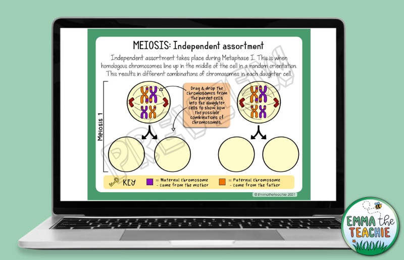
In addition, you can even add supplemental activities, such as videos, right into the Google Slides activity. Students can watch the video when completing the assignment, but also go back to it later to review.
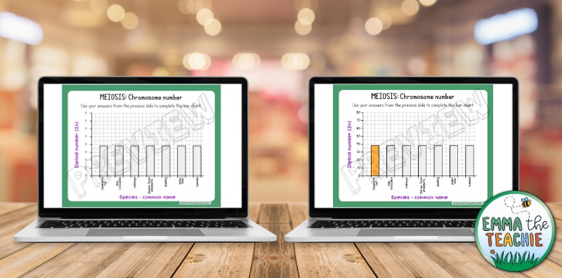
You can even use Google Slides to demonstrate how bar graphs can be relevant in this scientific unit.
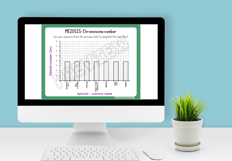
Students will begin by completing a table determining the diploid and haploid numbers of different species. Some of the species include a domesticated cat, broccoli, and a polar bear.
After completing the table, students click and drag the bars to their respective numbers. This bar graph creates a visual representation of how the diploid numbers vary across different species.
Differentiation in the classroom is extremely important. All you have to do is change the activity to give different students the extra help they need. You can assign students certain slides based on if they need more support or less support.
3 – Meiosis Lab
We all know that students love laboratory activities. You can take this opportunity to incorporate a lab into your meiosis lesson.
For this lab, students will be looking at an anther squash. Anthers are the part of a flower that produce pollen (a sex cell).
You can find more details on the anther squash lab here.
This lab provides the best educational outcomes when it is completed towards the end of the unit and all of the stages have been taught.
You can provide students with prepared slides or students can make up their own. In all honesty, I recommend determining which choice is best based on the students you have in your class this year.
Note: this lab is time-consuming and not always successful, but it is great for students to get familiar with using microscopes.
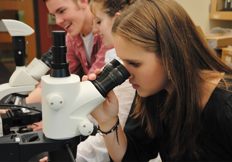
Students then stain the cells to make the chromosomes more visible. Once the stain is on, your students will place the slide under the microscope and search the cells. There are likely to be a mix of mature pollen grains, cells that not undergoing meiosis, and cells that are actively dividing.
Ask students to locate cells that are actively dividing. Students can then draw what they see, paying close attention to the location and appearance of the chromosomes.
Students should try to identify as many stages of meiosis as they can. They can then compare and discuss with other groups.
Tip: if you find a group that has any particularly clear examples of the phases of meiosis, ask other students to come and view these too.
The anther squash lab helps students gain a deeper understanding of meiosis, as well as develop their skills in microscopy and recording data.
4 – Create a flip book
An effective way for students to learn meiosis is by creating a flip book. This interactive visual will help students reinforce the multiple stages of meiosis.
You should begin by giving students an overview of meiosis and the different stages that are part of it. I recommend giving them a printout of the stages of meiosis, or leaving it projected on your board for reference.
Students will then need to get five pieces of paper, markers, crayons, and anything else they need to make a flip book that will suit their learning needs.
Each of the five papers should be folded to create ten flaps: prophase I, metaphase I, anaphase I, telophase I, cytokinesis, prophase II, metaphase II, anaphase II, telophase, and cytokinesis II.
On each flap, students will need to draw and label the appropriate stages of meiosis, as well write a brief description of what happens in each stage.
Now, students have their own personal reference guide!

5 – Use Doodle Notes
It is not a secret that there are many terms, steps, and information when it comes to meiosis. In order for students to fully grasp the concepts, they need to have a way to break down the information and see the big picture, which means…
DOODLE NOTES!
I have created an engaging set of Doodle Notes for meiosis.
Students begin by describing the purpose of meiosis and a basic overview of the process of meiosis.
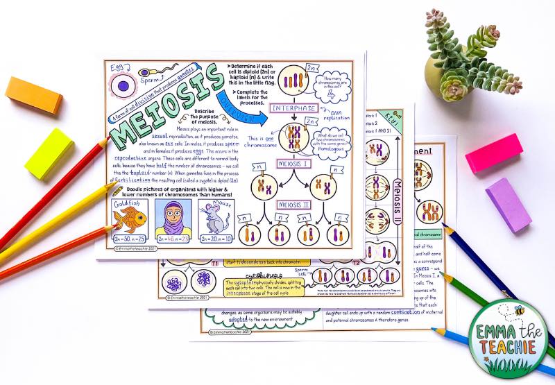
Once the basics are covered, more advanced topics are included within the Doodle Notes, such as crossing over.
The second page of the Doodle Notes help students compare meiosis I and meiosis II by having these side-by-side. Students describe each phase and then create a key. This helps them understand which events are common to both cellular divisions, and which are unique.
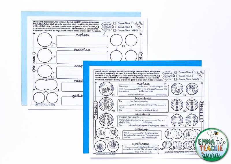
If the Doodle Notesare too complicated for some students, there are differentiated versions that will support students who need it. For instance, there are two versions of filling in the stages of meiosis. In one version, the descriptions are completely blank and the other includes a fill-in-the-blank option.
There are also blank versions for students to add their own doodles of what is happening inside the cell. This is great for students that love drawing!
How will you teach meiosis?
Hopefully, you have gained some ideas of how to teach meiosis to your students in a way that will make it a little less challenging and little more fun!
By using these diverse activities, you can help your students develop a deeper understanding of meiosis and its role in genetic diversity and evolution.
If you want an easy-to-implement bundle that you can use tomorrow – check out my bundle of meiosis resources here.
I hope you have a wonderful day,
Shop this post

Meiosis Bundle
Add to cart

Meiosis Google Slides

Meiosis Doodle Notes

- Study Guides
- Homework Questions
10-Meiosis Worksheet-23
Module 8: Cell Division
Assignment: mitosis and meiosis internet quests.
Use the two documents linked below to complete an internet hands-on activity involving mitosis and meiosis. During these activities you will demonstrate your understanding of cell division by identifying and drawing various stages of these events as well as answering questions about each.
Mitosis Internet Lesson
Download this lesson on Mitosis. Note that the last question on this document directs you to draw the four stages of mitosis. You should draw the five we studied in this course.
Meiosis Internet Lesson
Download this lesson on Meiosis. Compile your answers into a single document and submit it to complete this assessment.
You may chose to print these sheets, write your answers out and then rescan the documents to create your single submission file. Or you may chose to type your answers into a new Word document and submit that file. Do keep in mind that you must complete the drawing activities as well so in some fashion you have to get those images into your final file.
See this example for one way you may insert your images.
Basic Requirements (the assignment will not be accepted or assessed unless the follow criteria have been met):
- Assignment has been proofread and does not contain any major spelling or grammatical errors
- Assignment includes appropriate references
- Assignment includes answers to all questions (Mitosis: 25, Meiosis: 15) and required images (Mitosis: 8)
- Authored by : Shelli Carter. Provided by : Columbia Basin College. Located at : https://www.columbiabasin.edu/ . License : CC BY: Attribution
- Mitosis--Internet Lesson. Provided by : Biologycorner. Located at : https://www.biologycorner.com/worksheets/mitosis.html . License : CC BY-NC: Attribution-NonCommercial
- Meiosis--Internet Lesson. Provided by : Biologycorner. Located at : https://www.biologycorner.com//worksheets/meiosis_internet.html . License : CC BY-NC: Attribution-NonCommercial

IMAGES
VIDEO
COMMENTS
Rubric. Mitosis and Meiosis Internet Quests. Outcome: Describe and explain the various stages of cell division. Criteria. Ratings. Pts. Identify the stages of the cell cycle, by picture and description of major milestones. More than 22 questions answered correctly and all included images are correct/accurate. 5.0 pts.
Module 8 Assignment: Mitosis and Meiosis Worksheets Use the two documents linked below to complete an internet hands-on activity involving mitosis and meiosis. During these activities you will demonstrate your understanding of cell division by identifying and drawing various stages of these events as well as answering questions about each.
Meiosis but lacks complete information on the stages - or - the importance of the organelles involved. 13 points max. Information is well organized. While it is detailed, it lacks some clarity. 13 points max. Creative presentation and effective use of space. 8 points max. Includes diagrams on meiosis but the information is generalized. 11 ...
Meiosis produces haploid gametes from a diploid cell. DNA replicates once, but the cells divide twice. In biology, meiosis is the process where a cell replicates DNA once but divides twice, producing four cells that have half the genetic information of the original cell. It is how organisms produce gametes or sex cells, which are eggs in females and sperm in males.
This is the result of independent assortment and crossing over. Lessons on meiosis generally involve labeling drawings, viewing animations, or even making models, I have used pipecleaners, gummy worms, and popsicle sticks to guide students through meiosis I and meiosis II. You can even give students microscope slides to view cells undergoing ...
Young anthers in buds about six down from the top usually contain cells undergoing meiosis to yield microspores. Goals: a. find buds with anthers at the right stage of development to show meiosis; b. isolate and stain the cells undergoing meiosis; and c. see as many stages in meiosis as possible. A. Preparation of slides and coverslips:
To put that another way, meiosis in humans is a division process that takes us from a diploid cell—one with two sets of chromosomes—to haploid cells—ones with a single set of chromosomes. In humans, the haploid cells made in meiosis are sperm and eggs. When a sperm and an egg join in fertilization, the two haploid sets of chromosomes form a complete diploid set: a new genome.
Meiosis Worksheet. This worksheet is intended to reinforce concepts related to meiosis and sexual reproduction. Students compare terms such as diploid and haploid, mitosis and meiosis, and germ cells and somatic cells. Meiosis can be a difficult concept to understand because it is a reduction division that results in unique gametes due to ...
Meiosis I. Meiosis is preceded by an interphase consisting of G 1, S, and G 2 phases, which are nearly identical to the phases preceding mitosis. The G 1 phase (the "first gap phase") is focused on cell growth. During the S phase—the second phase of interphase—the cell copies or replicates the DNA of the chromosomes. Finally, in the G 2 phase (the "second gap phase") the cell ...
meiosis - Demonstrate the steps of mitosis and meiosis visually by creating a model of these processes. The model must be accurate, demonstrate knowledge of the information, and be a creative design. It must be a 3D model and cannot be a poster or drawing on paper. You must explain your model to the class. Research one of the following genetic
A sex cell (in humans: sperm for males, and eggs for females) Meiosis. A two-step process of cell division that is used to make gametes (sex cells) Crossing over. Process in which homologous chromosomes trade parts. Interphase. Phase of the cell cycle where the cell grows and makes a copy of its DNA. Homologous chromosomes.
This animation shows how meiosis, the form of cell division unique to egg and sperm production, can give rise to sperm that carry either an X or a Y chromosome. Meiosis starts with a diploid cell (a cell with two sets of chromosomes) and ends up with four haploid cells (cells with only one set of chromosomes), which are called gametes (eggs and ...
Assignment: Mitosis and Meiosis Internet Quests Use the two documents linked below to complete an internet hands-on activity involving mitosis and meiosis. During these activities you will demonstrate your understanding of cell division by identifying and drawing various stages of these events as well as answering questions about each.
Study with Quizlet and memorize flashcards containing terms like Identify the phases of meiosis I described below. Homologous chromosomes are separated. Homologous chromosome are paired. Nuclear envelopes form around separated chromosomes. The centrosome replicates., Identify the phases of Meiosis II described below. Chromosomes are lined up by spindle fibers. Nuclear envelope forms around ...
Assignment: Mitosis and Meiosis Worksheets. Use the two documents linked below to complete an internet hands-on activity involving mitosis and meiosis. During these activities you will demonstrate your understanding of cell division by identifying and drawing various stages of these events as well as answering questions about each.
Module 7 Assignment: Mitosis and Meiosis Worksheets Use the two documents linked below to complete an internet hands-on activity involving mitosis and meiosis. During these activities you will demonstrate your understanding of cell division by identifying and drawing various stages of these events as well as answering questions about each.
Meiosis is the process in eukaryotic, sexually-reproducing animals that reduces the number of chromosomes in a cell before reproduction. Many organisms package these cells into gametes, such as egg and sperm. The gametes can then meet, during reproduction, and fuse to create a new zygote. Because the number of alleles was reduced during meiosis ...
See meiosis; mitosis. Meiosis, division of a germ cell involving two fissions of the nucleus and giving rise to four gametes, or sex cells, each with half the number of chromosomes of the original cell. The process of meiosis is characteristic of organisms that reproduce sexually and have a diploid set of chromosomes in the nucleus.
1 - Gummy Worm Meiosis. Candy. That's all you need to say to engage students in biology class. One of my favorite activities to teach meiosis is using gummy worms. Not only does this activity involve candy, but it creates a visual picture for students that will help them retain the steps and details of meiosis.
10 - Meiosis Worksheet Student N ame: Answer the question below, draw a cell going through meiosis, and submit the worksheet and drawing through the assignment tab in iCollege. 1. What is the purpose of meiosis? Meiosis aims to produce gametes (sperm or egg cells) with half the number of chromosomes as the parent cell. 2.
Meiosis I. Meiosis is preceded by an interphase consisting of the G 1, S, and G 2 phases, which are nearly identical to the phases preceding mitosis. The G 1 phase, which is also called the first gap phase, is the first phase of the interphase and is focused on cell growth.
Step 1. A eukaryotic cell goes through a sequence of events called the cell cycle that results i... Meiosis Drawing Assignment: Please complete the chart below, drawing the chromosomes and celis as they are moved and describing the important events that are occurring during MEIOSIS. PLEASE NOTE THERE ARETWO PAGESTOTHIS DRAWINGIII Meiosis ...
Outcome: Describe and explain the various stages of cell division. Criteria. Ratings. Pts. Identify the stages of the cell cycle, by picture and description of major milestones. More than 22 questions answered correctly and all included images are correct/accurate. 5.0 pts. 20-22 mitosis questions answered correctly and 7 correct images included.