EJNMMI Research


Aims and scope
EJNMMI Research publishes new basic, translational and clinical research in the field of nuclear medicine and molecular imaging. Regular features include original research articles, short communication of preliminary data on innovative research, interesting case reports, editorials, and letters to the editor. Educational articles on basic sciences, fundamental aspects and controversy related to pre-clinical and clinical research or ethical aspects of research are also welcome. The journal also considers preliminary Research Articles or short communications that include novel findings, methods or radiopharmaceuticals with a high potential but limited data available, worth a preliminary information. Timely reviews provide updates on current applications, issues in imaging research and translational aspects of nuclear medicine and molecular imaging technologies. Topics include but not limited to innovative research covering all aspects of molecular imaging, e.g. radiopharmaceutical approaches with a high potential of clinical impact, important aspects in image reconstruction and accuracy, or within the theranostic field with therapy combinations or incorporation of artificial intelligence in image generation or interpretation.
Announcement Since January 2024: New Editor-in-Chief - Dr. Irene Burger, Chief Medical Officer, Kantonsspital Baden AG
- Most accessed
Mitochondrial complex I density is associated with IQ and cognition in cognitively healthy adults: an in vivo [ 18 F]BCPP-EF PET study
Authors: Ekaterina Shatalina, Thomas S. Whitehurst, Ellis Chika Onwordi, Barnabas J. Gilbert, Gaia Rizzo, Alex Whittington, Ayla Mansur, Hideo Tsukada, Tiago Reis Marques, Sridhar Natesan, Eugenii A. Rabiner, Matthew B. Wall and Oliver D. Howes
Short post-injection seizure duration is associated with reduced power of ictal brain perfusion SPECT to lateralize the seizure onset zone
Authors: Amir Karimzadeh, Kian Baradaran-Salimi, Berthold Voges, Ivayla Apostolova, Thomas Sauvigny, Michael Lanz, Susanne Klutmann, Stefan Stodieck, Philipp T. Meyer and Ralph Buchert
Non-invasive quantification of 18 F-florbetaben with total-body EXPLORER PET
Authors: Emily Nicole Holy, Elizabeth Li, Anjan Bhattarai, Evan Fletcher, Evelyn R. Alfaro, Danielle J. Harvey, Benjamin A. Spencer, Simon R. Cherry, Charles S. DeCarli and Audrey P. Fan
Performance and application of the total-body PET/CT scanner: a literature review
Authors: Yuanyuan Sun, Zhaoping Cheng, Jianfeng Qiu and Weizhao Lu
In-vivo inhibition of neutral endopeptidase 1 results in higher absorbed tumor doses of [ 177 Lu]Lu-PP-F11N in humans: the lumed phase 0b study
Authors: Christof Rottenburger, Michael Hentschel, Markus Fürstner, Lisa McDougall, Danijela Kottoros, Felix Kaul, Rosalba Mansi, Melpomeni Fani, A. Hans Vija, Roger Schibli, Susanne Geistlich, Martin Behe, Emanuel R. Christ and Damian Wild
Most recent articles RSS
View all articles
Nanobody: a promising toolkit for molecular imaging and disease therapy
Authors: Guangfa Bao, Ming Tang, Jun Zhao and Xiaohua Zhu
Anaesthesia and physiological monitoring during in vivo imaging of laboratory rodents: considerations on experimental outcomes and animal welfare
Authors: Jordi L Tremoleda, Angela Kerton and Willy Gsell
Gastrointestinal transit measurements in mice with 99m Tc-DTPA-labeled activated charcoal using NanoSPECT-CT
Authors: Parasuraman Padmanabhan, Johannes Grosse, Abu Bakar Md Ali Asad, George K Radda and Xavier Golay
About inflammation and infection
Authors: Alberto Signore
Comparative gallium-68 labeling of TRAP-, NOTA-, and DOTA-peptides: practical consequences for the future of gallium-68-PET
Authors: Johannes Notni, Karolin Pohle and Hans-Jürgen Wester
Most accessed articles RSS
Need help with APC funding?

We offer a free open access support service to make it easier for you to discover and apply for article-processing charge (APC) funding. Learn more here .
Check if your institution is a member

EJNMMI is part of the EJNMMI Journal Family. Highlights of our featured content and the latest developments from the EJNMMI group of journals can be found on the EJNMMI Journal Family webpage. There you find details about the annual EANM Springer Prize.
- Editorial Board
- Sign up for article alerts and news from this journal
Annual Journal Metrics
2022 Citation Impact 3.2 - 2-year Impact Factor 3.3 - 5-year Impact Factor 0.932 - SNIP (Source Normalized Impact per Paper) 0.853 - SJR (SCImago Journal Rank)
2023 Speed 12 days submission to first editorial decision for all manuscripts (Median) 87 days submission to accept (Median)
2023 Usage 604,325 downloads 175 Altmetric mentions
- More about our metrics
- Follow us on Twitter
- Follow us on Facebook
- ISSN: 2191-219X (electronic)
Affiliated with
European Association of Nuclear Medicine
Visit and follow the EANM on their social media channels:

- U.S. Department of Health & Human Services
- National Institutes of Health

En Español | Site Map | Staff Directory | Contact Us
- Science Education
- Science Topics
- Nuclear Medicine
What is nuclear medicine?
What are radioactive tracers, what is single photon emission computed tomography (spect), what is positron emission tomography (pet), what are nuclear medicine scans used for, are there risks, how are nibib-funded researchers advancing nuclear medicine.
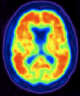
Nuclear medicine is a medical specialty that uses radioactive tracers (radiopharmaceuticals) to assess bodily functions and to diagnose and treat disease. Specially designed cameras allow doctors to track the path of these radioactive tracers. Single Photon Emission Computed Tomography or SPECT and Positron Emission Tomography or PET scans are the two most common imaging modalities in nuclear medicine.
Radioactive tracers are made up of carrier molecules that are bonded tightly to a radioactive atom. These carrier molecules vary greatly depending on the purpose of the scan. Some tracers employ molecules that interact with a specific protein or sugar in the body and can even employ the patient’s own cells. For example, in cases where doctors need to know the exact source of intestinal bleeding, they may radiolabel (add radioactive atoms) to a sample of red blood cells taken from the patient. They then reinject the blood and use a SPECT scan to follow the path of the blood in the patient. Any accumulation of radioactivity in the intestines informs doctors of where the problem lies.
For most diagnostic studies in nuclear medicine, the radioactive tracer is administered to a patient by intravenous injection. However a radioactive tracer may also be administered by inhalation, by oral ingestion, or by direct injection into an organ. The mode of tracer administration will depend on the disease process that is to be studied.
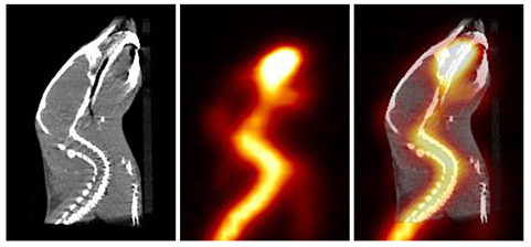
Approved tracers are called radiopharmaceuticals since they must meet FDA’s exacting standards for safety and appropriate performance for the approved clinical use. The nuclear medicine physician will select the tracer that will provide the most specific and reliable information for a patient’s particular problem. The tracer that is used determines whether the patient receives a SPECT or PET scan.
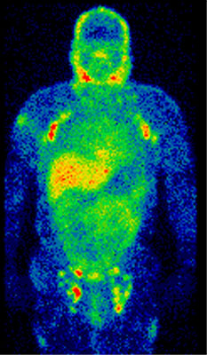
SPECT imaging instruments provide three-dimensional (tomographic) images of the distribution of radioactive tracer molecules that have been introduced into the patient’s body. The 3D images are computer generated from a large number of projection images of the body recorded at different angles. SPECT imagers have gamma camera detectors that can detect the gamma ray emissions from the tracers that have been injected into the patient. Gamma rays are a form of light that moves at a different wavelength than visible light. The cameras are mounted on a rotating gantry that allows the detectors to be moved in a tight circle around a patient who is lying motionless on a pallet.
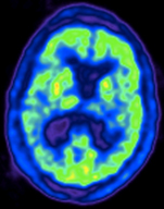
Click here to watch a short video about how PET scans work.
SPECT scans are primarily used to diagnose and track the progression of heart disease, such as blocked coronary arteries. There are also radiotracers to detect disorders in bone, gall bladder disease and intestinal bleeding. SPECT agents have recently become available for aiding in the diagnosis of Parkinson's disease in the brain, and distinguishing this malady from other anatomically-related movement disorders and dementias.
The major purpose of PET scans is to detect cancer and monitor its progression, response to treatment, and to detect metastases. Glucose utilization depends on the intensity of cellular and tissue activity so it is greatly increased in rapidly dividing cancer cells. In fact, the degree of aggressiveness for most cancers is roughly paralleled by their rate of glucose utilization. In the last 15 years, slightly modified radiolabeled glucose molecules (F-18 labeled deoxyglucose or FDG) have been shown to be the best available tracer for detecting cancer and its metastatic spread in the body.
A combination instrument that produces both PET and CT scans of the same body regions in one examination (PET/CT scanner) has become the primary imaging tool for the staging of most cancers worldwide.
Recently, a PET probe was approved by the FDA to aid in the accurate diagnosis of Alzheimer's disease, which previously could be diagnosed with accuracy only after a patient's death. In the absence of this PET imaging test, Alzheimer's disease can be difficult to distinguish from vascular dementia or other forms of dementia that affect older people.
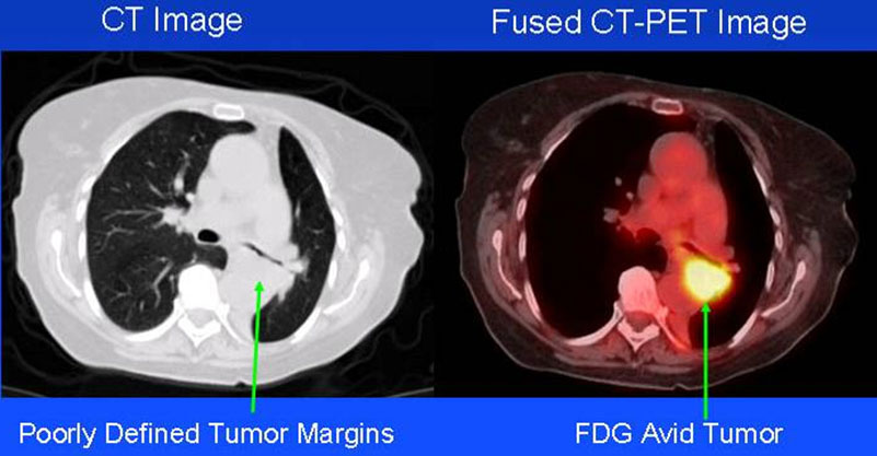
The total radiation dose conferred to patients by the majority of radiopharmaceuticals used in diagnostic nuclear medicine studies is no more than what is conferred during routine chest x-rays or CT exams. There are legitimate concerns about possible cancer induction even by low levels of radiation exposure from cumulative medical imaging examinations, but this risk is accepted to be quite small in contrast to the expected benefit derived from a medically needed diagnostic imaging study.
Like radiologists, nuclear medicine physicians are strongly committed to keeping radiation exposure to patients as low as possible, giving the least amount of radiotracer needed to provide a diagnostically useful examination.
Research in nuclear medicine involves developing new radio tracers as well as technologies that will help physicians produce clearer pictures.
Developing new tracers
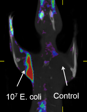
A bacterial infection is a common complication of implanting a medical device into the body. With more patients receiving device implants than ever before, infections from implants are a growing problem. Currently, these types of infections are diagnosed based on physical exam results and microbial cultures. However, such techniques are only useful for detecting late stage infections, which usually have already become difficult to treat. Conversely, medical devices may be needlessly removed when doctors mistake inflammation that is a normal consequence of surgery with inflammation due to an infection. NIBIB is currently supporting research to develop a new family of PET imaging contrast agents that are taken up specifically by bacterial cells, but not human cells. Such imaging agents would allow doctors to visualize early-stage bacterial infections so they can be easily treated, thereby reducing the number of implanted devices that are unnecessarily removed. They also have the potential to be used for diagnosing infections not associated with medical devices, for example, those affecting the heart or lungs.
Creating new technology
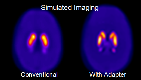
A SPECT tracer is currently available for accurate diagnosis of Parkinson's disease. However, the small region in the brain that must be imaged requires a dedicated brain SPECT imager with special gamma cameras to provide high resolution, which adds to the cost of the procedure. NIBIB is supporting research to create an inexpensive adapter for the conventional SPECT imagers that most hospitals already have. The adapter would allow standard clinical SPECT cameras to provide the s ame high resolution that currently only dedicated SPECT brain imaging systems can produce. These improvements would make Parkinson’s diagnosis less costly and more widely available.
Reviewed July 2016
Explore More
Heath Topics
- Alzheimer's
- Heart Disease
Research Topics
- Imaging tracer
- Optical Imaging
- PET imaging
PROGRAM AREAS
Inside NIBIB
- Director's Corner
- Funding Policies
- NIBIB Fact Sheets
- Press Releases
Thank you for visiting nature.com. You are using a browser version with limited support for CSS. To obtain the best experience, we recommend you use a more up to date browser (or turn off compatibility mode in Internet Explorer). In the meantime, to ensure continued support, we are displaying the site without styles and JavaScript.
- View all journals
- My Account Login
- Explore content
- About the journal
- Publish with us
- Sign up for alerts
- Open access
- Published: 28 June 2023
The effect of COVID-19 on nuclear medicine and radiopharmacy activities: A global survey
- Fatma Al-Saeedi 1 ,
- Peramaiyan Rajendran 2 , 3 ,
- Dnyanesh Tipre 4 ,
- Hassan Aladwani 5 ,
- Salem Alenezi 5 ,
- Maryam Alqabandi 1 ,
- Abdullah Alkhamis 5 ,
- Abdulmohsen Redha 5 ,
- Ahmed Mohammad 5 ,
- Fahad Ahmad 5 ,
- Yaaqoup Abdulnabi 5 ,
- Altaf Alfadhly 5 &
- Danah Alrasheedi 5
Scientific Reports volume 13 , Article number: 10489 ( 2023 ) Cite this article
1720 Accesses
1 Altmetric
Metrics details
- Health care
- Medical research
Globally, COVID-19 affected radiopharmaceutical laboratories. This study sought to determine the economic, service, and research impacts of COVID-19 on radiopharmacy. This online survey was conducted with the participation of employees from nuclear medicine and radiopharmaceutical companies. The socioeconomic status of the individuals was collected. The study was participated by 145 medical professionals from 25 different countries. From this work, it is evident that 2-deoxy-2-[18F]fluoro-D-glucose (2-[ 18 F]FDG), and 99m Tc-labeled macro aggregated albumin 99m Tc-MAA were necessary radiopharmaceuticals used by 57% (83/145and 34% (49/145;) respondents, respectively for determining how COVID infections affect a patient’s body. The normal scheduling procedure for the radiopharmacy laboratory was reduced by more than half (65%; 94/145). In COVID-19, 70% (102/145) of respondents followed the regulations established by the local departments. Throughout the pandemic, there was a 97% (141/145) decrease in all staffing recruitment efforts. The field of nuclear medicine research, as well as the radiopharmaceutical industry, were both adversely affected by COVID-19.
A reporting and analysis framework for structured evaluation of COVID-19 clinical and imaging data
Gabriel Alexander Salg, Maria-Katharina Ganten, … Jens Kleesiek
Indirect impact of Covid-19 on hospital care pathways in Italy
Teresa Spadea, Chiara Di Girolamo, … the Mimico-19 working group
Comparison of COVID-19 outcomes among shielded and non-shielded populations
Bhautesh D. Jani, Frederick K. Ho, … Jill P. Pell
Introduction
COVID-19 is an acute infectious disease since the “Spanish” influenza pandemic that occurred in 1918. The current COVID-19 pandemic is considered to be one of the most massive and chaotic challenges that are currently confronting global public health 1 , 2 , 3 , 4 , 5 . On a global scale, the COVID-19 outbreak has resulted in a significant number of deaths and economic losses, it affected life and lifestyle. Furthermore, the outbreak has caused uncertainty and it has posed a risk to the medical sectors and relevant persons 6 , 7 , 8 , 9 , 10 . Despite this, a survey that can provide a more detailed explanation of the pandemic, report on its effects, and forecast its potential future effects is needed, as very limited research reports are published in this domain. The current COVID-19 outbreak has introduced scientists to a pandemic challenge that necessitates optimal levels of information exchange about the current outbreak as well as any future outbreaks. This information exchange must take place outside of the limitations imposed by physical geography, climatology, and scientific inquiry. COVID-19 is a worldwide infection that can affect a wide range of medical professionals, also confronting the duties, activities, and work performed in radiopharmacy laboratories.
We were interested in determining how we could assess the impact of COVID-19 on the operations and services provided by the department of nuclear medicine, specifically the radiopharmacy lab, which is the department’s central division. The radiopharmacy laboratory is the site where many radiopharmaceuticals are produced, where their quality is monitored, and even where they are stocked in inventory. Our study aimed to develop a preliminary database for our international survey and to assess the impact of the COVID-19 pandemic on the research and commercial interactions of radiopharmaceutical companies.
Study design
This survey was conducted between the 15th of September 2021 and the 10th of October 2022 with nuclear medicine and radiopharmaceutical staff members from all over Kuwait and other countries. The survey was conducted after obtaining approval for the survey’s protocol (No. 1762/2021) from Kuwait’s ethics committee as well as the Ministry of Health-Kuwait. We confirm that all methods were performed in accordance with the relevant guidelines and regulations. We have collected the consent forms that were filled out by the participants, and they also signed it. Local governmental health areas in Kuwait, Saudi Arabia, the United Arab Emirates, Iraq, Jordan, Sudan, Morocco, Pakistan, India, the United States of America, the United Kingdom, France, Spain, Germany, Greece, Italy, Sweden, Brazil, Bosnia and Herzegovina, Turkey, South Africa, Tanzania, Colombia, Mexico, and Peru were represented by online survey respondents. The questionnaire was conducted in English. To achieve our objectives, we devised the simple idea of conducting a survey of the local residents. We searched for any fundamental information in different databases that could help us get started. We were astounded to discover so few studies on nuclear medicine procedures, particularly in radiopharmacy laboratories. The survey sought to learn more about COVID-19’s impact on the radiopharmacy industry and its activities.
During the pandemic, a web-based (online) questionnaire built on the Google platform was developed to investigate and collect data on the uses and activities involving radiopharmacy. The Centre for Research Support and Conferences of the University’s Faculty of Medicine and Health Sciences facilitated the production and distribution of email announcements. The participants were also provided with a link to a Google Form-generated web-based survey.
Data collection
The semi-structured questionnaire was divided into two parts: the first, which consisted of five questions, asked for demographic information, and the second, which inquired about respondents’ levels of knowledge, attitude, activities, and practises regarding radiopharmacy during a pandemic. The questionnaire was made up entirely of closed-ended questions that required a response in the form of one or more checkboxes to indicate the desired level of response. The questionnaire was designed in such a way that each individual participant had seen the same set of questions in the same order. Each question was intended to be succinct and to the point. To increase the dependability of the responses, they were all recorded using the identical method. Each questionnaire form was subjected to a series of repeated checks to determine its content's reliability. The link to Google Forms and Microsoft Teams was posted, distributed, and circulated via a variety of social media platforms.
Statistical analysis
We used descriptive statistics to characterise the population, their purchase preferences, and their scheduling activities. The paired sample t test was used to determine the reduction in the purchase of materials and radiopharmaceuticals in comparison to normal purchase status before the COVID-19 pandemic. A p-value of 0.05 was statistically significant.
The socio-demographic profile of the participants
Participants in the survey were classified based on where they worked, where they were from, what occupation they held (Fig. 1 ), and age. During the duration of the study, 145 medical professionals from 25 different countries participated and completed the questionnaire. The participants who had completed the survey included 20% (29/145) researchers, 15% (22/145) physicians, 10% (15/145) scientists, 2% (3/145) radiopharmacists, 0.7% (1/145) radiochemists, 0.7% (1/145) radiopharmaceutical chemists, 1.37% (2/145) physicists, 1.37% (2/145) cyclotron operators, and technologists who accounted for the remaining of 50% (73/145) of the survey responses.
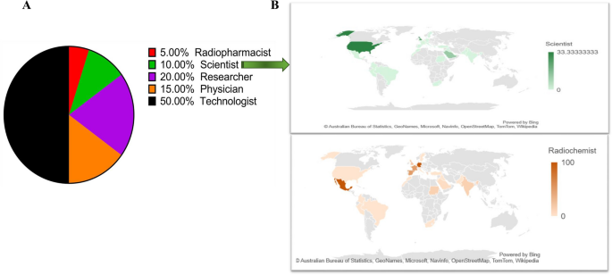
The socio-demographic pattern of participants surveyed. ( A ) profession ( B ) profession examples (scientist and radiochemist) among participants around the world.
Sixty percent (87/145) participants worked in radiopharmacy; and among them 26% (23/87) worked in PET centres; and 26% (23/87) worked in research. The majority of them were employed in the radiopharmacy section of the Nuclear Medicine Department.
The age distribution of the respondents shows that 25% (36/145) fall into the age range of 25–34 years and 25% (36/145) workers fall in the range of 35–44 years. The percentage of people who are employed is comparable across both age groups. The remaining 50% percent comprised of people aged 45 to 54 years (20%, 29/145, 55 to 64 years (20%, 29/145), and 65 years and above (10%; 15/145) as shown in Fig. 2 A. According to Fig. 2 B, the majority of the participants (50%; 73/145) had more than 6 years of experience, followed by 4 to 6 years (40%; 58/145) and 1 to 3 years (10%; 15/145).

The socio-demographic pattern of participants surveyed. ( A ) age ( B ) work experience.
The radiopharmacy activities and the measurements
Purchase and preparation.
Radiopharmaceutical preparation was prepared with more care than before pandemic; 75% (108/145) of participants prepared radiopharmaceuticals with greater care than they did before the pandemic (Fig. 3 A). The purchase of materials and radiopharmaceuticals were significantly reduced (p < 0.001), in comparison to the normal purchase orders. Purchase was reduced to more than half the normal situation before the pandemic (64%; 64/145). According to 25% respondents, purchase was not affected and remained same as it was before pandemic; however, 5% and 6% reported that processing of some purchase orders was halted and/or put on hold respectively (Fig. 3 B).
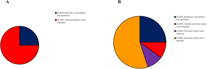
Radiopharmaceuticals preparation and purchase. ( A ) radiopharmaceuticals preparation ( B ) radiopharmaceuticals purchase.
The procedure for scheduling work in the radiopharmacy lab was decided to be simplified. As a result, 45% (65/145) of all respondents have simplified their appointment scheduling procedures. However, 40% (58/145) respondents claimed that scheduling procedures remained unchanged since the pandemic, while 15% (21/145) claimed that the use of certain radiopharmaceuticals was no longer permitted.
Most used radiopharmaceutical
This survey showed that the Positron Emission Tomography (PET) radiopharmaceutical 2-deoxy-2-[18F]fluoro-D-glucose (2-[ 18 F]FDG) was the most common (57%, 83/145) imaging radiopharmaceutical used during COVID–19, followed by respiratory (lung) 99m Tc-labeled macro aggregated albumin ( 99m Tc-MAA) of 34% (49/145). Then, the use of the cardiovascular (myocardial) systems (5%; 7/145) kits was reported followed using the musculoskeletal (bone) system (4%; 6/145) (Fig. 4 ). Although only a small percentage of respondents mentioned renal agents, the difference was not statistically significant.
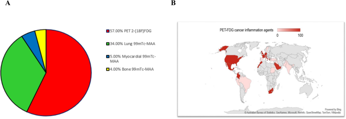
The most common imaging radiopharmaceutical used during COVID–19. ( A ) most used radiopharmaceuticals ( B ) 2-[ 18 F]FDG among participants worldwide.
Staffing levels in the radiopharmacy
The staffing level was reported in this survey. There was a 97% reduction (140/145) in staffing during the pandemic. All sections and professions almost stopped to recruit staff during COVID-19.
Compiling data about Covid-19
70% (101/145) of participants consulted local departmental regulations while collecting measures and learning about COVID-19, compared to 30% who used social media.
According to the findings, the vast majority of respondents worked in the radiopharmacy of the Nuclear Medicine Department. Technologists, who are considered members of the medical profession, comprised the majority of respondents to this survey. Technologists were present and actively involved in providing services to the nuclear medicine department and radiopharmacy operations. Most of the respondents had an experience of more than 6 years in nuclear medicine and radiopharmacy activities. The pandemic has already affected the process of preparing radiopharmaceuticals, so all nuclear departments have instructed their radiopharmacy lab employees to take additional precautions when preparing radiopharmaceuticals.
The results showed the interesting observation that 2-[ 18 F]FDG was reported to have been more widely used than normal during the pandemic. COVID–19 exemplifies a well-known principle underlying the use of 2-[ 18 F]FDG in oncology and during inflammatory processes, such as infections. A high uptake on an image is correlated with a high number of viable cancerous or inflammatory cells, an increased glucose metabolism, high levels of GLUT transporter expression, and the activity levels of various glycolytic enzymes or metabolic activity.
Numerous studies have demonstrated that 2-[ 18 F]FDG is effective for diagnosis, elucidating acute pulmonary-extrapulmonary manifestations, tracking treatment efficacy, recognising disease at an early stage, and directing medical management. These studies supported previous findings that 2-[ 18 F]FDG was effective in all of these areas 11 , 12 and concurred with studies that emphasised the importance of 2-[ 18 F]FDG during COVID–19 13 , 14 , 15 , 16 , 17 , 18 , 19 .
People exposed to the SARS-CoV-2 developed respiratory complications, a healthy respiratory system is required for healthy life and to investigate the COVID-19-related complications, the respiratory 99m Tc-MAA represented the second order of radiopharmaceuticals widely used during COVID–19. One of the most important diagnostic procedures that nuclear medicine can perform is an imaging test that uses 99m Tc-MAA radiopharmaceutical. Findings of several studies supported the observation that the frequency of this usage increased during pandemic 20 , 21 .
The myocardial and bone systems radiopharmaceuticals were performed almost equally during the pandemic due to the importance to exclude the causes of myocardial or skeletal. According to our results and other studies’ findings, more than 28% of COVID-19-infected patients exhibit signs of cardiac injury. This is associated with a poor prognosis, a sore throat, a newly developed loss of taste or smell, fever, chills, bone and muscle pain, and other symptoms 22 , 23 , 24 .
Although radiopharmacy has many factors, including location, funding, financial and technical factors, and departmental conditions, 2-[ 18 F]FDG and 99m Tc-MAA radiopharmaceuticals were the most prominent used during COVID–19.
The extraordinary presence of radiopharmaceuticals such as 2-[ 18 F]FDG and 99m Tc-MAA at COVID-19 was unaffected by geographical location, radiopharmaceutical production sources, or radiopharmaceutical production types. There was no variation in how individuals responded to the virus. Our survey proved the importance of these radiopharmaceuticals. We will expand on this knowledge to enhance the research that has already been conducted on the application of radiopharmaceuticals, and we will prioritise new features.
The process of scheduling was revealed to be reduced during the pandemic, this added support to the lack of staff and the reduced staffing levels. In addition, the use of certain radiopharmaceuticals was discontinued during the pandemic, which further supports the reduced pattern of purchasing. Similarly, Giammarile et al. 25 , also reported a significant reduction in nuclear medicine diagnostic and therapeutic procedures in June 2020 (73%) and October 2020 (56.9%) due to pandemic-related changes. The study also found that out of different nuclear medicine procedures, oncological PET tests, exhibited lower reduction in utilisation than the conventional nuclear medicine, especially nuclear cardiology. Moreover, in high-income nations, the detrimental effect was less evident. Gradually the situation of the supply chains of radioisotopes, generators and other essential materials is improving and showing the trend same as pre-Covid-19 time 25 . A European study reported that most of the European countries included in the study did not face any issue with the supply of radiopharmaceuticals 26 .
In the radiopharmacy lab, there was no standardised schedule to follow for the preparation of radiopharmaceuticals or specific uses. As reported by Moreira et al. 26 in most European nuclear medicine departments the most common organizational changes were alteration of scheduling practices, like rotating cohort teams of personnel to avoid widespread quarantine 26 . Additionally, the production schedule was modified to account for decreased demand resulting from staff reductions, fewer purchases, transportation issues, and additional hygienic precautions and regulations. COVID-19 impacted purchase orders, supply chains, how individuals work, and what they purchase. Numerous pandemics have been suggested to be cured by virtual reality 27 , 28 , 29 , 30 .
The regional lockdown, increased border controls, cancellation of most commercial passenger flights, and increases in cargo costs have reduced, delayed, or halted the availability and supply of vital medical radioisotopes. These radioisotopes are used in a variety of applications, including radiopharmacy, nuclear medicine, and research. This is true in light of the findings, which indicate that the supply and availability of these radioisotopes have been reduced, postponed, or completely halted 25 , 31 , 32 , 33 , 34 , 35 .
A significant proportion of respondents followed the regional department's rules. More people were gathering information about COVID-19 via various social media platforms. This is similar to the findings of multiple studies that cited World Health Organization, National Energy Commission, and International Atomic Energy Agency 36 , 37 , 38 , 39 . According to the findings of this study, the rules regarding this pandemic need to be updated in order to remain inside the existing local regulations 40 , 41 . The guidelines for the clinical practise of nuclear medicine and radiopharmacy should be made available as soon as possible during COVID-19. This action will benefit the management of future COVID-19 variants such as late Delta and Omicron 42 , 43 , 44 .
We determined that the most efficient way for us to accomplish our goals here would be to conduct a simple local survey. We searched for and located basic data or databases that we could use to initiate this project. We discovered few relevant studies and surveys on COVID-19 for the radiopharmacy lab work of the nuclear medicine department. This created a challenge for us because we required more information on the subject. We have presented evidence in this survey that the nuclear medicine industry, lacks some kind of standardisation, particularly with regard to the surveys presented here. When we talk about standardisation, we're talking about a set of agreements or procedures that ensures a method or piece of work meets a standard. All relevant groups, divisions, service providers, and organisations in an industrial society must adhere to predetermined quality, agreement, uniformity, and comparability guidelines. This guarantees the success of design, production, and service.
The COVID-19 pandemic has had a significant negative impact on nuclear medicine therapeutics, clinical/imaging settings and research activities. Despite this significant impact, the researchers are embracing an attitude that “the show must go on”. At the global level, the continuation of vital nuclear medicine services has been given priority. In the pandemic time, education on nuclear medicine was adapted. The research activity on nuclear medicine was not stopped during pandemic, rather the research activities continued significantly leading to emerging indications, innovative radiopharmaceuticals, and new imaging/data analysis techniques 45 .
Conclusions
According to the findings of this survey, COVID-19 made it difficult to conduct radiopharmacy business, duties, and activities, which hampered nuclear medicine research. The outstanding presence of 2-[ 18 F]FDG and 99m Tc-MAA radiopharmaceuticals during COVID-19 provide an opportunity and a scientific priority to study and research. There is a need for creative, quantitatively sound, and context-appropriate action plans. During and after the pandemic, standard regulations, instructions, patient care, recommendations, and protection should be implemented. There is currently a shortage of personnel in the highly specialised fields of nuclear medicine staffing and radiopharmacy, which may be responsible for at least some of the challenges. The number of people involved in radiopharmacy activities is expected to increase in the future. As a result, both the existence of a reliable information database and the standardisation of this process will be possible to avoid any other hurdles in the nuclear medicine and radiopharmaceuticals.
Data availability
The data are available within the paper.
Chen, Y. et al. Clinical characteristics and outcomes of patients with diabetes and COVID-19 in association with glucose-lowering medication. Diabetes Care 43 , 1399–1407. https://doi.org/10.2337/dc20-0660 (2020).
Article CAS PubMed Google Scholar
Huang, Y., Chen, D., Fietze, I. & Penzel, T. Obstructive sleep apnea with COVID-19. Adv. Exp. Med. Biol. 1384 , 281–293. https://doi.org/10.1007/978-3-031-06413-5_17 (2022).
Article PubMed Google Scholar
Wang, C., Havewala, M. & Zhu, Q. COVID-19 stressful life events and mental health: Personality and coping styles as moderators. J. Am. Coll. Health https://doi.org/10.1080/07448481.2022.2066977 (2022).
Gao, Q. et al. Perceived stress and stress responses during COVID-19: The multiple mediating roles of coping style and resilience. PLoS One 17 , e0279071. https://doi.org/10.1371/journal.pone.0279071 (2022).
Article CAS PubMed PubMed Central Google Scholar
Xu, S. et al. Impact of the COVID-19 on electricity consumption of open university campus buildings—The case of Twente University in the Netherlands. Energy Build 279 , 112723. https://doi.org/10.1016/j.enbuild.2022.112723 (2023).
Ahn, S. N. The potential impact of COVID-19 on health-related quality of life in children and adolescents: A systematic review. Int. J. Environ. Res. Public Health https://doi.org/10.3390/ijerph192214740 (2022).
Article PubMed PubMed Central Google Scholar
Juengling, F. D., Maldonado, A., Wuest, F. & Schindler, T. H. The role of nuclear medicine for COVID-19: Time to act now. J. Nucl. Med. 61 , 781–782. https://doi.org/10.2967/jnumed.120.246611 (2020).
Juengling, F. D., Maldonado, A., Wuest, F. & Schindler, T. H. Identify. Quantify. Predict. Why immunologists should widely use molecular imaging for coronavirus disease 2019. Front. Immunol. 12 , 568959. https://doi.org/10.3389/fimmu.2021.568959 (2021).
Guedj, E. et al. The impact of COVID-19 lockdown on brain metabolism. Hum. Brain. Mapp. 43 , 593–597. https://doi.org/10.1002/hbm.25673 (2022).
Mboowa, G. Current and emerging diagnostic tests available for the novel COVID-19 global pandemic. AAS Open Res. 3 , 8. https://doi.org/10.12688/aasopenres.13059.1 (2020).
Dietz, M. et al. COVID-19 pneumonia: Relationship between inflammation assessed by whole-body FDG PET/CT and short-term clinical outcome. Eur. J. Nucl. Med. Mol. Imaging 48 , 260–268 (2021).
Eibschutz, L. S. et al. Seminar in Nuclear Medicine 61–70 (Elsevier, 2022).
Google Scholar
Lütje, S. et al. Nuclear medicine in SARS-CoV-2 pandemia: 18F-FDG-PET/CT to visualize COVID-19. Nuklearmedizin 59 , 276–280. https://doi.org/10.1055/a-1152-2341 (2020).
Qin, C., Liu, F., Yen, T. C. & Lan, X. (18)F-FDG PET/CT findings of COVID-19: A series of four highly suspected cases. Eur. J. Nucl. Med. Mol. Imaging 47 , 1281–1286. https://doi.org/10.1007/s00259-020-04734-w (2020).
Annunziata, S. et al. Role of 2-[(18)F]FDG as a radiopharmaceutical for PET/CT in patients with COVID-19: A systematic review. Pharmaceuticals https://doi.org/10.3390/ph13110377 (2020).
Scarlattei, M. et al. Unknown SARS-CoV-2 pneumonia detected by PET/CT in patients with cancer. Tumori 106 , 325–332. https://doi.org/10.1177/0300891620935983 (2020).
Setti, L. et al. Increased incidence of interstitial pneumonia detected on [(18)F]-FDG-PET/CT in asymptomatic cancer patients during COVID-19 pandemic in Lombardy: A casualty or COVID-19 infection?. Eur. J. Nucl. Med. Mol. Imaging 48 , 777–785. https://doi.org/10.1007/s00259-020-05027-y (2021).
Treglia, G. et al. Prevalence and significance of hypermetabolic lymph nodes detected by 2-[(18)F]FDG PET/CT after COVID-19 vaccination: A systematic review and a meta-analysis. Pharmaceuticals https://doi.org/10.3390/ph14080762 (2021).
Zuckier, L. S. & Gordon, S. R. COVID-19 in the nuclear medicine department, be prepared for ventilation scans as well!. Nucl. Med. Commun. 41 , 494–495. https://doi.org/10.1097/mnm.0000000000001196 (2020).
Burger, I. A. et al. Lung perfusion [(99m)Tc]-MAA SPECT/CT to rule out pulmonary embolism in COVID-19 patients with contraindications for iodine contrast. Eur. J. Nucl. Med. Mol. Imaging 47 , 2209–2210. https://doi.org/10.1007/s00259-020-04862-3 (2020).
Acuña Hernandez, M., Morales Avellaneda, T., Narvaez Gomez, J. A. & Sanchez Orduz, L. Findings in [99mTc]MAA SPECT/CT in the diagnosis and follow-up of pulmonary embolism after infection by SARS-CoV-2 (COVID-19). Nucl. Med. Rev. Cent. East Eur. 24 , 120–121. https://doi.org/10.5603/nmr.2021.0029 (2021).
Alam, S. R. et al. Cardiovascular mechanisms in Covid-19: Methodology of a prospective observational multimodality imaging study (COSMIC-19 study). BMC Cardiovasc. Disord. 21 , 1–6 (2021).
Article Google Scholar
Singh, A. et al. Covid19, beyond just the lungs: A review of multisystemic involvement by Covid19. Pathol. Res. Pract. 224 , 153384 (2021).
Cichocki, P., Adamczewski, Z., Kuśmierek, J. & Płachcińska, A. Mask-related motion artifact on 99mTc-MIBI SPECT: Unexpected pitfalls of SARS-CoV-2 countermeasures. Diagnostics 11 , 1426 (2021).
Giammarile, F. et al. Changes in the global impact of COVID-19 on nuclear medicine departments during 2020: An international follow-up survey. Eur. J. Nucl. Med. Mol. Imaging 48 , 4318–4330. https://doi.org/10.1007/s00259-021-05444-7 (2021).
Moreira, A. P. et al. Impact of the COVID-19 pandemic on nuclear medicine departments in Europe. Eur. J. Nucl. Med. Mol. Imaging 48 , 3361–3364. https://doi.org/10.1007/s00259-021-05484-z (2021).
Ball, S. et al. Monitoring indirect impact of COVID-19 pandemic on services for cardiovascular diseases in the UK. Heart 106 , 1890–1897. https://doi.org/10.1136/heartjnl-2020-317870 (2020).
Bulchand-Gidumal, J. & Melián-González, S. Post-COVID-19 behavior change in purchase of air tickets. Ann. Tour. Res. 87 , 103129. https://doi.org/10.1016/j.annals.2020.103129 (2021).
Liu, S. et al. A novel hybrid multi-criteria group decision-making approach with intuitionistic fuzzy sets to design reverse supply chains for COVID-19 medical waste recycling channels. Comput. Ind. Eng. 169 , 108228. https://doi.org/10.1016/j.cie.2022.108228 (2022).
Schuster, A. M. & Cotten, S. R. COVID-19’s influence on information and communication technologies in long-term care: Results from a web-based survey with long-term care administrators. JMIR Aging 5 , e32442. https://doi.org/10.2196/32442 (2022).
Chatzipavlidou, V. The modifications brought about by the COVID-19 pandemic to nuclear medicine practice. Hell. J. Nucl. Med. 23 (Suppl), 6–7 (2020).
PubMed Google Scholar
Exadaktylou, P., Papadopoulos, N., Chatzipavlidou, V. & Giammarile, F. Non-oncological PET/CT imaging during SARS-CoV-2 pandemic. Hell. J. Nucl. Med. 23 (Suppl), 51–56 (2020).
Freudenberg, L. S. et al. Global impact of COVID-19 on nuclear medicine departments: An international survey in April 2020. J. Nucl. Med. 61 , 1278–1283. https://doi.org/10.2967/jnumed.120.249821 (2020).
Giannoula, E. et al. Nuclear thyroidology in pandemic times: The paradigm shift of COVID-19. Hell. J. Nucl. Med. 23 (Suppl), 41–50 (2020).
Kudo, T. et al. Impact of COVID-19 pandemic on cardiovascular testing in Asia: The IAEA INCAPS-COVID study. JACC Asia 1 , 187–199. https://doi.org/10.1016/j.jacasi.2021.06.002 (2021).
Fan, J. & Smith, A. P. Information overload, wellbeing and COVID-19: A survey in China. Behav. Sci. (Basel) https://doi.org/10.3390/bs11050062 (2021).
Rabiller, G. et al. Hospital radiopharmacy in Argentina during the COVID-19 pandemic. Criteria and rationale for the organization of work. Rev. Esp. Med. Nucl. Imagen. Mol. (Engl Ed) 39 , 375–379. https://doi.org/10.1016/j.remn.2020.08.015 (2020).
Revell, L. J. Covid19.explorer: A web application and R package to explore United States COVID-19 data. PeerJ 9 , e11489. https://doi.org/10.7717/peerj.11489 (2021).
Scrima, G. et al. Safety measures and clinical outcome of nuclear cardiology department during Covid-19 lockdown pandemic: Northern Italy experience. J. Nucl. Cardiol. 28 , 331–335. https://doi.org/10.1007/s12350-020-02286-y (2021).
Lam, W.W.-C., Loke, K.S.-H., Wong, W. Y. & Ng, D.C.-E. Facing a disruptive threat: How can a nuclear medicine service be prepared for the coronavirus outbreak 2020?. Eur. J. Nucl. Med. Mol. Imaging 47 , 1645–1648 (2020).
Zhang, X., Shao, F. & Lan, X. Suggestions for safety and protection control in department of nuclear medicine during the outbreak of COVID-19. Eur. J. Nucl. Med. Mol. Imaging 47 , 1632–1633 (2020).
Aggarwal, A. et al. SARS-CoV-2 omicron BA.5: Evolving tropism and evasion of potent humoral responses and resistance to clinical immunotherapeutics relative to viral variants of concern. EBioMedicine 84 , 104270. https://doi.org/10.1016/j.ebiom.2022.104270 (2022).
Chia, T. R. T., Young, B. E. & Chia, P. Y. The omicron-transformer: Rise of the subvariants in the age of vaccines. Ann. Acad. Med. Singap. 51 , 712–729. https://doi.org/10.47102/annals-acadmedsg.2022294 (2022).
Rubin, R. COVID-19 vaccines vs variants-determining how much immunity is enough. JAMA 325 , 1241–1243. https://doi.org/10.1001/jama.2021.3370 (2021).
Treglia, G. Nuclear medicine during the COVID-19 pandemic: The show must go on. Front. Med. (Lausanne) 9 , 896069. https://doi.org/10.3389/fmed.2022.896069 (2022).
Download references
Acknowledgements
We would like to thank and acknowledge all nuclear medicine departments personnel who assist filling the survey and questionnaire provided.
Author information
Authors and affiliations.
The Department of Nuclear Medicine, College of Medicine, Kuwait University, P.O. box: 24923, 13110, Safat, Kuwait
Fatma Al-Saeedi & Maryam Alqabandi
Department of Biological Sciences, College of Science, King Faisal University, AlAhsa, Saudi Arabia
Peramaiyan Rajendran
Department of Biochemistry, Saveetha Dental College & Hospitals, Saveetha Institute of Medical and Technical Sciences, Chennai, 600 077, Tamil Nadu, India
Translational Biomedical Imaging Laboratory, Department of Radiology, Children’s Hospital Los Angeles, Los Angeles, California, USA
Dnyanesh Tipre
College of Medicine, Kuwait University, P.O. box: 24923, 13110, Safat, Kuwait
Hassan Aladwani, Salem Alenezi, Abdullah Alkhamis, Abdulmohsen Redha, Ahmed Mohammad, Fahad Ahmad, Yaaqoup Abdulnabi, Altaf Alfadhly & Danah Alrasheedi
You can also search for this author in PubMed Google Scholar
Contributions
F.A. established the idea, survey, and questionnaire questions. F.A. wrote the conception, design, acquisition, seek the ethical approval, wrote the main manuscript text and prepared figures of the work. P.R. and D.T. assisted in the acquisition, analysis, preparing graphs and figures, interpretation of data, and writing. H.A., S.A., A.A., A.R., A.M., F.A., Y.A., A.L.A. and D.A. revised the figures. M.A. helped in references at the beginning. All authors read, revised, and reviewed the manuscript.
Corresponding author
Correspondence to Fatma Al-Saeedi .

Ethics declarations
Competing interests.
The authors declare no competing interests.
Additional information
Publisher's note.
Springer Nature remains neutral with regard to jurisdictional claims in published maps and institutional affiliations.
Rights and permissions
Open Access This article is licensed under a Creative Commons Attribution 4.0 International License, which permits use, sharing, adaptation, distribution and reproduction in any medium or format, as long as you give appropriate credit to the original author(s) and the source, provide a link to the Creative Commons licence, and indicate if changes were made. The images or other third party material in this article are included in the article's Creative Commons licence, unless indicated otherwise in a credit line to the material. If material is not included in the article's Creative Commons licence and your intended use is not permitted by statutory regulation or exceeds the permitted use, you will need to obtain permission directly from the copyright holder. To view a copy of this licence, visit http://creativecommons.org/licenses/by/4.0/ .
Reprints and permissions
About this article
Cite this article.
Al-Saeedi, F., Rajendran, P., Tipre, D. et al. The effect of COVID-19 on nuclear medicine and radiopharmacy activities: A global survey. Sci Rep 13 , 10489 (2023). https://doi.org/10.1038/s41598-023-36925-4
Download citation
Received : 23 December 2022
Accepted : 12 June 2023
Published : 28 June 2023
DOI : https://doi.org/10.1038/s41598-023-36925-4
Share this article
Anyone you share the following link with will be able to read this content:
Sorry, a shareable link is not currently available for this article.
Provided by the Springer Nature SharedIt content-sharing initiative
By submitting a comment you agree to abide by our Terms and Community Guidelines . If you find something abusive or that does not comply with our terms or guidelines please flag it as inappropriate.
Quick links
- Explore articles by subject
- Guide to authors
- Editorial policies
Sign up for the Nature Briefing newsletter — what matters in science, free to your inbox daily.
We've detected unusual activity from your computer network
To continue, please click the box below to let us know you're not a robot.
Why did this happen?
Please make sure your browser supports JavaScript and cookies and that you are not blocking them from loading. For more information you can review our Terms of Service and Cookie Policy .
For inquiries related to this message please contact our support team and provide the reference ID below.
Application of Particle Physics Detector in Medical Science
- 28th International Conference on Nuclear Tracks and Radiation Measurements
- Published: 17 April 2024
Cite this article
- M. Verma 1 ,
- P. Kumar 1 ,
- B. Kumari 1 &
- M. K. Singh ORCID: orcid.org/0000-0002-5451-6622 1
Initially, the growth of particle detectors was focused on the identification of particles, but new detector designs have made it possible to build new diagnostic techniques. Clinical settings and medical research facilities utilize cutting-edge methods developed from particle accelerators, detectors, and physics computers, including imaging technologies, accelerators specifically designed for cancer therapy, simulations, data analysis, and nuclear medicine. It is still obvious that research in nuclear and basic particle physics is what motivates and propels the development of particle detectors for medical applications. The applications of the particle physics detector in medical science will be the main topic of this paper.
This is a preview of subscription content, log in via an institution to check access.
Access this article
Price includes VAT (Russian Federation)
Instant access to the full article PDF.
Rent this article via DeepDyve
Institutional subscriptions
M. Dosanjh, M. Cirilli, S. Myers, and S. Navin, Medical applications at CERN and the ENLIGHT network. Front. Oncol. 6, 9 (2016).
Article PubMed PubMed Central Google Scholar
N.A. Pavel, Particle detectors for biomedical applications-demands and trends. Nucl. Instrum. Methods Phys. Res. A 478, 1 (2002).
Article CAS Google Scholar
S. Braccini, Particle Accelerators and Detectors for Medical Diagnostics and Therapy . PhD Thesis, University of Bern (2016).
R.-D. Heuer, The future of the large hadron collider and CERN. Philos. Trans. R. Soc. A 370, 986 (2012).
Article Google Scholar
M. Dosanjh, From Particle Physics to Medical Application (Bristol: IOP Publishing Ltd, 2017), p.1.
Book Google Scholar
D.K. Bewley, The 8 MeV linear accelerator at the MRC Cyclotron Unit, Hammersmith Hospital, London. Br. J. Radiol. 58, 213 (1985).
Article CAS PubMed Google Scholar
http://cerncourier.com/cws/article/cern/44361 .
J.H. Lawrence, C.A. Tobias, J.L. Born, R.K. Mccombs, J.E. Roberts, H.O. Anger, B.V. Low-Beer, and C.B. Huggins, Pituitary irradiation with high-energy proton beams: a preliminary report. Cancer Res. 18, 121 (1958).
CAS PubMed Google Scholar
Download references
Author information
Authors and affiliations.
Department of Physics, Institute of Applied Sciences and Humanities, GLA University, Mathura, 281406, India
M. Verma, P. Kumar, B. Kumari & M. K. Singh
You can also search for this author in PubMed Google Scholar
Corresponding author
Correspondence to M. K. Singh .
Ethics declarations
Conflict of interest.
The authors declare that they have no conflict of interest.
Additional information
Publisher's note.
Springer Nature remains neutral with regard to jurisdictional claims in published maps and institutional affiliations.
Rights and permissions
Springer Nature or its licensor (e.g. a society or other partner) holds exclusive rights to this article under a publishing agreement with the author(s) or other rightsholder(s); author self-archiving of the accepted manuscript version of this article is solely governed by the terms of such publishing agreement and applicable law.
Reprints and permissions
About this article
Verma, M., Kumar, P., Kumari, B. et al. Application of Particle Physics Detector in Medical Science. J. Electron. Mater. (2024). https://doi.org/10.1007/s11664-024-11062-4
Download citation
Received : 21 November 2023
Accepted : 21 March 2024
Published : 17 April 2024
DOI : https://doi.org/10.1007/s11664-024-11062-4
Share this article
Anyone you share the following link with will be able to read this content:
Sorry, a shareable link is not currently available for this article.
Provided by the Springer Nature SharedIt content-sharing initiative
- Particle physics
- accelerators
- hadron therapy
Advertisement
- Find a journal
- Publish with us
- Track your research
- Frontiers in Medicine
- Nuclear Medicine
- Research Topics
Expert Opinions & Viewpoints in Nuclear Medicine
Total Downloads
Total Views and Downloads
About this Research Topic
This editorial initiative of particular relevance, led by Dr Giorgio Treglia, Specialty Chief Editor of the Nuclear Medicine section, is focused on new insights, novel developments, current challenges, latest discoveries, recent advances, and future perspectives in the field of Nuclear Medicine. In this Research Topic, hosted by the Speciality Chief Editor of Nuclear Medicine, leading researchers have been invited to contribute Expert Opinions & viewpoints assessing the current state of the field of nuclear medicine, evaluating future prospects and stimulating debate. This research topic only accepts Opinion, Mini-Review and Perspective articles.
Keywords : Nuclear Medicine, Radiology, Radiopharmaceuticals, Radionuclide therapy, imaging analysis
Important Note : All contributions to this Research Topic must be within the scope of the section and journal to which they are submitted, as defined in their mission statements. Frontiers reserves the right to guide an out-of-scope manuscript to a more suitable section or journal at any stage of peer review.
Topic Editors
Topic coordinators, submission deadlines, participating journals.
Manuscripts can be submitted to this Research Topic via the following journals:
total views
- Demographics
No records found
total views article views downloads topic views
Top countries
Top referring sites, about frontiers research topics.
With their unique mixes of varied contributions from Original Research to Review Articles, Research Topics unify the most influential researchers, the latest key findings and historical advances in a hot research area! Find out more on how to host your own Frontiers Research Topic or contribute to one as an author.
New partnership with King’s College London advances nuclear medicine research and training
BY Daniella Fiorentino, Office of the VP Research
April 12, 2024
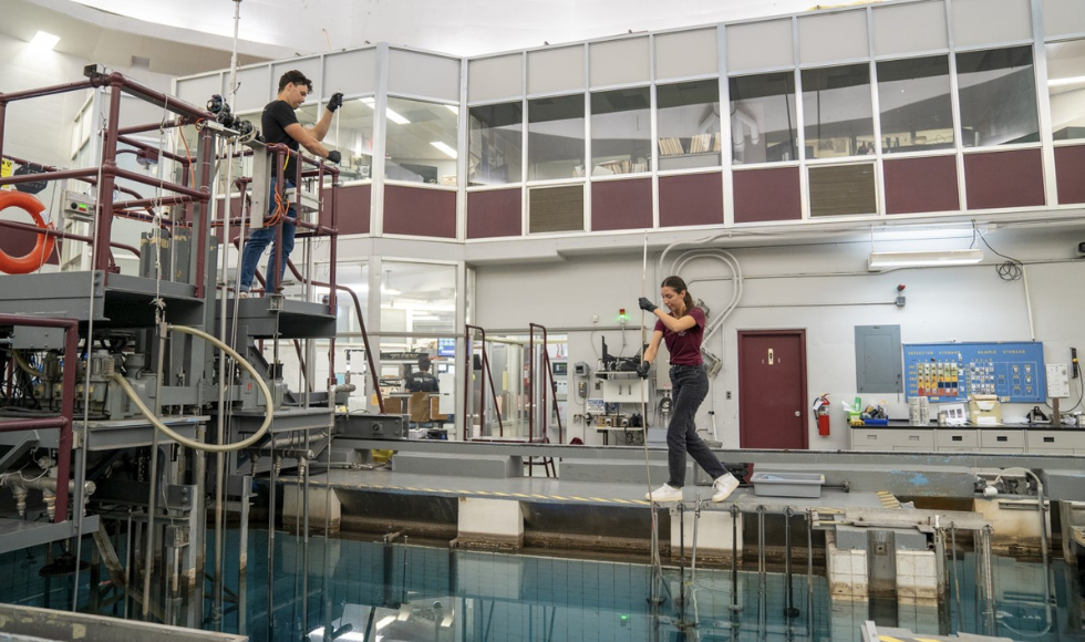
McMaster University and King’s College London’s School of Biomedical Engineering and Imaging Sciences (BMEIS) have formed a partnership to advance nuclear medicine research and education.
The two world-leading nuclear research institutions will work together to produce 94mTc — a radioisotope that can be used in PET scans of tissue and organs, to help diagnose cancer and heart disease, among others.
The partnership builds on McMaster and King’s global leadership in nuclear medicine, says Karin Stephenson, McMaster’s Director of Nuclear Research and Education Support.
“McMaster is thrilled to work with King’s to advance research in nuclear medicine — a field that continues to play an essential role in global health care,” Stephenson says.
“Together, we will use our expertise and infrastructure to develop new radiation-based diagnostic technologies and provide training to the next generation of nuclear scientists in Canada and the U.K.”
There is an increasing global need for new radiopharmaceuticals and a shortage of skilled scientists, says Professor Steve Archibald, Head of Department for Imaging Chemistry and Biology at King’s College London.
“King’s has developed a strategy of integration in nuclear medicine research with collaborative groups of scientists and clinicians that complements the expert scientists and capabilities at McMaster,” he says.
“We are excited to work together to drive innovation in nuclear medicine and unique training opportunities.”
The two institutions will also collaborate to deliver unique learning opportunities for students and professionals interested or involved in the field of nuclear medicine.
Available to senior graduate students and early-career professionals, McMaster and King’s are hosting the Next Generation in Nuclear Medicine Workshop from June 12 – 14 at McMaster.
Learn more about the workshop and submit an expression of interest form.
About King’s College of London
King’s is an international leader in the delivery of fundamental and translational nuclear medicine research. BMEIS trains the next generation of biomedical engineers, imaging scientists and radiochemists and prioritizes collaboration between researchers, clinicians and industry.
About McMaster University
McMaster is a world-leading supplier of medical isotopes and home to a unique suite of nuclear research facilities, including the McMaster Nuclear Reactor, a 16.5 MeV cyclotron, and a high-level laboratory facility, where scientists and graduate students perform radioisotope processing, radiotracer production, radiopharmaceutical development and radiation biology research.

Republish this Article
All republished articles must be attributed in the following way and contain links to both the site and original article: “This article was first published on Brighter World . Read the original article. ”
Media Enquiries
The Communications and Public Affairs Office is staffed from 8:30 a.m. to 4:30 p.m. Monday to Friday.
The University has a broadcast quality television studio to facilitate live and pre-recorded interviews with media. Learn more about our experts.
Related Stories

Celebrating 65 years of nuclear research and innovation
Since 1959, the McMaster Nuclear Reactor has driven groundbreaking discoveries in energy, medicine and materials; fuelled job creation and the economy; and provided unique training opportunities for future generations of nuclear leaders.
Analysis: New electrochemical technology could de-acidify the oceans – and even remove carbon dioxide in the process
Global warming is making the oceans more acidic. A new electrochemical approach could reduce this acidity and remove carbon from the atmosphere in the process.

- Nuclear Physics
- NP Highlights
- Nuclear Science Advisory Committee
The Science
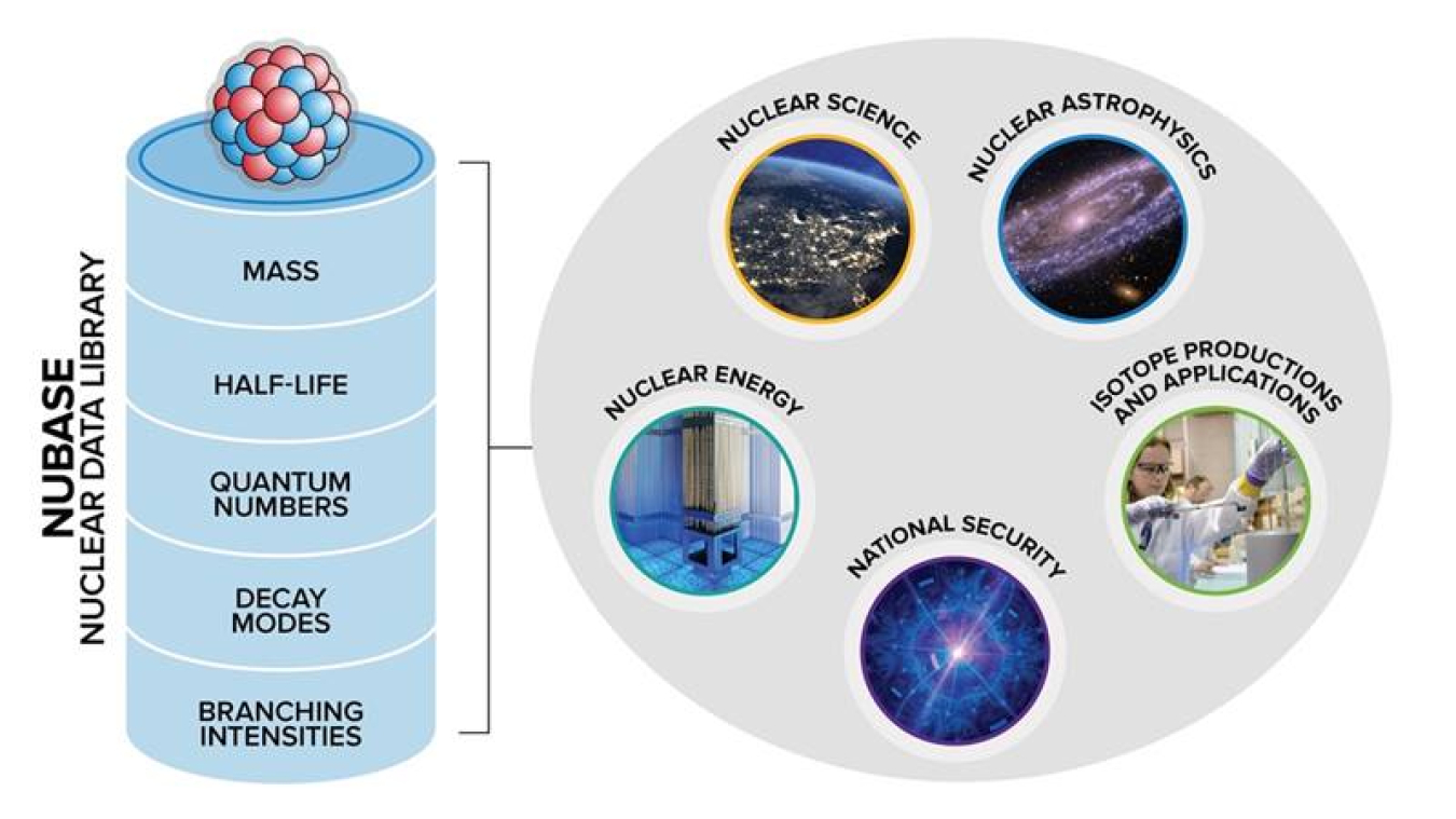
Atomic nuclei exist in the form of isotopes that differ in how many neutrons and protons they have. Today, scientists know of more than 3,300 isotopes. While most isotopes are produced artificially in scientific laboratories, many are created in stars and stellar explosions . A research team has now compiled experimental data for all known nuclei. This includes the isotopes’ main nuclear physics properties, such as mass, quantum numbers, half-life, decay modes, and branching intensities. The researchers evaluated all the data and reported recommended values and their uncertainties. The results are available in the NUBASE library, which also includes estimates for isotopes that are predicted by theoretical models but that scientists have not yet observed.
The data in the NUBASE library cover each nucleus in its ground state (its lowest energy level) and its isomeric states (higher energy levels that live longer than what is typical for other excited levels). Well-defined and credible nuclear data for these states are key to scientists’ understanding of the Universe. The recommended nuclear data are also important in many applications. They are relevant to nearly every field of nuclear research, from basic science and astrophysics to energy, national security, and medicine.
Reliable information on the basic properties of nuclei is a fundamental building block for research in modern nuclear structure and astrophysics. Researchers need good-quality nuclear data formulated and recommended through a speedy assessment and incorporation of new and improved measurements. A sound understanding and accurate quantification of basic nuclear properties help drive advances in many areas.
The current NUBASE nuclear data library contains recommended values and uncertainties for nuclear physics characteristics of all nuclei in their ground and isomeric states. It incorporates a variety of experimental data produced at world-wide nuclear physics facilities. The entries are drawn from primary sources such as journal articles and from secondary sources such as laboratory reports and conference proceedings. Each data point includes pertinent bibliographic details. The entry data are critically evaluated, discrepant results are dismissed, and statistical analysis is applied when recommending the final values. Where experimental data are lacking, estimates are given based on trends in the behavior of the properties for neighboring nuclei. The recommended data are useful for applications in fundamental science, astrophysics, power production, space exploration, national security, human health, and environmental protection.
Filip G. Kondev Argonne National Laboratory [email protected]
This research was supported by the Department of Energy Office of Science, Office of Nuclear Physics.
Publications
Kondev, F.G., et al. , The NUBASE2020 evaluation of nuclear physics properties . Chinese Physics C 45 , 030001 (2021). [DOI: 10.1088/1674-1137/abddae]
Related Links
Argonne physicist recognized for “Top Cited Paper” by Institute of Physics , Argonne National Laboratory news.
Facility for Rare Isotope Beams
At michigan state university, frib researchers lead team to merge nuclear physics experiments and astronomical observations to advance equation-of-state research, world-class particle-accelerator facilities and recent advances in neutron-star observation give physicists a new toolkit for describing nuclear interactions at a wide range of densities..
For most stars, neutron stars and black holes are their final resting places. When a supergiant star runs out of fuel, it expands and then rapidly collapses on itself. This act creates a neutron star—an object denser than our sun crammed into a space 13 to 18 miles wide. In such a heavily condensed stellar environment, most electrons combine with protons to make neutrons, resulting in a dense ball of matter consisting mainly of neutrons. Researchers try to understand the forces that control this process by creating dense matter in the laboratory through colliding neutron-rich nuclei and taking detailed measurements.
A research team—led by William Lynch and Betty Tsang at FRIB—is focused on learning about neutrons in dense environments. Lynch, Tsang, and their collaborators used 20 years of experimental data from accelerator facilities and neutron-star observations to understand how particles interact in nuclear matter under a wide range of densities and pressures. The team wanted to determine how the ratio of neutrons to protons influences nuclear forces in a system. The team recently published its findings in Nature Astronomy .
“In nuclear physics, we are often confined to studying small systems, but we know exactly what particles are in our nuclear systems. Stars provide us an unbelievable opportunity, because they are large systems where nuclear physics plays a vital role, but we do not know for sure what particles are in their interiors,” said Lynch, professor of nuclear physics at FRIB and in the Michigan State University (MSU) Department of Physics and Astronomy. “They are interesting because the density varies greatly within such large systems. Nuclear forces play a dominant role within them, yet we know comparatively little about that role.”
When a star with a mass that is 20-30 times that of the sun exhausts its fuel, it cools, collapses, and explodes in a supernova. After this explosion, only the matter in the deepest part of the star’s interior coalesces to form a neutron star. This neutron star has no fuel to burn and over time, it radiates its remaining heat into the surrounding space. Scientists expect that matter in the outer core of a cold neutron star is roughly similar to the matter in atomic nuclei but with three differences: neutron stars are much larger, they are denser in their interiors, and a larger fraction of their nucleons are neutrons. Deep within the inner core of a neutron star, the composition of neutron star matter remains a mystery.
“If experiments could provide more guidance about the forces that act in their interiors, we could make better predictions of their interior composition and of phase transitions within them. Neutron stars present a great research opportunity to combine these disciplines,” said Lynch.
Accelerator facilities like FRIB help physicists study how subatomic particles interact under exotic conditions that are more common in neutron stars. When researchers compare these experiments to neutron-star observations, they can calculate the equation of state (EOS) of particles interacting in low-temperature, dense environments. The EOS describes matter in specific conditions, and how its properties change with density. Solving EOS for a wide range of settings helps researchers understand the strong nuclear force’s effects within dense objects, like neutron stars, in the cosmos. It also helps us learn more about neutron stars as they cool.
“This is the first time that we pulled together such a wealth of experimental data to explain the equation of state under these conditions, and this is important,” said Tsang, professor of nuclear science at FRIB. “Previous efforts have used theory to explain the low-density and low-energy end of nuclear matter. We wanted to use all the data we had available to us from our previous experiences with accelerators to obtain a comprehensive equation of state.”
Researchers seeking the EOS often calculate it at higher temperatures or lower densities. They then draw conclusions for the system across a wider range of conditions. However, physicists have come to understand in recent years that an EOS obtained from an experiment is only relevant for a specific range of densities. As a result, the team needed to pull together data from a variety of accelerator experiments that used different measurements of colliding nuclei to replace those assumptions with data. “In this work, we asked two questions,” said Lynch. “For a given measurement, what density does that measurement probe? After that, we asked what that measurement tells us about the equation of state at that density.”
In its recent paper, the team combined its own experiments from accelerator facilities in the United States and Japan. It pulled together data from 12 different experimental constraints and three neutron-star observations. The researchers focused on determining the EOS for nuclear matter ranging from half to three times a nuclei’s saturation density—the density found at the core of all stable nuclei. By producing this comprehensive EOS, the team provided new benchmarks for the larger nuclear physics and astrophysics communities to more accurately model interactions of nuclear matter.
The team improved its measurements at intermediate densities that neutron star observations do not provide through experiments at the GSI Helmholtz Centre for Heavy Ion Research in Germany, the RIKEN Nishina Center for Accelerator-Based Science in Japan, and the National Superconducting Cyclotron Laboratory (FRIB’s predecessor). To enable key measurements discussed in this article, their experiments helped fund technical advances in data acquisition for active targets and time projection chambers that are being employed in many other experiments world-wide.
In running these experiments at FRIB, Tsang and Lynch can continue to interact with MSU students who help advance the research with their own input and innovation. MSU operates FRIB as a scientific user facility for the U.S. Department of Energy Office of Science (DOE-SC), supporting the mission of the DOE-SC Office of Nuclear Physics. FRIB is the only accelerator-based user facility on a university campus as one of 28 DOE-SC user facilities . Chun Yen Tsang, the first author on the Nature Astronomy paper, was a graduate student under Betty Tsang during this research and is now a researcher working jointly at Brookhaven National Laboratory and Kent State University.
“Projects like this one are essential for attracting the brightest students, which ultimately makes these discoveries possible, and provides a steady pipeline to the U.S. workforce in nuclear science,” Tsang said.
The proposed FRIB energy upgrade ( FRIB400 ), supported by the scientific user community in the 2023 Nuclear Science Advisory Committee Long Range Plan , will allow the team to probe at even higher densities in the years to come. FRIB400 will double the reach of FRIB along the neutron dripline into a region relevant for neutron-star crusts and to allow study of extreme, neutron-rich nuclei such as calcium-68.
Eric Gedenk is a freelance science writer.
Michigan State University operates the Facility for Rare Isotope Beams (FRIB) as a user facility for the U.S. Department of Energy Office of Science (DOE-SC), supporting the mission of the DOE-SC Office of Nuclear Physics. Hosting what is designed to be the most powerful heavy-ion accelerator, FRIB enables scientists to make discoveries about the properties of rare isotopes in order to better understand the physics of nuclei, nuclear astrophysics, fundamental interactions, and applications for society, including in medicine, homeland security, and industry.
The U.S. Department of Energy Office of Science is the single largest supporter of basic research in the physical sciences in the United States and is working to address some of today’s most pressing challenges. For more information, visit energy.gov/science.
An official website of the United States government
The .gov means it’s official. Federal government websites often end in .gov or .mil. Before sharing sensitive information, make sure you’re on a federal government site.
The site is secure. The https:// ensures that you are connecting to the official website and that any information you provide is encrypted and transmitted securely.
- Publications
- Account settings
Preview improvements coming to the PMC website in October 2024. Learn More or Try it out now .
- Advanced Search
- Journal List
- Asia Ocean J Nucl Med Biol
- v.2(2); Autumn 2014

Quality Assessment of Research Articles in Nuclear Medicine Using STARD and QUADAS-2 Tools
Krisana roysri.
1 Department of Biostatistics, Faculty of Public Health, Khon Kaen University, Khon Kaen, Thailand
Chanisa Chotipanich
3 National Cyclotron and PET Center, Chulabhorn Hospital, Bangkok, Thailand
Vallop Laopaiboon
2 Department of Radiology, Faculty of Medicine, Khon Kaen University, Khon Kaen, Thailand
Jiraporn Khiewyoo
Objective(s):.
Diagnostic nuclear medicine is being increasingly employed in clinical practice with the advent of new technologies and radiopharmaceuticals. The report of the prevalence of a certain disease is important for assessing the quality of that article. Therefore, this study was performed to evaluate the quality of published nuclear medicine articles and determine the frequency of reporting the prevalence of studied diseases.
We used Standards for Reporting of Diagnostic Accuracy (STARD) and Quality Assessment of Diagnostic Accuracy Studies (QUADAS-2) checklists for evaluating the quality of articles published in five nuclear medicine journals with the highest impact factors in 2012. The articles were retrieved from Scopus database and were selected and assessed independently by two nuclear medicine physicians. Decision concerning equivocal data was made by consensus between the reviewers.
The average STARD score was approximately 17 points, and the highest score was 17.19±2.38 obtained by the European Journal of Nuclear Medicine. QUADAS-2 tool showed that all journals had low bias regarding study population. The Journal of Nuclear Medicine had the highest score in terms of index test, reference standard, and time interval. Lack of clarity regarding the index test, reference standard, and time interval was frequently observed in all journals including Clinical Nuclear Medicine, in which 64% of the studies were unclear regarding the index test. Journal of Nuclear Cardiology had the highest number of articles with appropriate reference standard (83.3%), though it had the lowest frequency of reporting disease prevalence (zero reports). All five journals had the same STARD score, while index test, reference standard, and time interval were very unclear according to QUADAS-2 tool. Unfortunately, data were too limited to determine which journal had the lowest risk of bias. In fact, it is the author's responsibility to provide details of research methodology so that the reader can assess the quality of research articles.
Conclusion:
Five nuclear medicine journals with the highest impact factor were comparable in terms of STARD score, although they all showed lack of clarity regarding index test, reference standard, and time interval, according to QUADAS-2. The current data were too limited to determine the journal with the lowest bias. Thus, a comprehensive overview of the research methodology of each article is of paramount importance to enable the reader to assess the quality of articles.
Introduction
Nuclear medicine imaging as well as diagnostic radiological studies including computed tomography (CT) and magnetic resonance imaging (MRI) are important for patient management particularly for making an accurate diagnosis and staging/or restaging of a disease. Even though nuclear medicine studies tend to be less specific, compared to diagnostic radiological imaging, they mostly have high sensitivity, which makes them suitable for early diagnosis, staging, and restaging of diseases.
Some diagnostic nuclear medicine tests are helpful because of their high negative predictive value. Therefore, reporting the prevalence of a disease is essential for helping physicians make decisions based on test results.
The report of the prevalence of a certain disease or condition is very important in studies concerning diagnostic testing, since it affects the positive predictive value (PPV) of a diagnostic test. In fact, a test carried out in a population with a high prevalence of the disease would have a higher PPV, compared to a test performed in a population, where the disease occurrence is rare.
Various radioisotopes are used in nuclear medicine imaging studies so that the patient must receive an appropriate radiation dose. Physicians, who refer patients for nuclear medicine tests, as well as diagnostic radiology, should be concerned about the radiation risks for the patients.
Findings of many studies in diagnostic nuclear medicine provide new insights into this field and help clinicians make decisions for patient management. Standards for Reporting of Diagnostic Accuracy (STARD), as a well-established tool for assessing the value of diagnostic studies, has been adopted by many journals and can be found at www.stard-statement.org. If researchers report their study methods and findings according to STARD checklist, readers will be able to assess the validity of the publication.
Developed from Quality Assessment of Diagnostic Accuracy Studies (QUADAS) ( 1 , 2 ), QUADAS-2 is used for the assessment of studies, which are planned to be included in the systematic reviews of Cochrane library. This tool focuses on the methodology of a study since the value of study results is dependent on methodology. This checklist assesses the presence of bias (high/low/unclear), although it does not appraise the results or discussion section.
The quality of reporting in diagnostic studies is evaluated by STARD checklist ( 3 - 10 ). Some systematic review articles also use STARD for bias assessment ( 11 ), whereas QUADAS is applied to evaluate the reporting of specific diseases in systematic reviews ( 11 - 15 ). Some articles are assessed by both STARD and QUADAS tools ( 16 - 18 ) to determine the quality of research. In fact, both tools can help readers evaluate the quality of a certain article.
Reference standard is of high significance in diagnostic studies. In clinical imaging studies, readers must be familiar with gold standards for each specific disease (such as histopathology report, angiogram, and culture). Some studies may use other imaging modalities, follow up with the same study or other methods.
Research articles published in journals with high impact factors usually have high quality; therefore, readers may be inclined to use the information of a certain article, based on the impact factor of the journal in which that article is published. Reporting the prevalence of a specific disease, which is an essential part of diagnostic radiology and nuclear medicine research, is sometimes not included in some articles.
This study was carried out to determine the frequency of reporting the prevalence of diseases in nuclear medicine articles. We determined the frequency of reporting disease prevalence in nuclear medicine journals with high impact factor, according to STARD stagnant title, each signal question in QUADAS-2 as well as the quality of reference standard.
The journals were sorted according to their impact factors in year 2012, provided by the website: www.medical-journals-links.com/radiology-journals-nuclear-medicine-imaging.php. Then, original and clinical research articles were selected from the Journal of Nuclear Medicine, European Journal of Nuclear Medicine and Molecular Imaging, Clinical Nuclear Medicine, Journal of Nuclear Cardiology, and Nuclear Medicine Communications.
Diagnostic clinical studies, published in 2012, were included in the current study. However, studies with similar objectives and populations were excluded. We searched the articles in Scopus database and limited the results of each journal to studies published in 2012.
The search terms were limited to “sensitivity” and “specificity” in order to obtain comprehensive search results; then, articles consisting of diagnostic nuclear medicine tests were determined and included in the study. Two researchers read the abstracts of the articles separately and selected the diagnostic studies for further evaluation. In case of disagreement, the full article was read and a consensus between the two reviewers was reached.
To evaluate the quality of research articles, we assessed each article and recorded the results in a form including STARD, QUADAS-2, disease prevalence report, and a check list concerning the quality of reference standard.
Descriptive statistics were used to analyze and describe the results. Frequency of each STARD and QUADAS-2 item was reported and mean and standard deviation of STARD scores were also calculated. Analysis of the data was carried out using SPSS version 17, and graphs were generated by Microsoft Excel version 2007.
Our search yielded 212 articles from 5 nuclear medicine journals, among which 101 articles were diagnostic studies. The number of the articles is presented in Figure 1 .

The number of articles in each journal
Overall, the average STARD score was approximately 17±2.39 points, and the European Journal of Nuclear Medicine had the highest score (17.19±2.38). Some items in the STARD checklist were absent in all journals such as item No.13 (describing the methods for calculating test reproducibility, if done) and No. 24 (reporting the estimates of test reproducibility, if done). Item No. 20 (reporting any adverse events due to performing index tests or reference standard) was found only in the Journal of Nuclear Medicine and Nuclear Medicine Communications. The frequency and proportion of reporting each item in all journals are shown in Table 1 , and the average scores are reported in Table 2 .
The presence of each item of STARD checklist (%) in the articles of 5 nuclear medicine journals (CNM=Clinical Nuclear Medicine, EJNMMI= European Journal of Nuclear Medicine and molecular imaging, JNC= Journal of Nuclear Cardiology, JNM=Journal of Nuclear Medicine, NMC= Nuclear Medicine Communications)
The average STARD scores of 5 nuclear medicine journals with the highest impact factors
According to QUADAS-2 checklist, there was a low risk of bias in many studies of all journals, regarding the study population; however, the index test and reference standard were highly unclear. The European Journal of Nuclear Medicine had the largest number of studies with a high risk of bias regarding reference standard. On the other hand, there was a low risk of bias concerning both reference standard and time interval in articles of the Journal of Nuclear Cardiology. The results obtained from QUADAS-2 tool are shown in Table 3 and Figures Figures2 2 - -5 5 .
The results of QUADAS-2 for 5 nuclear medicine journals with the highest impact factors

Risk assessment of population, using QUADAS-2

Risk assessment of time interval

Risk assessment of index test, using QUADAS-2

Risk assessment of reference standard, using QUADAS-2
The rate of reporting the prevalence of the studied diseases and reference standard are shown in Table 4 . The journal with the most frequent reporting of disease prevalence was Nuclear Medicine Communications. The Journal of Nuclear Cardiology had the highest rate of appropriate reference standard (66.7%).
The report of disease prevalence and the appropriateness of reference standard
All journals of clinical nuclear medicine showed similar scores of STARD. This may be related to the researchers’ familiarity with STARD. Some items of STARD were not present or less frequently reported, e.g., reproducibility or adverse effect from the tests. This might be due to the fact that all articles were clinical studies (it is not possible to repeat a certain test on the same subject). Therefore, the details about the adverse effects are not presented since they might be reported to the ethical committee.
Although reporting confidence interval can be helpful for readers in decision-making process, according to statistical findings, the rate of such reports was low (only 42% in European Journal of Nuclear Medicine).
Another important item that should be reported in nearly all articles is describing whether or not the readers of the index test(s) and reference standard are blinded (masked) to the results; however, as the results indicated, the highest rate of reporting was 73%.
According to QUADAS-2 tool, all the studied journal articles were clear in terms of population and sample size. However, regarding index test, a high proportion of studies lacked clarity, e.g., 64% of the articles in Clinical Nuclear Medicine were unclear in this regard; it is not reported in the methodology section. There was also a high risk of bias because the cut-off point for diagnosis was not determined before the results of the standard test were known.
Regarding reference standard, some articles used other imaging modalities, which were not the true reference standard in the study. We found a high risk of bias in 23.1% of the articles in European Journal of Nuclear Medicine; the same was observed concerning time interval. This may be because the authors could not perform an invasive test or had to use more than one single reference standard; the main problem was a negative test.
All journal articles infrequently reported the disease prevalence, e.g., disease prevalence was mentioned only in 31% of the articles in Nuclear Medicine Communication. This may be related to patients’ referral from different parts of the country to nuclear medicine centers; therefore, the authors could not report (or ignored to report) the rate of disease prevalence of a certain disease.
Concerning the reference standard, the articles in the Journal of Nuclear Cardiology used the appropriate reference standard (reported in 66.7% of the articles, since coronary artery catheter is the only reference standard for the diagnosis of coronary heart disease). For other diseases, authors used histopathology, culture, or angiograms to denote a positive finding in the index test. Regarding negative results, researchers might have used other imaging studies or clinical follow-ups as the reference standard.
The average score of STARD was similar among all five nuclear medicine journals. According to QUADAS-2, there was a low risk of bias in terms of study population. However, there was a lack of clarity in other parts including index test, reference standard, and time interval, due to insufficient reporting the details in the articles.
Overall, STARD can familiarize the readers with the details of methodology and results of a study, and help them decide on the study bias. By using QUADAS-2, readers can know the risk of bias for the methodology as low risk, high risk or unclear from this assessment tool; however, they would not be informed about the bias of the results. We suggest that readers use both tools in the assessment of diagnostic research articles.

IMAGES
VIDEO
COMMENTS
The Journal of Nuclear Medicine (JNM)—self-published by the Society of Nuclear Medicine and Molecular Imaging (SNMMI), a nonprofit, international scientific and professional organization—offers readers around the globe clinical investigations, basic science reports, continuing education articles, book reviews, employment opportunities, and updates on rapidly changing issues in practice and ...
Wolfgang A. Weber and Johannes Czernin. Scientific discoveries published in The Journal of Nuclear Medicine over the past 60 years have shaped the practice of medicine and form the foundation for the future of nuclear medicine, molecular imaging, and theranostics. Nuclear medicine now provides diagnostic, prognostic, predictive, and intermediate endpoint biomarkers in oncology, cardiology ...
This article provides a perspective on the current and future state of nuclear medicine (NM) in the United States, based on data from the American Board of Nuclear Medicine (ABNM). It covers topics such as workforce, training, certification, and research in NM.
Nuclear Medicine has become an integral part of an efficient health system that acknowledges this shift with the management of various NCDs, in particular cardiovascular, oncological, and neurodegenerative diseases. Without a doubt, innovation in research and development is a driving force in nuclear medicine. New devices, radiopharmaceuticals ...
Nuclear Medicine is witnessing a revolution across a large spectrum of patient care applications, hardware, software and novel radiopharmaceuticals. We propose to offer a framework of the nuclear medicine practice of the future that incorporates multiple novelties and coined as the NEW (nu) Clear medicine. All these new developments offer a significant clarity and real clinical impact, and we ...
This authoritative journal provides up-to-date information on nuclear medicine that can be readily applied to clinical situations. Written for both generalists and specialists in nuclear medicine, Clinical Nuclear Medicine ensures timely dissemination of data on current developments that affect all aspects of the specialty. The most practice-oriented journal in the field of nuclear imaging ...
Official Journal of the Society of Radiopharmaceutical Sciences Nuclear Medicine and Biology publishes original research addressing all aspects of radiopharmaceutical science for imaging as well as therapeutic applications. More specifically the synthesis (automated and manual), in vitro and ex vivo studies, in vivo biodistribution by dissection or imaging, radiopharmacology, radiopharmacy of ...
Part c, this research was originally published in JNM. Müller, C. et al. Folic acid conjugates for nuclear imaging of folate receptor-positive cancer. J. Nucl. Med. 52, 1-4 (2011), ©SNMMI (ref ...
Timothée Marchal. Pierre Etienne Heudel. Frontiers in Nuclear Medicine. doi 10.3389/fnume.2023.1292676. 667 views. An exciting new journal which advances the use of nuclear medicine in diagnostics and therapeutics as an essential part of patient care.
Introduction. The field of nuclear medicine (NM) is unique regarding its intricate cum essential dependence on the use of radiopharmaceuticals (RPh) for every procedure. RPh comprises a radioisotope (radioisotopes [RI), produced in a research reactor [RR] or particle accelerator like medical cyclotron [MC] delivering radiation used for detection-based imaging, or for targeted therapy, and a ...
EJNMMI Research publishes original and review articles on basic, translational and clinical research in nuclear medicine and molecular imaging. The journal covers topics such as radiopharmaceuticals, image reconstruction, therapy combinations, artificial intelligence and ethical aspects of research.
Nuclear Medicine. This chapter provides an overview of the field of nuclear medicine for readers who are not familiar with the discipline. It includes a description of the history and major discoveries in this field, the challenges of conducting nuclear medicine research, and the foreseeable new technologies and opportunities for personalizing ...
Ming Xue. Xiaoyu Zhang. Xigang Xiao. Frontiers in Medicine. doi 10.3389/fmed.2024.1357981. 464 views. Part of a multidisciplinary journal which advances our medical knowledge, this section explores the use of nuclear medicine in diagnostics and therapeutics as an essential part of patient care.
The diagnosis and treatment of patients with cancer requires access to imaging to ensure accurate management decisions and optimal outcomes. Our global assessment of imaging and nuclear medicine resources identified substantial shortages in equipment and workforce, particularly in low-income and middle-income countries (LMICs). A microsimulation model of 11 cancers showed that the scale-up of ...
Nuclear medicine contributes greatly to the clinical management of patients and experimental medicine. This report aims to (1) outline the current landscape of nuclear medicine research in the UK, including current facilities and recent or ongoing clinical studies and (2) provide information about the available pathways for clinical adoption and NHS funding (commissioning) of radiopharmaceuticals.
Nuclear medicine in the United States has grown because of advances in technology, including hybrid imaging, the introduction of new radiopharmaceuticals for diagnosis and therapy, and the development of molecular imaging based on the tracer principle, which is not based on radioisotopes. Continued growth of the field will require cost-effectiveness data and evidence that nuclear medicine ...
Research in nuclear medicine involves developing new radio tracers as well as technologies that will help physicians produce clearer pictures. PET contrast agents designed to detect bacterial infections reveal E. coli in a rat. Developing new tracers. A bacterial infection is a common complication of implanting a medical device into the body.
The field of nuclear medicine research, as well as the radiopharmaceutical industry, were both adversely affected by COVID-19. Similar content being viewed by others.
The history of nuclear medicine over the past 50 years reflects the strong link between government investments in science and technology and advances in health care in the United States and worldwide. As a result of these investments, new nuclear medicine procedures have been developed that can diagnose diseases non-invasively, providing information that cannot be acquired with other imaging ...
Morgan Stanley estimates that the radiopharmaceutical market will grow to $39 billion by 2032 from about $7 billion in 2022. One such medicine has already reached pharmacy shelves. Developed by ...
Nuclear medicine (NM) in the United States is experiencing a manpower shortage that is steadily getting worse. It largely derives from inadequate production of well-trained NM physicians. It is different in the rest of the world, where NM is an independent specialty and training is more rigorous. Three suggestions are offered to help reverse the situation: (1) stop radiologists with inadequate ...
The advancement of particle detectors for use in medicine is still clearly influenced and driven by work in nuclear and basic particle physics. Modern clinical settings and medical research facilities frequently employ cutting-edge techniques generated from particle accelerators, detectors, and physics computers.
Keywords: Nuclear Medicine, Radiology, Radiopharmaceuticals, Radionuclide therapy, imaging analysis . Important Note: All contributions to this Research Topic must be within the scope of the section and journal to which they are submitted, as defined in their mission statements.Frontiers reserves the right to guide an out-of-scope manuscript to a more suitable section or journal at any stage ...
McMaster University and King's College London's School of Biomedical Engineering and Imaging Sciences (BMEIS) have formed a partnership to advance nuclear medicine research and education. The two world-leading nuclear research institutions will work together to produce 94mTc — a radioisotope that can be used in PET scans of tissue and organs, to help diagnose cancer and heart disease ...
Apr 16, 2024. The Society of Nuclear Medicine and Molecular Imaging (SNMMI) has declared an advocacy win, with language it crafted to promote nuclear medicine research included in the recently approved 2024 Defense Appropriations Act. "In 2023, SNMMI worked closely with Congress for legislative language that increases recognition for the ...
Materials and methods. Based on the Essential Science Indicators, this study employed a bibliometric method to examine the highly cited papers in the subject category of Radiology, Nuclear Medicine & Medical Imaging in Web of Science (WoS) Categories, both quantitatively and qualitatively. In total, 1325 highly cited papers were retrieved and assessed spanning from the years of 2011 to 2021.
Choice Is Good at Times: The Emergence of [64 Cu]Cu-DOTATATE-Based Somatostatin Receptor Imaging in the Era of [68 Ga]Ga-DOTATATE
A research team has now compiled experimental data for all known nuclei. This includes the isotopes' main nuclear physics properties, such as mass, quantum numbers, half-life, decay modes, and branching intensities. The researchers evaluated all the data and reported recommended values and their uncertainties.
World-class particle-accelerator facilities and recent advances in neutron-star observation give physicists a new toolkit for describing nuclear interactions at a wide range of densities.For most stars, neutron stars and black holes are their final resting places. When a supergiant star runs out of fuel, it expands and then rapidly collapses on itself.
Reporting the prevalence of a specific disease, which is an essential part of diagnostic radiology and nuclear medicine research, is sometimes not included in some articles. This study was carried out to determine the frequency of reporting the prevalence of diseases in nuclear medicine articles. We determined the frequency of reporting disease ...