- U.S. Department of Health & Human Services
- National Institutes of Health

En Español | Site Map | Staff Directory | Contact Us
- Science Education
- Science Topics
- Magnetic Resonance Imaging (MRI)

What is MRI?
How does mri work, what is mri used for, are there risks, what are examples of nibib-funded projects in mri.
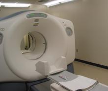
Magnetic Resonance Imaging (MRI) is a non-invasive imaging technology that produces three dimensional detailed anatomical images. It is often used for disease detection, diagnosis, and treatment monitoring. It is based on sophisticated technology that excites and detects the change in the direction of the rotational axis of protons found in the water that makes up living tissues.

MRIs employ powerful magnets which produce a strong magnetic field that forces protons in the body to align with that field. When a radiofrequency current is then pulsed through the patient, the protons are stimulated, and spin out of equilibrium, straining against the pull of the magnetic field. When the radiofrequency field is turned off, the MRI sensors are able to detect the energy released as the protons realign with the magnetic field. The time it takes for the protons to realign with the magnetic field, as well as the amount of energy released, changes depending on the environment and the chemical nature of the molecules. Physicians are able to tell the difference between various types of tissues based on these magnetic properties.
To obtain an MRI image, a patient is placed inside a large magnet and must remain very still during the imaging process in order not to blur the image. Contrast agents (often containing the element Gadolinium) may be given to a patient intravenously before or during the MRI to increase the speed at which protons realign with the magnetic field. The faster the protons realign, the brighter the image.
MRI scanners are particularly well suited to image the non-bony parts or soft tissues of the body. They differ from computed tomography (CT), in that they do not use the damaging ionizing radiation of x-rays. The brain, spinal cord and nerves, as well as muscles, ligaments, and tendons are seen much more clearly with MRI than with regular x-rays and CT; for this reason MRI is often used to image knee and shoulder injuries.
In the brain, MRI can differentiate between white matter and grey matter and can also be used to diagnose aneurysms and tumors. Because MRI does not use x-rays or other radiation, it is the imaging modality of choice when frequent imaging is required for diagnosis or therapy, especially in the brain. However, MRI is more expensive than x-ray imaging or CT scanning.
One kind of specialized MRI is functional Magnetic Resonance Imaging (fMRI.) This is used to observe brain structures and determine which areas of the brain “activate” (consume more oxygen) during various cognitive tasks. It is used to advance the understanding of brain organization and offers a potential new standard for assessing neurological status and neurosurgical risk.
Although MRI does not emit the ionizing radiation that is found in x-ray and CT imaging, it does employ a strong magnetic field. The magnetic field extends beyond the machine and exerts very powerful forces on objects of iron, some steels, and other magnetizable objects; it is strong enough to fling a wheelchair across the room. Patients should notify their physicians of any form of medical or implant prior to an MR scan.
When having an MRI scan, the following should be taken into consideration:
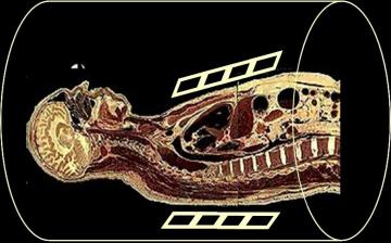
- People with implants, particularly those containing iron, — pacemakers, vagus nerve stimulators, implantable cardioverter- defibrillators, loop recorders, insulin pumps, cochlear implants, deep brain stimulators, and capsules from capsule endoscopy should not enter an MRI machine.
- Noise —loud noise commonly referred to as clicking and beeping, as well as sound intensity up to 120 decibels in certain MR scanners, may require special ear protection.
- Nerve Stimulation —a twitching sensation sometimes results from the rapidly switched fields in the MRI.
- Contrast agents —patients with severe renal failure who require dialysis may risk a rare but serious illness called nephrogenic systemic fibrosis that may be linked to the use of certain gadolinium-containing agents, such as gadodiamide and others. Although a causal link has not been established, current guidelines in the United States recommend that dialysis patients should only receive gadolinium agents when essential, and that dialysis should be performed as soon as possible after the scan to remove the agent from the body promptly.
- Pregnancy —while no effects have been demonstrated on the fetus, it is recommended that MRI scans be avoided as a precaution especially in the first trimester of pregnancy when the fetus’ organs are being formed and contrast agents, if used, could enter the fetal bloodstream.
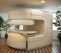
- Claustrophobia —people with even mild claustrophobia may find it difficult to tolerate long scan times inside the machine. Familiarization with the machine and process, as well as visualization techniques, sedation, and anesthesia provide patients with mechanisms to overcome their discomfort. Additional coping mechanisms include listening to music or watching a video or movie, closing or covering the eyes, and holding a panic button. The open MRI is a machine that is open on the sides rather than a tube closed at one end, so it does not fully surround the patient. It was developed to accommodate the needs of patients who are uncomfortable with the narrow tunnel and noises of the traditional MRI and for patients whose size or weight make the traditional MRI impractical. Newer open MRI technology provides high quality images for many but not all types of examinations.
Replacing Biopsies with Sound Chronic liver disease and cirrhosis affect more than 5.5 million people in the United States. NIBIB-funded researchers have developed a method to turn sound waves into images of the liver, which provides a new non-invasive, pain-free approach to find tumors or tissue damaged by liver disease. The Magnetic Resonance Elastography (MRE) device is placed over the liver of the patient before he enters the MRI machine. It then pulses sound waves through the liver, which the MRI is able to detect and use to determine the density and health of the liver tissue. This technique is safer and more comfortable for the patient as well as being less expensive than a traditional biopsy. Since MRE is able to recognize very slight differences in tissue density, there is the potential that it could also be used to detect cancer.
New MRI just for Kids MRI is potentially one of the best imaging modalities for children since unlike CT, it does not have any ionizing radiation that could potentially be harmful. However, one of the most difficult challenges that MRI technicians face is obtaining a clear image, especially when the patient is a child or has some kind of ailment that prevents them from staying still for extended periods of time. As a result, many young children require anesthesia, which increases the health risk for the patient. NIBIB is funding research that is attempting to develop a robust pediatric body MRI. By creating a pediatric coil made specifically for smaller bodies, the image can be rendered more clearly and quickly and will demand less MR operator skill. This will make MRIs cheaper, safer, and more available to children. The faster imaging and motion compensation could also potentially benefit adult patients as well.
Another NIBIB-funded researcher is trying to solve this problem from a different angle. He is developing a motion correction system that could greatly improve image quality for MR exams. Researchers are developing an optical tracking system that would be able to match and adapt the MRI pulses to changes in the patient’s pose in real time. This improvement could reduce cost (since less repeat MR exams will have to take place due to poor quality) as well as make MRI a viable option for many patients who are unable to remain still for the exam and reduce the amount of anesthesia used for MR exams.
Determining the aggressiveness of a tumor Traditional MRI, unlike PET or SPECT, cannot measure metabolic rates. However, researchers funded by NIBIB have discovered a way to inject specialized compounds (hyperpolarized carbon 13) into prostate cancer patients to measure the metabolic rate of a tumor. This information can provide a fast and accurate picture of the tumor’s aggressiveness. Monitoring disease progression can improve risk prediction, which is critical for prostate cancer patients who often adopt a wait and watch approach.
Explore More
Heath Topics
- Brain Disorders
- Liver Disease
- Musculoskeletal Disease
Research Topics
Scientific Program Areas
- Division of Applied Science & Technology (Bioimaging)
- Division of Discovery Science & Technology (Bioengineering)
- Division of Health Informatics Technologies (Informatics)
- Division of Interdisciplinary Training (DIDT)
Inside NIBIB
- Director's Corner
- Funding Policies
- NIBIB Fact Sheets
- Press Releases
Emerging ethical issues raised by highly portable MRI research in remote and resource-limited international settings
Affiliations.
- 1 Professor of Law and Faculty Member, Graduate Program in Neuroscience, University of Minnesota; Instructor in Psychology, Harvard Medical School; Executive Director, MGH Center for Law, Brain & Behavior USA. Electronic address: [email protected].
- 2 McKnight Presidential Professor of Law, Medicine & Public Policy; Faegre Baker Daniels Professor of Law; Professor of Medicine; Chair, Consortium on Law and Values in Health, Environment & the Life Sciences, University of Minnesota USA.
- 3 Co-Principal Investigator, Child Development Group, Sangath, New Delhi, India.
- 4 Associate Professor of Pediatrics (Research), Associate Professor of Diagnostic Imaging (Research), Brown University; Senior Program Officer, Maternal, Newborn & Child Health Discovery & Tools, Discovery & Translational Sciences, Bill & Melinda Gates Foundation USA.
- 5 Associate Professor, Institute of Child Development, Department of Pediatrics, University of Minnesota USA.
- 6 Redleaf Endowed Director, Masonic Institute for the Developing Brain; Professor, Institute of Child Development, College of Education and Human Development; Professor, Department of Pediatrics, Medical School, University of Minnesota USA.
- 7 Malcolm B. Hanson Professor of Radiology, Department of Radiology, Center for Magnetic Resonance Research, University of Minnesota USA.
- 8 Vice-Chair of Clinical Operations, Chief of Pediatric Radiology, Pediatric Imaging Research Center Director, Massachusetts General Hospital; Co-Director, Mass General Imaging Global Health Educational Programs USA.
- 9 Associate Research Scientist, Columbia Magnetic Resonance Research Center, Columbia University USA.
- 10 Assistant Professor, Center for Magnetic Resonance Research, Department of Radiology, University of Minnesota USA.
- 11 Professor, Vice-Chair of Research, Drs. T. J. and Ella M. Arneson Land-Grant Chair in Human Behavior, Department of Psychiatry and Behavioral Sciences, University of Minnesota USA.
- 12 Sir Henry Wellcome Postdoctoral Research Fellow, Centre for Brain and Cognitive Development, Department of Psychological Sciences, Birkbeck College, University of London UK.
- 13 Professor, Department of Psychology; Adjunct Faculty Member, Institute of Child Development; Core Faculty Member, Center for Neurobehavioral Development, University of Minnesota USA.
- 14 Associate, Ropes & Gray LLP USA.
- 15 Director, Neuroethics Program, Center for Ethics; Associate Professor, Departments of Neurology and Psychiatry and Behavioral Sciences, School of Medicine, Emory University USA.
- 16 Technical Officer, Brain Health Unit, World Health Organization Switzerland.
- 17 Chief Medical Officer and Chief Strategy Officer, Hyperfine USA.
- 18 PhD Candidate in the Department of Biomedical Engineering, NSF GRFP Fellow, University of Minnesota; Garwood Lab member USA.
- 19 Professor in the Departments of Biomedical Engineering and Radiology, Director of the Columbia Magnetic Resonance Research Center; Principal and Investigator and MR Platform Director of the Zuckerman Institute, Columbia University; Director of the High Field Imaging Lab, Nathan Kline Institute USA.
- PMID: 34062266
- PMCID: PMC8382487
- DOI: 10.1016/j.neuroimage.2021.118210
Smaller, more affordable, and more portable MRI brain scanners offer exciting opportunities to address unmet research needs and long-standing health inequities in remote and resource-limited international settings. Field-based neuroimaging research in low- and middle-income countries (LMICs) can improve local capacity to conduct both structural and functional neuroscience studies, expand knowledge of brain injury and neuropsychiatric and neurodevelopmental disorders, and ultimately improve the timeliness and quality of clinical diagnosis and treatment around the globe. Facilitating MRI research in remote settings can also diversify reference databases in neuroscience, improve understanding of brain development and degeneration across the lifespan in diverse populations, and help to create reliable measurements of infant and child development. These deeper understandings can lead to new strategies for collaborating with communities to mitigate and hopefully overcome challenges that negatively impact brain development and quality of life. Despite the potential importance of research using highly portable MRI in remote and resource-limited settings, there is little analysis of the attendant ethical, legal, and social issues (ELSI). To begin addressing this gap, this paper presents findings from the first phase of an envisioned multi-staged and iterative approach for creating ethical and legal guidance in a complex global landscape. Section 1 provides a brief introduction to the emerging technology for field-based MRI research. Section 2 presents our methodology for generating plausible use cases for MRI research in remote and resource-limited settings and identifying associated ELSI issues. Section 3 analyzes core ELSI issues in designing and conducting field-based MRI research in remote, resource-limited settings and offers recommendations. We argue that a guiding principle for field-based MRI research in these contexts should be including local communities and research participants throughout the research process in order to create sustained local value. Section 4 presents a recommended path for the next phase of work that could further adapt these use cases, address ethical and legal issues, and co-develop guidance in partnership with local communities.
Keywords: International research; Low- and middle-income countries (LMICs); Neuroethics; Neuroimaging; Portable MRI.
Copyright © 2021. Published by Elsevier Inc.
Publication types
- Research Support, N.I.H., Extramural
- Research Support, Non-U.S. Gov't
- Developing Countries
- Ethics, Research
- Magnetic Resonance Imaging / ethics*
- Neuroimaging / ethics*
Grants and funding
- RF1 MH123698/MH/NIMH NIH HHS/United States
- 204706/Z/16/Z/WT_/Wellcome Trust/United Kingdom
- WT_/Wellcome Trust/United Kingdom
- 001/WHO_/World Health Organization/International
- U01 EB025153/EB/NIBIB NIH HHS/United States
Head Start Your Radiology Residency [Online] ↗️
- Radiology Thesis – More than 400 Research Topics (2022)!
Please login to bookmark

Introduction
A thesis or dissertation, as some people would like to call it, is an integral part of the Radiology curriculum, be it MD, DNB, or DMRD. We have tried to aggregate radiology thesis topics from various sources for reference.
Not everyone is interested in research, and writing a Radiology thesis can be daunting. But there is no escape from preparing, so it is better that you accept this bitter truth and start working on it instead of cribbing about it (like other things in life. #PhilosophyGyan!)
Start working on your thesis as early as possible and finish your thesis well before your exams, so you do not have that stress at the back of your mind. Also, your thesis may need multiple revisions, so be prepared and allocate time accordingly.
Tips for Choosing Radiology Thesis and Research Topics
Keep it simple silly (kiss).
Retrospective > Prospective
Retrospective studies are better than prospective ones, as you already have the data you need when choosing to do a retrospective study. Prospective studies are better quality, but as a resident, you may not have time (, energy and enthusiasm) to complete these.
Choose a simple topic that answers a single/few questions
Original research is challenging, especially if you do not have prior experience. I would suggest you choose a topic that answers a single or few questions. Most topics that I have listed are along those lines. Alternatively, you can choose a broad topic such as “Role of MRI in evaluation of perianal fistulas.”
You can choose a novel topic if you are genuinely interested in research AND have a good mentor who will guide you. Once you have done that, make sure that you publish your study once you are done with it.
Get it done ASAP.
In most cases, it makes sense to stick to a thesis topic that will not take much time. That does not mean you should ignore your thesis and ‘Ctrl C + Ctrl V’ from a friend from another university. Thesis writing is your first step toward research methodology so do it as sincerely as possible. Do not procrastinate in preparing the thesis. As soon as you have been allotted a guide, start researching topics and writing a review of the literature.
At the same time, do not invest a lot of time in writing/collecting data for your thesis. You should not be busy finishing your thesis a few months before the exam. Some people could not appear for the exam because they could not submit their thesis in time. So DO NOT TAKE thesis lightly.
Do NOT Copy-Paste
Reiterating once again, do not simply choose someone else’s thesis topic. Find out what are kind of cases that your Hospital caters to. It is better to do a good thesis on a common topic than a crappy one on a rare one.
Books to help you write a Radiology Thesis
Event country/university has a different format for thesis; hence these book recommendations may not work for everyone.

- Amazon Kindle Edition
- Gupta, Piyush (Author)
- English (Publication Language)
- 206 Pages - 10/12/2020 (Publication Date) - Jaypee Brothers Medical Publishers (P) Ltd. (Publisher)
In A Hurry? Download a PDF list of Radiology Research Topics!
Sign up below to get this PDF directly to your email address.
100% Privacy Guaranteed. Your information will not be shared. Unsubscribe anytime with a single click.
List of Radiology Research /Thesis / Dissertation Topics
- State of the art of MRI in the diagnosis of hepatic focal lesions
- Multimodality imaging evaluation of sacroiliitis in newly diagnosed patients of spondyloarthropathy
- Multidetector computed tomography in oesophageal varices
- Role of positron emission tomography with computed tomography in the diagnosis of cancer Thyroid
- Evaluation of focal breast lesions using ultrasound elastography
- Role of MRI diffusion tensor imaging in the assessment of traumatic spinal cord injuries
- Sonographic imaging in male infertility
- Comparison of color Doppler and digital subtraction angiography in occlusive arterial disease in patients with lower limb ischemia
- The role of CT urography in Haematuria
- Role of functional magnetic resonance imaging in making brain tumor surgery safer
- Prediction of pre-eclampsia and fetal growth restriction by uterine artery Doppler
- Role of grayscale and color Doppler ultrasonography in the evaluation of neonatal cholestasis
- Validity of MRI in the diagnosis of congenital anorectal anomalies
- Role of sonography in assessment of clubfoot
- Role of diffusion MRI in preoperative evaluation of brain neoplasms
- Imaging of upper airways for pre-anaesthetic evaluation purposes and for laryngeal afflictions.
- A study of multivessel (arterial and venous) Doppler velocimetry in intrauterine growth restriction
- Multiparametric 3tesla MRI of suspected prostatic malignancy.
- Role of Sonography in Characterization of Thyroid Nodules for differentiating benign from
- Role of advances magnetic resonance imaging sequences in multiple sclerosis
- Role of multidetector computed tomography in evaluation of jaw lesions
- Role of Ultrasound and MR Imaging in the Evaluation of Musculotendinous Pathologies of Shoulder Joint
- Role of perfusion computed tomography in the evaluation of cerebral blood flow, blood volume and vascular permeability of cerebral neoplasms
- MRI flow quantification in the assessment of the commonest csf flow abnormalities
- Role of diffusion-weighted MRI in evaluation of prostate lesions and its histopathological correlation
- CT enterography in evaluation of small bowel disorders
- Comparison of perfusion magnetic resonance imaging (PMRI), magnetic resonance spectroscopy (MRS) in and positron emission tomography-computed tomography (PET/CT) in post radiotherapy treated gliomas to detect recurrence
- Role of multidetector computed tomography in evaluation of paediatric retroperitoneal masses
- Role of Multidetector computed tomography in neck lesions
- Estimation of standard liver volume in Indian population
- Role of MRI in evaluation of spinal trauma
- Role of modified sonohysterography in female factor infertility: a pilot study.
- The role of pet-CT in the evaluation of hepatic tumors
- Role of 3D magnetic resonance imaging tractography in assessment of white matter tracts compromise in supratentorial tumors
- Role of dual phase multidetector computed tomography in gallbladder lesions
- Role of multidetector computed tomography in assessing anatomical variants of nasal cavity and paranasal sinuses in patients of chronic rhinosinusitis.
- magnetic resonance spectroscopy in multiple sclerosis
- Evaluation of thyroid nodules by ultrasound elastography using acoustic radiation force impulse (ARFI) imaging
- Role of Magnetic Resonance Imaging in Intractable Epilepsy
- Evaluation of suspected and known coronary artery disease by 128 slice multidetector CT.
- Role of regional diffusion tensor imaging in the evaluation of intracranial gliomas and its histopathological correlation
- Role of chest sonography in diagnosing pneumothorax
- Role of CT virtual cystoscopy in diagnosis of urinary bladder neoplasia
- Role of MRI in assessment of valvular heart diseases
- High resolution computed tomography of temporal bone in unsafe chronic suppurative otitis media
- Multidetector CT urography in the evaluation of hematuria
- Contrast-induced nephropathy in diagnostic imaging investigations with intravenous iodinated contrast media
- Comparison of dynamic susceptibility contrast-enhanced perfusion magnetic resonance imaging and single photon emission computed tomography in patients with little’s disease
- Role of Multidetector Computed Tomography in Bowel Lesions.
- Role of diagnostic imaging modalities in evaluation of post liver transplantation recipient complications.
- Role of multislice CT scan and barium swallow in the estimation of oesophageal tumour length
- Malignant Lesions-A Prospective Study.
- Value of ultrasonography in assessment of acute abdominal diseases in pediatric age group
- Role of three dimensional multidetector CT hysterosalpingography in female factor infertility
- Comparative evaluation of multi-detector computed tomography (MDCT) virtual tracheo-bronchoscopy and fiberoptic tracheo-bronchoscopy in airway diseases
- Role of Multidetector CT in the evaluation of small bowel obstruction
- Sonographic evaluation in adhesive capsulitis of shoulder
- Utility of MR Urography Versus Conventional Techniques in Obstructive Uropathy
- MRI of the postoperative knee
- Role of 64 slice-multi detector computed tomography in diagnosis of bowel and mesenteric injury in blunt abdominal trauma.
- Sonoelastography and triphasic computed tomography in the evaluation of focal liver lesions
- Evaluation of Role of Transperineal Ultrasound and Magnetic Resonance Imaging in Urinary Stress incontinence in Women
- Multidetector computed tomographic features of abdominal hernias
- Evaluation of lesions of major salivary glands using ultrasound elastography
- Transvaginal ultrasound and magnetic resonance imaging in female urinary incontinence
- MDCT colonography and double-contrast barium enema in evaluation of colonic lesions
- Role of MRI in diagnosis and staging of urinary bladder carcinoma
- Spectrum of imaging findings in children with febrile neutropenia.
- Spectrum of radiographic appearances in children with chest tuberculosis.
- Role of computerized tomography in evaluation of mediastinal masses in pediatric
- Diagnosing renal artery stenosis: Comparison of multimodality imaging in diabetic patients
- Role of multidetector CT virtual hysteroscopy in the detection of the uterine & tubal causes of female infertility
- Role of multislice computed tomography in evaluation of crohn’s disease
- CT quantification of parenchymal and airway parameters on 64 slice MDCT in patients of chronic obstructive pulmonary disease
- Comparative evaluation of MDCT and 3t MRI in radiographically detected jaw lesions.
- Evaluation of diagnostic accuracy of ultrasonography, colour Doppler sonography and low dose computed tomography in acute appendicitis
- Ultrasonography , magnetic resonance cholangio-pancreatography (MRCP) in assessment of pediatric biliary lesions
- Multidetector computed tomography in hepatobiliary lesions.
- Evaluation of peripheral nerve lesions with high resolution ultrasonography and colour Doppler
- Multidetector computed tomography in pancreatic lesions
- Multidetector Computed Tomography in Paediatric abdominal masses.
- Evaluation of focal liver lesions by colour Doppler and MDCT perfusion imaging
- Sonographic evaluation of clubfoot correction during Ponseti treatment
- Role of multidetector CT in characterization of renal masses
- Study to assess the role of Doppler ultrasound in evaluation of arteriovenous (av) hemodialysis fistula and the complications of hemodialysis vasular access
- Comparative study of multiphasic contrast-enhanced CT and contrast-enhanced MRI in the evaluation of hepatic mass lesions
- Sonographic spectrum of rheumatoid arthritis
- Diagnosis & staging of liver fibrosis by ultrasound elastography in patients with chronic liver diseases
- Role of multidetector computed tomography in assessment of jaw lesions.
- Role of high-resolution ultrasonography in the differentiation of benign and malignant thyroid lesions
- Radiological evaluation of aortic aneurysms in patients selected for endovascular repair
- Role of conventional MRI, and diffusion tensor imaging tractography in evaluation of congenital brain malformations
- To evaluate the status of coronary arteries in patients with non-valvular atrial fibrillation using 256 multirow detector CT scan
- A comparative study of ultrasonography and CT – arthrography in diagnosis of chronic ligamentous and meniscal injuries of knee
- Multi detector computed tomography evaluation in chronic obstructive pulmonary disease and correlation with severity of disease
- Diffusion weighted and dynamic contrast enhanced magnetic resonance imaging in chemoradiotherapeutic response evaluation in cervical cancer.
- High resolution sonography in the evaluation of non-traumatic painful wrist
- The role of trans-vaginal ultrasound versus magnetic resonance imaging in diagnosis & evaluation of cancer cervix
- Role of multidetector row computed tomography in assessment of maxillofacial trauma
- Imaging of vascular complication after liver transplantation.
- Role of magnetic resonance perfusion weighted imaging & spectroscopy for grading of glioma by correlating perfusion parameter of the lesion with the final histopathological grade
- Magnetic resonance evaluation of abdominal tuberculosis.
- Diagnostic usefulness of low dose spiral HRCT in diffuse lung diseases
- Role of dynamic contrast enhanced and diffusion weighted magnetic resonance imaging in evaluation of endometrial lesions
- Contrast enhanced digital mammography anddigital breast tomosynthesis in early diagnosis of breast lesion
- Evaluation of Portal Hypertension with Colour Doppler flow imaging and magnetic resonance imaging
- Evaluation of musculoskeletal lesions by magnetic resonance imaging
- Role of diffusion magnetic resonance imaging in assessment of neoplastic and inflammatory brain lesions
- Radiological spectrum of chest diseases in HIV infected children High resolution ultrasonography in neck masses in children
- with surgical findings
- Sonographic evaluation of peripheral nerves in type 2 diabetes mellitus.
- Role of perfusion computed tomography in the evaluation of neck masses and correlation
- Role of ultrasonography in the diagnosis of knee joint lesions
- Role of ultrasonography in evaluation of various causes of pelvic pain in first trimester of pregnancy.
- Role of Magnetic Resonance Angiography in the Evaluation of Diseases of Aorta and its Branches
- MDCT fistulography in evaluation of fistula in Ano
- Role of multislice CT in diagnosis of small intestine tumors
- Role of high resolution CT in differentiation between benign and malignant pulmonary nodules in children
- A study of multidetector computed tomography urography in urinary tract abnormalities
- Role of high resolution sonography in assessment of ulnar nerve in patients with leprosy.
- Pre-operative radiological evaluation of locally aggressive and malignant musculoskeletal tumours by computed tomography and magnetic resonance imaging.
- The role of ultrasound & MRI in acute pelvic inflammatory disease
- Ultrasonography compared to computed tomographic arthrography in the evaluation of shoulder pain
- Role of Multidetector Computed Tomography in patients with blunt abdominal trauma.
- The Role of Extended field-of-view Sonography and compound imaging in Evaluation of Breast Lesions
- Evaluation of focal pancreatic lesions by Multidetector CT and perfusion CT
- Evaluation of breast masses on sono-mammography and colour Doppler imaging
- Role of CT virtual laryngoscopy in evaluation of laryngeal masses
- Triple phase multi detector computed tomography in hepatic masses
- Role of transvaginal ultrasound in diagnosis and treatment of female infertility
- Role of ultrasound and color Doppler imaging in assessment of acute abdomen due to female genetal causes
- High resolution ultrasonography and color Doppler ultrasonography in scrotal lesion
- Evaluation of diagnostic accuracy of ultrasonography with colour Doppler vs low dose computed tomography in salivary gland disease
- Role of multidetector CT in diagnosis of salivary gland lesions
- Comparison of diagnostic efficacy of ultrasonography and magnetic resonance cholangiopancreatography in obstructive jaundice: A prospective study
- Evaluation of varicose veins-comparative assessment of low dose CT venogram with sonography: pilot study
- Role of mammotome in breast lesions
- The role of interventional imaging procedures in the treatment of selected gynecological disorders
- Role of transcranial ultrasound in diagnosis of neonatal brain insults
- Role of multidetector CT virtual laryngoscopy in evaluation of laryngeal mass lesions
- Evaluation of adnexal masses on sonomorphology and color Doppler imaginig
- Role of radiological imaging in diagnosis of endometrial carcinoma
- Comprehensive imaging of renal masses by magnetic resonance imaging
- The role of 3D & 4D ultrasonography in abnormalities of fetal abdomen
- Diffusion weighted magnetic resonance imaging in diagnosis and characterization of brain tumors in correlation with conventional MRI
- Role of diffusion weighted MRI imaging in evaluation of cancer prostate
- Role of multidetector CT in diagnosis of urinary bladder cancer
- Role of multidetector computed tomography in the evaluation of paediatric retroperitoneal masses.
- Comparative evaluation of gastric lesions by double contrast barium upper G.I. and multi detector computed tomography
- Evaluation of hepatic fibrosis in chronic liver disease using ultrasound elastography
- Role of MRI in assessment of hydrocephalus in pediatric patients
- The role of sonoelastography in characterization of breast lesions
- The influence of volumetric tumor doubling time on survival of patients with intracranial tumours
- Role of perfusion computed tomography in characterization of colonic lesions
- Role of proton MRI spectroscopy in the evaluation of temporal lobe epilepsy
- Role of Doppler ultrasound and multidetector CT angiography in evaluation of peripheral arterial diseases.
- Role of multidetector computed tomography in paranasal sinus pathologies
- Role of virtual endoscopy using MDCT in detection & evaluation of gastric pathologies
- High resolution 3 Tesla MRI in the evaluation of ankle and hindfoot pain.
- Transperineal ultrasonography in infants with anorectal malformation
- CT portography using MDCT versus color Doppler in detection of varices in cirrhotic patients
- Role of CT urography in the evaluation of a dilated ureter
- Characterization of pulmonary nodules by dynamic contrast-enhanced multidetector CT
- Comprehensive imaging of acute ischemic stroke on multidetector CT
- The role of fetal MRI in the diagnosis of intrauterine neurological congenital anomalies
- Role of Multidetector computed tomography in pediatric chest masses
- Multimodality imaging in the evaluation of palpable & non-palpable breast lesion.
- Sonographic Assessment Of Fetal Nasal Bone Length At 11-28 Gestational Weeks And Its Correlation With Fetal Outcome.
- Role Of Sonoelastography And Contrast-Enhanced Computed Tomography In Evaluation Of Lymph Node Metastasis In Head And Neck Cancers
- Role Of Renal Doppler And Shear Wave Elastography In Diabetic Nephropathy
- Evaluation Of Relationship Between Various Grades Of Fatty Liver And Shear Wave Elastography Values
- Evaluation and characterization of pelvic masses of gynecological origin by USG, color Doppler and MRI in females of reproductive age group
- Radiological evaluation of small bowel diseases using computed tomographic enterography
- Role of coronary CT angiography in patients of coronary artery disease
- Role of multimodality imaging in the evaluation of pediatric neck masses
- Role of CT in the evaluation of craniocerebral trauma
- Role of magnetic resonance imaging (MRI) in the evaluation of spinal dysraphism
- Comparative evaluation of triple phase CT and dynamic contrast-enhanced MRI in patients with liver cirrhosis
- Evaluation of the relationship between carotid intima-media thickness and coronary artery disease in patients evaluated by coronary angiography for suspected CAD
- Assessment of hepatic fat content in fatty liver disease by unenhanced computed tomography
- Correlation of vertebral marrow fat on spectroscopy and diffusion-weighted MRI imaging with bone mineral density in postmenopausal women.
- Comparative evaluation of CT coronary angiography with conventional catheter coronary angiography
- Ultrasound evaluation of kidney length & descending colon diameter in normal and intrauterine growth-restricted fetuses
- A prospective study of hepatic vein waveform and splenoportal index in liver cirrhosis: correlation with child Pugh’s classification and presence of esophageal varices.
- CT angiography to evaluate coronary artery by-pass graft patency in symptomatic patient’s functional assessment of myocardium by cardiac MRI in patients with myocardial infarction
- MRI evaluation of HIV positive patients with central nervous system manifestations
- MDCT evaluation of mediastinal and hilar masses
- Evaluation of rotator cuff & labro-ligamentous complex lesions by MRI & MRI arthrography of shoulder joint
- Role of imaging in the evaluation of soft tissue vascular malformation
- Role of MRI and ultrasonography in the evaluation of multifidus muscle pathology in chronic low back pain patients
- Role of ultrasound elastography in the differential diagnosis of breast lesions
- Role of magnetic resonance cholangiopancreatography in evaluating dilated common bile duct in patients with symptomatic gallstone disease.
- Comparative study of CT urography & hybrid CT urography in patients with haematuria.
- Role of MRI in the evaluation of anorectal malformations
- Comparison of ultrasound-Doppler and magnetic resonance imaging findings in rheumatoid arthritis of hand and wrist
- Role of Doppler sonography in the evaluation of renal artery stenosis in hypertensive patients undergoing coronary angiography for coronary artery disease.
- Comparison of radiography, computed tomography and magnetic resonance imaging in the detection of sacroiliitis in ankylosing spondylitis.
- Mr evaluation of painful hip
- Role of MRI imaging in pretherapeutic assessment of oral and oropharyngeal malignancy
- Evaluation of diffuse lung diseases by high resolution computed tomography of the chest
- Mr evaluation of brain parenchyma in patients with craniosynostosis.
- Diagnostic and prognostic value of cardiovascular magnetic resonance imaging in dilated cardiomyopathy
- Role of multiparametric magnetic resonance imaging in the detection of early carcinoma prostate
- Role of magnetic resonance imaging in white matter diseases
- Role of sonoelastography in assessing the response to neoadjuvant chemotherapy in patients with locally advanced breast cancer.
- Role of ultrasonography in the evaluation of carotid and femoral intima-media thickness in predialysis patients with chronic kidney disease
- Role of H1 MRI spectroscopy in focal bone lesions of peripheral skeleton choline detection by MRI spectroscopy in breast cancer and its correlation with biomarkers and histological grade.
- Ultrasound and MRI evaluation of axillary lymph node status in breast cancer.
- Role of sonography and magnetic resonance imaging in evaluating chronic lateral epicondylitis.
- Comparative of sonography including Doppler and sonoelastography in cervical lymphadenopathy.
- Evaluation of Umbilical Coiling Index as Predictor of Pregnancy Outcome.
- Computerized Tomographic Evaluation of Azygoesophageal Recess in Adults.
- Lumbar Facet Arthropathy in Low Backache.
- “Urethral Injuries After Pelvic Trauma: Evaluation with Uretrography
- Role Of Ct In Diagnosis Of Inflammatory Renal Diseases
- Role Of Ct Virtual Laryngoscopy In Evaluation Of Laryngeal Masses
- “Ct Portography Using Mdct Versus Color Doppler In Detection Of Varices In
- Cirrhotic Patients”
- Role Of Multidetector Ct In Characterization Of Renal Masses
- Role Of Ct Virtual Cystoscopy In Diagnosis Of Urinary Bladder Neoplasia
- Role Of Multislice Ct In Diagnosis Of Small Intestine Tumors
- “Mri Flow Quantification In The Assessment Of The Commonest CSF Flow Abnormalities”
- “The Role Of Fetal Mri In Diagnosis Of Intrauterine Neurological CongenitalAnomalies”
- Role Of Transcranial Ultrasound In Diagnosis Of Neonatal Brain Insults
- “The Role Of Interventional Imaging Procedures In The Treatment Of Selected Gynecological Disorders”
- Role Of Radiological Imaging In Diagnosis Of Endometrial Carcinoma
- “Role Of High-Resolution Ct In Differentiation Between Benign And Malignant Pulmonary Nodules In Children”
- Role Of Ultrasonography In The Diagnosis Of Knee Joint Lesions
- “Role Of Diagnostic Imaging Modalities In Evaluation Of Post Liver Transplantation Recipient Complications”
- “Diffusion-Weighted Magnetic Resonance Imaging In Diagnosis And
- Characterization Of Brain Tumors In Correlation With Conventional Mri”
- The Role Of PET-CT In The Evaluation Of Hepatic Tumors
- “Role Of Computerized Tomography In Evaluation Of Mediastinal Masses In Pediatric patients”
- “Trans Vaginal Ultrasound And Magnetic Resonance Imaging In Female Urinary Incontinence”
- Role Of Multidetector Ct In Diagnosis Of Urinary Bladder Cancer
- “Role Of Transvaginal Ultrasound In Diagnosis And Treatment Of Female Infertility”
- Role Of Diffusion-Weighted Mri Imaging In Evaluation Of Cancer Prostate
- “Role Of Positron Emission Tomography With Computed Tomography In Diagnosis Of Cancer Thyroid”
- The Role Of CT Urography In Case Of Haematuria
- “Value Of Ultrasonography In Assessment Of Acute Abdominal Diseases In Pediatric Age Group”
- “Role Of Functional Magnetic Resonance Imaging In Making Brain Tumor Surgery Safer”
- The Role Of Sonoelastography In Characterization Of Breast Lesions
- “Ultrasonography, Magnetic Resonance Cholangiopancreatography (MRCP) In Assessment Of Pediatric Biliary Lesions”
- “Role Of Ultrasound And Color Doppler Imaging In Assessment Of Acute Abdomen Due To Female Genital Causes”
- “Role Of Multidetector Ct Virtual Laryngoscopy In Evaluation Of Laryngeal Mass Lesions”
- MRI Of The Postoperative Knee
- Role Of Mri In Assessment Of Valvular Heart Diseases
- The Role Of 3D & 4D Ultrasonography In Abnormalities Of Fetal Abdomen
- State Of The Art Of Mri In Diagnosis Of Hepatic Focal Lesions
- Role Of Multidetector Ct In Diagnosis Of Salivary Gland Lesions
- “Role Of Virtual Endoscopy Using Mdct In Detection & Evaluation Of Gastric Pathologies”
- The Role Of Ultrasound & Mri In Acute Pelvic Inflammatory Disease
- “Diagnosis & Staging Of Liver Fibrosis By Ultraso Und Elastography In
- Patients With Chronic Liver Diseases”
- Role Of Mri In Evaluation Of Spinal Trauma
- Validity Of Mri In Diagnosis Of Congenital Anorectal Anomalies
- Imaging Of Vascular Complication After Liver Transplantation
- “Contrast-Enhanced Digital Mammography And Digital Breast Tomosynthesis In Early Diagnosis Of Breast Lesion”
- Role Of Mammotome In Breast Lesions
- “Role Of MRI Diffusion Tensor Imaging (DTI) In Assessment Of Traumatic Spinal Cord Injuries”
- “Prediction Of Pre-eclampsia And Fetal Growth Restriction By Uterine Artery Doppler”
- “Role Of Multidetector Row Computed Tomography In Assessment Of Maxillofacial Trauma”
- “Role Of Diffusion Magnetic Resonance Imaging In Assessment Of Neoplastic And Inflammatory Brain Lesions”
- Role Of Diffusion Mri In Preoperative Evaluation Of Brain Neoplasms
- “Role Of Multidetector Ct Virtual Hysteroscopy In The Detection Of The
- Uterine & Tubal Causes Of Female Infertility”
- Role Of Advances Magnetic Resonance Imaging Sequences In Multiple Sclerosis Magnetic Resonance Spectroscopy In Multiple Sclerosis
- “Role Of Conventional Mri, And Diffusion Tensor Imaging Tractography In Evaluation Of Congenital Brain Malformations”
- Role Of MRI In Evaluation Of Spinal Trauma
- Diagnostic Role Of Diffusion-weighted MR Imaging In Neck Masses
- “The Role Of Transvaginal Ultrasound Versus Magnetic Resonance Imaging In Diagnosis & Evaluation Of Cancer Cervix”
- “Role Of 3d Magnetic Resonance Imaging Tractography In Assessment Of White Matter Tracts Compromise In Supra Tentorial Tumors”
- Role Of Proton MR Spectroscopy In The Evaluation Of Temporal Lobe Epilepsy
- Role Of Multislice Computed Tomography In Evaluation Of Crohn’s Disease
- Role Of MRI In Assessment Of Hydrocephalus In Pediatric Patients
- The Role Of MRI In Diagnosis And Staging Of Urinary Bladder Carcinoma
- USG and MRI correlation of congenital CNS anomalies
- HRCT in interstitial lung disease
- X-Ray, CT and MRI correlation of bone tumors
- “Study on the diagnostic and prognostic utility of X-Rays for cases of pulmonary tuberculosis under RNTCP”
- “Role of magnetic resonance imaging in the characterization of female adnexal pathology”
- “CT angiography of carotid atherosclerosis and NECT brain in cerebral ischemia, a correlative analysis”
- Role of CT scan in the evaluation of paranasal sinus pathology
- USG and MRI correlation on shoulder joint pathology
- “Radiological evaluation of a patient presenting with extrapulmonary tuberculosis”
- CT and MRI correlation in focal liver lesions”
- Comparison of MDCT virtual cystoscopy with conventional cystoscopy in bladder tumors”
- “Bleeding vessels in life-threatening hemoptysis: Comparison of 64 detector row CT angiography with conventional angiography prior to endovascular management”
- “Role of transarterial chemoembolization in unresectable hepatocellular carcinoma”
- “Comparison of color flow duplex study with digital subtraction angiography in the evaluation of peripheral vascular disease”
- “A Study to assess the efficacy of magnetization transfer ratio in differentiating tuberculoma from neurocysticercosis”
- “MR evaluation of uterine mass lesions in correlation with transabdominal, transvaginal ultrasound using HPE as a gold standard”
- “The Role of power Doppler imaging with trans rectal ultrasonogram guided prostate biopsy in the detection of prostate cancer”
- “Lower limb arteries assessed with doppler angiography – A prospective comparative study with multidetector CT angiography”
- “Comparison of sildenafil with papaverine in penile doppler by assessing hemodynamic changes”
- “Evaluation of efficacy of sonosalphingogram for assessing tubal patency in infertile patients with hysterosalpingogram as the gold standard”
- Role of CT enteroclysis in the evaluation of small bowel diseases
- “MRI colonography versus conventional colonoscopy in the detection of colonic polyposis”
- “Magnetic Resonance Imaging of anteroposterior diameter of the midbrain – differentiation of progressive supranuclear palsy from Parkinson disease”
- “MRI Evaluation of anterior cruciate ligament tears with arthroscopic correlation”
- “The Clinicoradiological profile of cerebral venous sinus thrombosis with prognostic evaluation using MR sequences”
- “Role of MRI in the evaluation of pelvic floor integrity in stress incontinent patients” “Doppler ultrasound evaluation of hepatic venous waveform in portal hypertension before and after propranolol”
- “Role of transrectal sonography with colour doppler and MRI in evaluation of prostatic lesions with TRUS guided biopsy correlation”
- “Ultrasonographic evaluation of painful shoulders and correlation of rotator cuff pathologies and clinical examination”
- “Colour Doppler Evaluation of Common Adult Hepatic tumors More Than 2 Cm with HPE and CECT Correlation”
- “Clinical Relevance of MR Urethrography in Obliterative Posterior Urethral Stricture”
- “Prediction of Adverse Perinatal Outcome in Growth Restricted Fetuses with Antenatal Doppler Study”
- Radiological evaluation of spinal dysraphism using CT and MRI
- “Evaluation of temporal bone in cholesteatoma patients by high resolution computed tomography”
- “Radiological evaluation of primary brain tumours using computed tomography and magnetic resonance imaging”
- “Three dimensional colour doppler sonographic assessment of changes in volume and vascularity of fibroids – before and after uterine artery embolization”
- “In phase opposed phase imaging of bone marrow differentiating neoplastic lesions”
- “Role of dynamic MRI in replacing the isotope renogram in the functional evaluation of PUJ obstruction”
- Characterization of adrenal masses with contrast-enhanced CT – washout study
- A study on accuracy of magnetic resonance cholangiopancreatography
- “Evaluation of median nerve in carpal tunnel syndrome by high-frequency ultrasound & color doppler in comparison with nerve conduction studies”
- “Correlation of Agatston score in patients with obstructive and nonobstructive coronary artery disease following STEMI”
- “Doppler ultrasound assessment of tumor vascularity in locally advanced breast cancer at diagnosis and following primary systemic chemotherapy.”
- “Validation of two-dimensional perineal ultrasound and dynamic magnetic resonance imaging in pelvic floor dysfunction.”
- “Role of MR urethrography compared to conventional urethrography in the surgical management of obliterative urethral stricture.”
Search Diagnostic Imaging Research Topics
You can also search research-related resources on our custom search engine .
Free Resources for Preparing Radiology Thesis
- Radiology thesis topics- Benha University – Free to download thesis
- Radiology thesis topics – Faculty of Medical Science Delhi
- Radiology thesis topics – IPGMER
- Fetal Radiology thesis Protocols
- Radiology thesis and dissertation topics
- Radiographics
Proofreading Your Thesis:
Make sure you use Grammarly to correct your spelling , grammar , and plagiarism for your thesis. Grammarly has affordable paid subscriptions, windows/macOS apps, and FREE browser extensions. It is an excellent tool to avoid inadvertent spelling mistakes in your research projects. It has an extensive built-in vocabulary, but you should make an account and add your own medical glossary to it.

Guidelines for Writing a Radiology Thesis:
These are general guidelines and not about radiology specifically. You can share these with colleagues from other departments as well. Special thanks to Dr. Sanjay Yadav sir for these. This section is best seen on a desktop. Here are a couple of handy presentations to start writing a thesis:
Read the general guidelines for writing a thesis (the page will take some time to load- more than 70 pages!
A format for thesis protocol with a sample patient information sheet, sample patient consent form, sample application letter for thesis, and sample certificate.
Resources and References:
- Guidelines for thesis writing.
- Format for thesis protocol
- Thesis protocol writing guidelines DNB
- Informed consent form for Research studies from AIIMS
- Radiology Informed consent forms in local Indian languages.
- Sample Informed Consent form for Research in Hindi
- Guide to write a thesis by Dr. P R Sharma
- Guidelines for thesis writing by Dr. Pulin Gupta.
- Preparing MD/DNB thesis by A Indrayan
- Another good thesis reference protocol
Hopefully, this post will make the tedious task of writing a Radiology thesis a little bit easier for you. Best of luck with writing your thesis and your residency too!
More guides for residents :
- Guide for the MD/DMRD/DNB radiology exam!
- Guide for First-Year Radiology Residents
- FRCR Exam: THE Most Comprehensive Guide (2022)!
- Radiology Practical Exams Questions compilation for MD/DNB/DMRD !
Radiology Exam Resources (Oral Recalls, Instruments, etc )!
- Tips and Tricks for DNB/MD Radiology Practical Exam
- FRCR 2B exam- Tips and Tricks !
- FRCR exam preparation – An alternative take!
- Why did I take up Radiology?
- Radiology Conferences – A comprehensive guide!
- ECR (European Congress Of Radiology)
- European Diploma in Radiology (EDiR) – The Complete Guide!
- Radiology NEET PG guide – How to select THE best college for post-graduation in Radiology (includes personal insights)!
- Interventional Radiology – All Your Questions Answered!
What It Means To Be A Radiologist: A Guide For Medical Students!
- Radiology Mentors for Medical Students (Post NEET-PG)
- MD vs DNB Radiology: Which Path is Right for Your Career?
- DNB Radiology OSCE – Tips and Tricks
More radiology resources here: Radiology resources This page will be updated regularly. Kindly leave your feedback in the comments or send us a message here . Also, you can comment below regarding your department’s thesis topics.
Note: All topics have been compiled from available online resources. If anyone has an issue with any radiology thesis topics displayed here, you can message us here , and we can delete them. These are only sample guidelines. Thesis guidelines differ from institution to institution.
Image source: Thesis complete! (2018). Flickr. Retrieved 12 August 2018, from https://www.flickr.com/photos/cowlet/354911838 by Victoria Catterson
About The Author
Dr. amar udare, md, related posts ↓.
7 thoughts on “Radiology Thesis – More than 400 Research Topics (2022)!”
Amazing & The most helpful site for Radiology residents…
Thank you for your kind comments 🙂
Dr. I saw your Tips is very amazing and referable. But Dr. Can you help me with the thesis of Evaluation of Diagnostic accuracy of X-ray radiograph in knee joint lesion.
Wow! These are excellent stuff. You are indeed a teacher. God bless
Glad you liked these!
happy to see this
Glad I could help :).
Leave a Comment Cancel Reply
Your email address will not be published. Required fields are marked *
Get Radiology Updates to Your Inbox!
This site is for use by medical professionals. To continue, you must accept our use of cookies and the site's Terms of Use. Learn more Accept!
Wish to be a BETTER Radiologist? Join 14000 Radiology Colleagues !
Enter your email address below to access HIGH YIELD radiology content, updates, and resources.
No spam, only VALUE! Unsubscribe anytime with a single click.
- Brownell Lab
- Brugarolas Lab
- Elmaleh Lab
- Johnson Lab
- Normandin Lab
- Santarnecchi Lab
- Sepulcre Lab
- Technical Staff
- Administrative Staff
- Collaborators
- Publications
Image Generation and Clinical Assessment
- PET and SPECT Instrumentation
Multimodal MRI Research
Quantitative pet-mr imaging.
- Quantitative PET/SPECT
Machine Learning for Anatomical imaging
- Therapy Imaging Program
Pharmaco-Kinetic Modeling
Monitoring radiotherapy with pet.
- Radiochemistry Discovery
- Precision Neuroscience & Neuromodulation Program
- Video Lectures
- T32 Postgraduate Training Program in Medical Imaging (PTPMI)
- The Gordon Speaker Series
- Translation
- Covid-19 response
- Recent Seminars
- Awards and Honors
- Recent Publications
- Other News Items
- Grant Proposals
- Grant Related FAQs
- Travel Policies
- Purchasing and Other Daily Operations
- Human Resources
- Contact Forms
- Science Resources
- Computing Resources
- File Repository
- Seminar Sign Up

Research Topics
The Gordon Center is conducting work at the forefront of quantitative PET in brain, oncologic and cardiac imaging. This webpage summarizes some of the research performed in the Center in kinetic modeling of neurotransmission, cardiac perfusion and mitochondrial function, simultaneous PET-MR imaging, in-room PET monitoring of proton therapy, high sensitivity/resolution brain imaging, quantitative dual tracer PET and SPECT, and objective assessment of image quality for estimation and detection tasks. More information about our research areas can be found in the links below.
PET-CT and SPECT Instrumentation
Quantitative PET-CT and SPECT-CT
Therapy Imaging Program (TIP)
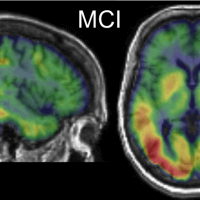
Radiochemistry Discovery and Multifunctional Nanomaterials
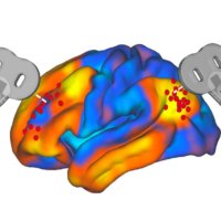
Precision Neuroscience & Neuromodulation Program
Radiology Research Paper Topics

Radiology research paper topics encompass a wide range of fascinating areas within the field of medical imaging. This page aims to provide students studying health sciences with a comprehensive collection of radiology research paper topics to inspire and guide their research endeavors. By delving into various categories and exploring ten thought-provoking topics within each, students can gain insights into the diverse research possibilities in radiology. From advancements in imaging technology to the evaluation of diagnostic accuracy and the impact of radiological interventions, these topics offer a glimpse into the exciting world of radiology research. Additionally, expert advice is provided to help students choose the most suitable research topics and navigate the process of writing a research paper in radiology. By leveraging iResearchNet’s writing services, students can further enhance their research papers with professional assistance, ensuring the highest quality and adherence to academic standards. Explore the realm of radiology research paper topics and unleash your potential to contribute to the advancement of medical imaging and patient care.
100 Radiology Research Paper Topics
Radiology encompasses a broad spectrum of imaging techniques used to diagnose diseases, monitor treatment progress, and guide interventions. This comprehensive list of radiology research paper topics serves as a valuable resource for students in the field of health sciences who are seeking inspiration and guidance for their research endeavors. The following ten categories highlight different areas within radiology, each containing ten thought-provoking topics. Exploring these topics will provide students with a deeper understanding of the diverse research possibilities and current trends within the field of radiology.
Academic Writing, Editing, Proofreading, And Problem Solving Services
Get 10% off with 24start discount code.
Diagnostic Imaging Techniques
- Comparative analysis of imaging modalities: CT, MRI, and PET-CT.
- The role of artificial intelligence in radiological image interpretation.
- Advancements in digital mammography for breast cancer screening.
- Emerging techniques in nuclear medicine imaging.
- Image-guided biopsy: Enhancing accuracy and safety.
- Application of radiomics in predicting treatment response.
- Dual-energy CT: Expanding diagnostic capabilities.
- Radiological evaluation of traumatic brain injuries.
- Imaging techniques for evaluating cardiovascular diseases.
- Radiographic evaluation of pulmonary nodules: Challenges and advancements.
Interventional Radiology
- Minimally invasive treatments for liver tumors: Embolization techniques.
- Radiofrequency ablation in the management of renal cell carcinoma.
- Role of interventional radiology in the treatment of peripheral artery disease.
- Transarterial chemoembolization in hepatocellular carcinoma.
- Evaluation of uterine artery embolization for the treatment of fibroids.
- Percutaneous vertebroplasty and kyphoplasty: Efficacy and complications.
- Endovascular repair of abdominal aortic aneurysms: Long-term outcomes.
- Interventional radiology in the management of deep vein thrombosis.
- Transcatheter aortic valve replacement: Imaging considerations.
- Emerging techniques in interventional oncology.
Radiation Safety and Dose Optimization
- Strategies for reducing radiation dose in pediatric imaging.
- Imaging modalities with low radiation exposure: Current advancements.
- Effective use of dose monitoring systems in radiology departments.
- The impact of artificial intelligence on radiation dose optimization.
- Optimization of radiation therapy treatment plans: Balancing efficacy and safety.
- Radioprotective measures for patients and healthcare professionals.
- The role of radiology in addressing radiation-induced risks.
- Evaluating the long-term effects of radiation exposure in diagnostic imaging.
- Radiation dose tracking and reporting: Implementing best practices.
- Patient education and communication regarding radiation risks.
Radiology in Oncology
- Imaging techniques for early detection and staging of lung cancer.
- Quantitative imaging biomarkers for predicting treatment response in solid tumors.
- Radiogenomics: Linking imaging features to genetic profiles in cancer.
- The role of imaging in assessing tumor angiogenesis.
- Radiological evaluation of lymphoma: Challenges and advancements.
- Imaging-guided interventions in the treatment of hepatocellular carcinoma.
- Assessment of tumor heterogeneity using functional imaging techniques.
- Radiomics and machine learning in predicting treatment outcomes in cancer.
- Multimodal imaging in the evaluation of brain tumors.
- Imaging surveillance after cancer treatment: Optimizing follow-up protocols.
Radiology in Musculoskeletal Disorders
- Imaging modalities in the evaluation of sports-related injuries.
- The role of imaging in diagnosing and monitoring rheumatoid arthritis.
- Assessment of bone health using dual-energy X-ray absorptiometry (DXA).
- Imaging techniques for evaluating osteoarthritis progression.
- Imaging-guided interventions in the management of musculoskeletal tumors.
- Role of imaging in diagnosing and managing spinal disorders.
- Evaluation of traumatic injuries using radiography, CT, and MRI.
- Imaging of joint prostheses: Complications and assessment techniques.
- Imaging features and classifications of bone fractures.
- Musculoskeletal ultrasound in the diagnosis of soft tissue injuries.
Neuroradiology
- Advanced neuroimaging techniques for early detection of neurodegenerative diseases.
- Imaging evaluation of acute stroke: Current guidelines and advancements.
- Role of functional MRI in mapping brain functions.
- Imaging of brain tumors: Classification and treatment planning.
- Diffusion tensor imaging in assessing white matter integrity.
- Neuroimaging in the evaluation of multiple sclerosis.
- Imaging techniques for the assessment of epilepsy.
- Radiological evaluation of neurovascular diseases.
- Imaging of cranial nerve disorders: Diagnosis and management.
- Radiological assessment of developmental brain abnormalities.
Pediatric Radiology
- Radiation dose reduction strategies in pediatric imaging.
- Imaging evaluation of congenital heart diseases in children.
- Role of imaging in the diagnosis and management of pediatric oncology.
- Imaging of pediatric gastrointestinal disorders.
- Evaluation of developmental hip dysplasia using ultrasound and radiography.
- Imaging features and management of pediatric musculoskeletal infections.
- Neuroimaging in the assessment of pediatric neurodevelopmental disorders.
- Radiological evaluation of pediatric respiratory conditions.
- Imaging techniques for the evaluation of pediatric abdominal emergencies.
- Imaging-guided interventions in pediatric patients.
Breast Imaging
- Advances in digital mammography for early breast cancer detection.
- The role of tomosynthesis in breast imaging.
- Imaging evaluation of breast implants: Complications and assessment.
- Radiogenomic analysis of breast cancer subtypes.
- Contrast-enhanced mammography: Diagnostic benefits and challenges.
- Emerging techniques in breast MRI for high-risk populations.
- Evaluation of breast density and its implications for cancer risk.
- Role of molecular breast imaging in dense breast tissue evaluation.
- Radiological evaluation of male breast disorders.
- The impact of artificial intelligence on breast cancer screening.
Cardiac Imaging
- Imaging evaluation of coronary artery disease: Current techniques and challenges.
- Role of cardiac CT angiography in the assessment of structural heart diseases.
- Imaging of cardiac tumors: Diagnosis and treatment considerations.
- Advanced imaging techniques for assessing myocardial viability.
- Evaluation of valvular heart diseases using echocardiography and MRI.
- Cardiac magnetic resonance imaging in the evaluation of cardiomyopathies.
- Role of nuclear cardiology in the assessment of cardiac function.
- Imaging evaluation of congenital heart diseases in adults.
- Radiological assessment of cardiac arrhythmias.
- Imaging-guided interventions in structural heart diseases.
Abdominal and Pelvic Imaging
- Evaluation of hepatobiliary diseases using imaging techniques.
- Imaging features and classification of renal masses.
- Radiological assessment of gastrointestinal bleeding.
- Imaging evaluation of pancreatic diseases: Challenges and advancements.
- Evaluation of pelvic floor disorders using MRI and ultrasound.
- Role of imaging in diagnosing and staging gynecological cancers.
- Imaging of abdominal and pelvic trauma: Current guidelines and techniques.
- Radiological evaluation of genitourinary disorders.
- Imaging features of abdominal and pelvic infections.
- Assessment of abdominal and pelvic vascular diseases using imaging techniques.
This comprehensive list of radiology research paper topics highlights the vast range of research possibilities within the field of medical imaging. Each category offers unique insights and avenues for exploration, enabling students to delve into various aspects of radiology. By choosing a topic of interest and relevance, students can contribute to the advancement of medical imaging and patient care. The provided topics serve as a starting point for students to engage in in-depth research and produce high-quality research papers.
Radiology: Exploring the Range of Research Paper Topics
Introduction: Radiology plays a crucial role in modern healthcare, providing valuable insights into the diagnosis, treatment, and monitoring of various medical conditions. As a dynamic and rapidly evolving field, radiology offers a wide range of research opportunities for students in the health sciences. This article aims to explore the diverse spectrum of research paper topics within radiology, shedding light on the current trends, innovations, and challenges in the field.
Radiology in Diagnostic Imaging : Diagnostic imaging is one of the core areas of radiology, encompassing various modalities such as X-ray, computed tomography (CT), magnetic resonance imaging (MRI), ultrasound, and nuclear medicine. Research topics in this domain may include advancements in imaging techniques, comparative analysis of modalities, radiomics, and the integration of artificial intelligence in image interpretation. Students can explore how these technological advancements enhance diagnostic accuracy, improve patient outcomes, and optimize radiation exposure.
Interventional Radiology : Interventional radiology focuses on minimally invasive procedures performed under image guidance. Research topics in this area can cover a wide range of interventions, such as angioplasty, embolization, radiofrequency ablation, and image-guided biopsies. Students can delve into the latest techniques, outcomes, and complications associated with interventional procedures, as well as explore the emerging role of interventional radiology in managing various conditions, including vascular diseases, cancer, and pain management.
Radiation Safety and Dose Optimization : Radiation safety is a critical aspect of radiology practice. Research in this field aims to minimize radiation exposure to patients and healthcare professionals while maintaining optimal diagnostic image quality. Topics may include strategies for reducing radiation dose in pediatric imaging, dose monitoring systems, the impact of artificial intelligence on radiation dose optimization, and radioprotective measures. Students can investigate how to strike a balance between effective imaging and patient safety, exploring advancements in dose reduction techniques and the implementation of best practices.
Radiology in Oncology : Radiology plays a vital role in the diagnosis, staging, and treatment response assessment in cancer patients. Research topics in this area can encompass the use of imaging techniques for early detection, tumor characterization, response prediction, and treatment planning. Students can explore the integration of radiomics, machine learning, and molecular imaging in oncology research, as well as advancements in functional imaging and image-guided interventions.
Radiology in Neuroimaging : Neuroimaging is a specialized field within radiology that focuses on imaging the brain and central nervous system. Research topics in neuroimaging can cover areas such as stroke imaging, neurodegenerative diseases, brain tumors, neurovascular disorders, and functional imaging for mapping brain functions. Students can explore the latest imaging techniques, image analysis tools, and their clinical applications in understanding and diagnosing various neurological conditions.
Radiology in Musculoskeletal Imaging : Musculoskeletal imaging involves the evaluation of bone, joint, and soft tissue disorders. Research topics in this area can encompass imaging techniques for sports-related injuries, arthritis, musculoskeletal tumors, spinal disorders, and trauma. Students can explore the role of advanced imaging modalities such as MRI and ultrasound in diagnosing and managing musculoskeletal conditions, as well as the use of imaging-guided interventions for treatment.
Pediatric Radiology : Pediatric radiology focuses on imaging children, who have unique anatomical and physiological considerations. Research topics in this field may include radiation dose reduction strategies in pediatric imaging, imaging evaluation of congenital anomalies, pediatric oncology imaging, and imaging assessment of developmental disorders. Students can explore how to tailor imaging protocols for children, minimize radiation exposure, and improve diagnostic accuracy in pediatric patients.
Breast Imaging : Breast imaging is essential for the early detection and diagnosis of breast cancer. Research topics in this area can cover advancements in mammography, tomosynthesis, breast MRI, and molecular imaging. Students can explore topics related to breast density, imaging-guided biopsies, breast cancer screening, and the impact of artificial intelligence in breast imaging. Additionally, they can investigate the use of imaging techniques for evaluating breast implants and assessing high-risk populations.
Cardiac Imaging : Cardiac imaging focuses on the evaluation of heart structure and function. Research topics in this field may include imaging techniques for coronary artery disease, valvular heart diseases, cardiomyopathies, and cardiac tumors. Students can explore the role of cardiac CT, MRI, nuclear cardiology, and echocardiography in diagnosing and managing various cardiac conditions. Additionally, they can investigate the use of imaging in guiding interventional procedures and assessing treatment outcomes.
Abdominal and Pelvic Imaging : Abdominal and pelvic imaging involves the evaluation of organs and structures within the abdominal and pelvic cavities. Research topics in this area can encompass imaging of the liver, kidneys, gastrointestinal tract, pancreas, genitourinary system, and pelvic floor. Students can explore topics related to imaging techniques, evaluation of specific diseases or conditions, and the role of imaging in guiding interventions. Additionally, they can investigate emerging modalities such as elastography and diffusion-weighted imaging in abdominal and pelvic imaging.
Radiology offers a vast array of research opportunities for students in the field of health sciences. The topics discussed in this article provide a glimpse into the breadth and depth of research possibilities within radiology. By exploring these research areas, students can contribute to advancements in diagnostic accuracy, treatment planning, and patient care. With the rapid evolution of imaging technologies and the integration of artificial intelligence, the future of radiology research holds immense potential for improving healthcare outcomes.
Choosing Radiology Research Paper Topics
Introduction: Selecting a research topic is a crucial step in the journey of writing a radiology research paper. It determines the focus of your study and influences the impact your research can have in the field. To help you make an informed choice, we have compiled expert advice on selecting radiology research paper topics. By following these tips, you can identify a relevant and engaging research topic that aligns with your interests and contributes to the advancement of radiology knowledge.
- Identify Your Interests : Start by reflecting on your own interests within the field of radiology. Consider which subspecialties or areas of radiology intrigue you the most. Are you interested in diagnostic imaging, interventional radiology, radiation safety, oncology imaging, or any other specific area? Identifying your interests will guide you in selecting a topic that excites you and keeps you motivated throughout the research process.
- Stay Updated on Current Trends : Keep yourself updated on the latest advancements, breakthroughs, and emerging trends in radiology. Read scientific journals, attend conferences, and engage in discussions with experts in the field. By staying informed, you can identify gaps in knowledge or areas that require further investigation, providing you with potential research topics that are timely and relevant.
- Consult with Faculty or Mentors : Seek guidance from your faculty members or mentors who are experienced in the field of radiology. They can provide valuable insights into potential research areas, ongoing projects, and research gaps. Discuss your research interests with them and ask for their suggestions and recommendations. Their expertise and guidance can help you narrow down your research topic and refine your research question.
- Conduct a Literature Review : Conducting a thorough literature review is an essential step in choosing a research topic. It allows you to familiarize yourself with the existing body of knowledge, identify research gaps, and build a strong foundation for your study. Analyze recent research papers, systematic reviews, and meta-analyses related to radiology to identify areas that need further investigation or where controversies exist.
- Brainstorm Research Questions : Once you have gained an understanding of the current state of research in radiology, brainstorm potential research questions. Consider the gaps or controversies you identified during your literature review. Develop research questions that address these gaps and contribute to the existing knowledge. Ensure that your research questions are clear, focused, and answerable within the scope of your study.
- Consider the Practicality and Feasibility : When selecting a research topic, consider the practicality and feasibility of conducting the study. Evaluate the availability of resources, access to data, research facilities, and ethical considerations. Assess the time frame and potential constraints that may impact your research. Choosing a topic that is feasible within your given resources and time frame will ensure a successful and manageable research experience.
- Collaborate with Peers : Consider collaborating with your peers or forming a research group to enhance your research experience. Collaborative research allows for a sharing of ideas, resources, and expertise, fostering a supportive environment. By working together, you can explore more complex research topics, conduct multicenter studies, and generate more impactful findings.
- Seek Multidisciplinary Perspectives : Radiology intersects with various other medical disciplines. Consider exploring interdisciplinary research topics that integrate radiology with fields such as oncology, cardiology, neurology, or orthopedics. By incorporating multidisciplinary perspectives, you can address complex healthcare challenges and contribute to a broader understanding of patient care.
- Choose a Topic with Clinical Relevance : Select a research topic that has direct clinical relevance. Focus on topics that can potentially influence patient outcomes, improve diagnostic accuracy, optimize treatment strategies, or enhance patient safety. By choosing a clinically relevant topic, you can contribute to the advancement of radiology practice and have a positive impact on patient care.
- Seek Ethical Considerations : Ensure that your research topic adheres to ethical considerations in radiology research. Patient privacy, confidentiality, and informed consent should be prioritized when conducting studies involving human subjects. Familiarize yourself with the ethical guidelines and regulations specific to radiology research and ensure that your study design and data collection methods are in line with these principles.
Choosing a radiology research paper topic requires careful consideration and alignment with your interests, expertise, and the current trends in the field. By following the expert advice provided in this section, you can select a research topic that is engaging, relevant, and contributes to the advancement of radiology knowledge. Remember to consult with mentors, conduct a thorough literature review, and consider practicality and feasibility. With a well-chosen research topic, you can embark on an exciting journey of exploration, innovation, and contribution to the field of radiology.
How to Write a Radiology Research Paper
Introduction: Writing a radiology research paper requires a systematic approach and attention to detail. It is essential to effectively communicate your research findings, methodology, and conclusions to contribute to the body of knowledge in the field. In this section, we will provide you with valuable tips on how to write a successful radiology research paper. By following these guidelines, you can ensure that your paper is well-structured, informative, and impactful.
- Define the Research Question : Start by clearly defining your research question or objective. It serves as the foundation of your research paper and guides your entire study. Ensure that your research question is specific, focused, and relevant to the field of radiology. Clearly articulate the purpose of your study and its potential implications.
- Conduct a Thorough Literature Review : Before diving into writing, conduct a comprehensive literature review to familiarize yourself with the existing body of knowledge in your research area. Identify key studies, seminal papers, and relevant research articles that will support your research. Analyze and synthesize the literature to identify gaps, controversies, or areas for further investigation.
- Develop a Well-Structured Outline : Create a clear and well-structured outline for your research paper. An outline serves as a roadmap and helps you organize your thoughts, arguments, and evidence. Divide your paper into logical sections such as introduction, literature review, methodology, results, discussion, and conclusion. Ensure a logical flow of ideas and information throughout the paper.
- Write an Engaging Introduction : The introduction is the opening section of your research paper and should capture the reader’s attention. Start with a compelling hook that introduces the importance of the research topic. Provide background information, context, and the rationale for your study. Clearly state the research question or objective and outline the structure of your paper.
- Conduct Rigorous Methodology : Describe your research methodology in detail, ensuring transparency and reproducibility. Explain your study design, data collection methods, sample size, inclusion/exclusion criteria, and statistical analyses. Clearly outline the steps you took to ensure scientific rigor and address potential biases. Include any ethical considerations and institutional review board approvals, if applicable.
- Present Clear and Concise Results : Present your research findings in a clear, concise, and organized manner. Use tables, figures, and charts to visually represent your data. Provide accurate and relevant statistical analyses to support your results. Explain the significance and implications of your findings and their alignment with your research question.
- Analyze and Interpret Results : In the discussion section, analyze and interpret your research results in the context of existing literature. Compare and contrast your findings with previous studies, highlighting similarities, differences, and potential explanations. Discuss any limitations or challenges encountered during the study and propose areas for future research.
- Ensure Clear and Coherent Writing : Maintain clarity, coherence, and precision in your writing. Use concise and straightforward language to convey your ideas effectively. Avoid jargon or excessive technical terms that may hinder understanding. Clearly define any acronyms or abbreviations used in your paper. Ensure that each paragraph has a clear topic sentence and flows smoothly into the next.
- Citations and References : Properly cite all the sources used in your research paper. Follow the citation style recommended by your institution or the journal you intend to submit to (e.g., APA, MLA, or Chicago). Include in-text citations for direct quotes, paraphrased information, or any borrowed ideas. Create a comprehensive reference list at the end of your paper, following the formatting guidelines.
- Revise and Edit : Take the time to revise and edit your research paper before final submission. Review the content, structure, and organization of your paper. Check for grammatical errors, spelling mistakes, and typos. Ensure that your paper adheres to the specified word count and formatting guidelines. Seek feedback from colleagues or mentors to gain valuable insights and suggestions for improvement.
Conclusion: Writing a radiology research paper requires careful planning, attention to detail, and effective communication. By following the tips provided in this section, you can write a well-structured and impactful research paper in the field of radiology. Define a clear research question, conduct a thorough literature review, develop a strong outline, and present your findings with clarity. Remember to adhere to proper citation guidelines and revise your paper before submission. With these guidelines in mind, you can contribute to the advancement of radiology knowledge and make a meaningful impact in the field.
iResearchNet’s Writing Services
Introduction: At iResearchNet, we understand the challenges faced by students in the field of health sciences when it comes to writing research papers, including those in radiology. Our writing services are designed to provide you with expert assistance and support throughout your research paper journey. With our team of experienced writers, in-depth research capabilities, and commitment to excellence, we offer a range of services that will help you achieve your academic goals and ensure the success of your radiology research papers.
- Expert Degree-Holding Writers : Our team consists of expert writers who hold advanced degrees in various fields, including radiology and health sciences. They possess extensive knowledge and expertise in their respective areas, allowing them to deliver high-quality and well-researched papers.
- Custom Written Works : We understand that each research paper is unique, and we tailor our services to meet your specific requirements. Our writers craft custom-written research papers that align with your research objectives, ensuring originality and authenticity in every piece.
- In-Depth Research : Research is at the core of any high-quality paper. Our writers conduct comprehensive and in-depth research to gather relevant literature, scientific articles, and other credible sources to support your research paper. They have access to reputable databases and libraries to ensure that your paper is backed by the latest and most reliable information.
- Custom Formatting : Formatting your research paper according to the specified guidelines can be a challenging task. Our writers are well-versed in various formatting styles, including APA, MLA, Chicago/Turabian, and Harvard. They ensure that your paper adheres to the required formatting standards, including citations, references, and overall document structure.
- Top Quality : We prioritize delivering top-quality research papers that meet the highest academic standards. Our writers pay attention to detail, ensuring accurate information, logical flow, and coherence in your paper. We conduct thorough editing and proofreading to eliminate any errors and improve the overall quality of your work.
- Customized Solutions : We understand that every student has unique research requirements. Our services are tailored to provide customized solutions that address your specific needs. Whether you need assistance with topic selection, literature review, methodology, data analysis, or any other aspect of your research paper, we are here to support you at every step.
- Flexible Pricing : We strive to make our services affordable and accessible to students. Our pricing structure is flexible, allowing you to choose the package that suits your budget and requirements. We offer competitive rates without compromising on the quality of our work.
- Short Deadlines : We recognize the importance of meeting deadlines. Our team is equipped to handle urgent orders with short turnaround times. Whether you have a tight deadline or need assistance in a time-sensitive situation, we can deliver high-quality research papers within as little as three hours.
- Timely Delivery : Punctuality is a priority for us. We understand the significance of submitting your research papers on time. Our writers work diligently to ensure that your paper is delivered within the agreed-upon timeframe, allowing you ample time for review and submission.
- 24/7 Support : We provide round-the-clock support to address any queries or concerns you may have. Our customer support team is available 24/7 to assist you with any questions related to our services, order status, or any other inquiries you may have.
- Absolute Privacy : We prioritize your privacy and confidentiality. Rest assured that all your personal information and research paper details are handled with the utmost discretion. We adhere to strict privacy policies to protect your identity and ensure confidentiality throughout the process.
- Easy Order Tracking : We provide a user-friendly platform that allows you to easily track the progress of your order. You can stay updated on the status of your research paper, communicate with your assigned writer, and receive notifications regarding the completion and delivery of your paper.
- Money Back Guarantee : We are committed to your satisfaction. In the rare event that you are not satisfied with the delivered research paper, we offer a money back guarantee. Our aim is to ensure that you are fully content with the final product and receive the value you expect.
At iResearchNet, we understand the challenges students face when it comes to writing research papers in radiology and other health sciences. Our comprehensive range of writing services is designed to provide you with expert assistance, customized solutions, and top-quality research papers. With our team of experienced writers, in-depth research capabilities, and commitment to excellence, we are dedicated to helping you succeed in your academic endeavors. Place your order with iResearchNet and experience the benefits of our professional writing services for your radiology research papers.
Unlock Your Research Potential with iResearchNet
Are you ready to take your radiology research papers to the next level? Look no further than iResearchNet. Our team of expert writers, in-depth research capabilities, and commitment to excellence make us the perfect partner for your academic success. With our range of comprehensive writing services, you can unlock your research potential and achieve outstanding results in your radiology studies.
Why settle for average when you can have exceptional? Our team of expert degree-holding writers is ready to work with you, providing custom-written research papers that meet your specific requirements. We delve deep into the world of radiology, conducting in-depth research and crafting well-structured papers that showcase your knowledge and expertise.
Don’t let the complexities of choosing a research topic hold you back. Our expert advice on selecting radiology research paper topics will guide you through the process, ensuring that you choose a topic that aligns with your interests and has the potential to make a meaningful contribution to the field of radiology.
It’s time to unleash your potential and achieve academic excellence in your radiology studies. Place your trust in iResearchNet and experience the exceptional quality and support that our writing services offer. Let us be your partner in success as you embark on your journey of writing remarkable radiology research papers.
Take the first step towards elevating your radiology research papers by contacting us today. Our dedicated support team is available 24/7 to assist you with any inquiries and guide you through the ordering process. Don’t settle for mediocrity when you can achieve greatness with iResearchNet. Unlock your research potential and exceed your academic expectations.
ORDER HIGH QUALITY CUSTOM PAPER

University of Michigan Functional MRI Laboratory
- Research Topics of Lab Faculty Arterial Spin Labeling
Transcranial Magnetic Stimulation
- fMRI Primer
- Publications
- Associated Labs & Departments
- Animal MRI Facility
- Michigan Institute for Imaging Technology and Translation
Research Topics of Lab Faculty
Arterial spin labeling, parallel excitation, reconstruction methods, real-time fmri, functional connectivity.
Arterial Spin Labeling (ASL) allows us to acquire quantitative images of brain perfusion without injecting contrast agents. Instead, the blood is magnetically labeled upstream from the tissue of interest before acquiring MR images. At the FMRI laboratory, we are developing new techniques to improve the quality of ASL images. Our aim is to use ASL as a quantitative alternative for functional imaging. We are developing new pulse sequences to improve the ASL signal, and new processing strategies for ASL-based functional MRI data in order to reduce noise, and detect and quantify brain activity.
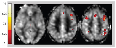
Quantitative images depicting the perfusion changes produced by two weeks of training on the cerebral activation pattern during a working memory task. The changes are overlaid on a map of the resting perfusion. The data are expressed in units of ml/min/100g of tissue.
Researcher: Luis Hernandez-Garcia
Two parts of every MRI acquisition are excitation and reception. One new technology being pursued is parallel excitation, in which multiple independent channels are used to rapidly excite customized patterns (the standard procedure uses a single excitation channel) for:
- Selection of specific regions of the body to allow for a more focused imaging study,
- Correction of intensity variations across the images,
- Correction of magnetic field inhomogeneities by pre-encoding specific compensation patterns to reduce distortions in fMRI.
Our group focuses on excitation pulse design (signal processing), system integration, and applications of parallel excitation.
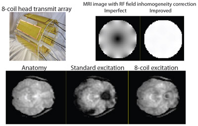
Main field inhomogeneity correction for functional MRI with parallel excitation.
Researchers: Douglas Noll and Jon-Fredrik Nielsen
In MRI, ideal image reconstruction uses a simple Fourier transform. However, there are a number of cases where this approach does not work well. For example, when magnetic field variations distort the simple Fourier relationship leading to image distortion, and in parallel imaging, where multiple independent channels are acquired using reduced sampling. Our lab, in collaboration with Prof. Fessler (EECS & BME), develops new image reconstruction algorithms that use statistical estimation approaches and models of the MRI physics to produce more stable and artifact-free images as well as estimates of physically and physiologically relevant parameters.
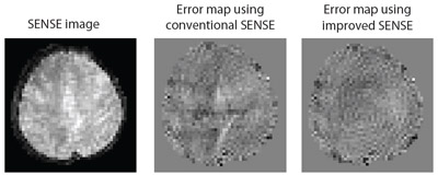
SENSE image and error maps showing reduction of aliasing artifact when using an improved SENSE with a new regularization method.
Conventional functional MRI collects the BOLD-sensitive MR images of a subject performing a cognitive task, with subsequent image reconstruction and analysis performed offline. Recent advances in fMRI processing allow real-time applications (within same scan, with minimal lag): online data quality control, real-time functional activation monitoring, and interactive paradigms based on the subjects dynamic functional activity. At the FMRI laboratory, we are developing pattern classification techniques to investigate their use in real-time fMRI neurofeedback to modulate craving.
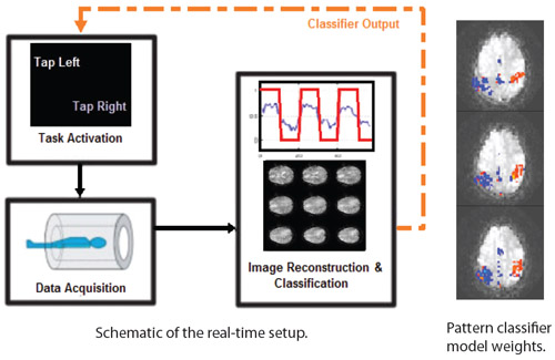
Researchers: Scott Peltier and Luis Hernandez-Garcia
Recent fMRI have shown slowly varying timecourse fluctuations that are temporally correlated between functionally related areas (e.g. motor, visual, attentional networks), even at rest. Further studies have demonstrated altered connectivity for various neurological states. Thus, functional connectivity is potentially important as an indicator of regular neuronal activity. Our current work involves investigating and characterizing these functional connectivity patterns in control and patient groups, including detection using model-free analysis methods.
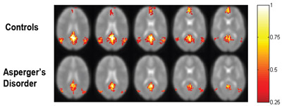
Figure showing reduced functional connectivity within the default mode network in subjects with Asperger's disorder as compared to healthy controls.
Researcher: Scott Peltier
Transcranial Magnetic Stimulation (TMS) is a technique to produce neuronal excitations in the brain non-invasively. A strong electric field is produced by a coil that is held next to the subject's head, and this field causes the neurons to fire. This is a useful research and therapy tool. At the FMRI laboratory, we are conducting several studies of cognitive function using TMS. We are also developing techniques to improve the targetting capabilities of the TMS technique, which is not very accurate in its current state.
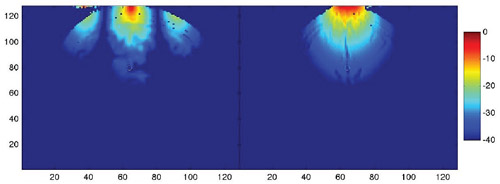
Calculated Electric Field Pattern produced on a human brain by a new TMS coil design.
Researchers: Luis Hernandez-Garcia
Site Index | Internal Resources | Contact Us | U-M Gateway
© Copyright 2024 The Regents of the University of Michigan Ann Arbor, MI 48109 USA All Rights Reserved - Non-Discrimination Policy
- Alzheimer's disease & dementia
- Arthritis & Rheumatism
- Attention deficit disorders
- Autism spectrum disorders
- Biomedical technology
- Diseases, Conditions, Syndromes
- Endocrinology & Metabolism
- Gastroenterology
- Gerontology & Geriatrics
- Health informatics
- Inflammatory disorders
- Medical economics
- Medical research
- Medications
- Neuroscience
- Obstetrics & gynaecology
- Oncology & Cancer
- Ophthalmology
- Overweight & Obesity
- Parkinson's & Movement disorders
- Psychology & Psychiatry
- Radiology & Imaging
- Sleep disorders
- Sports medicine & Kinesiology
- Vaccination
- Breast cancer
- Cardiovascular disease
- Chronic obstructive pulmonary disease
- Colon cancer
- Coronary artery disease
- Heart attack
- Heart disease
- High blood pressure
- Kidney disease
- Lung cancer
- Multiple sclerosis
- Myocardial infarction
- Ovarian cancer
- Post traumatic stress disorder
- Rheumatoid arthritis
- Schizophrenia
- Skin cancer
- Type 2 diabetes
- Full List »
share this!
May 1, 2024
This article has been reviewed according to Science X's editorial process and policies . Editors have highlighted the following attributes while ensuring the content's credibility:
fact-checked
peer-reviewed publication
trusted source
Brain imaging study reveals connections critical to human consciousness
by Massachusetts General Hospital
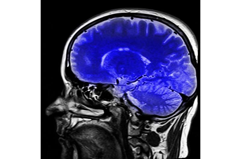
In a paper titled, "Multimodal MRI reveals brainstem connections that sustain wakefulness in human consciousness," published in Science Translational Medicine , a group of researchers at Massachusetts General Hospital and Boston Children's Hospital, created a connectivity map of a brain network that they propose is critical to human consciousness.
The study involved high-resolution scans that enabled the researchers to visualize brain connections at submillimeter spatial resolution. This technical advance allowed them to identify previously unseen pathways connecting the brainstem, thalamus, hypothalamus, basal forebrain, and cerebral cortex.
Together, these pathways form a "default ascending arousal network" that sustains wakefulness in the resting, conscious human brain. The concept of a "default" network is based on the idea that specific networks within the brain are most functionally active when the brain is in a resting state of consciousness. In contrast, other networks are more active when the brain is performing goal-directed tasks.
To investigate the functional properties of this default brain network, the researchers analyzed 7 Tesla resting-state functional MRI data from the Human Connectome Project . These analyses revealed functional connections between the subcortical default ascending arousal network and the cortical default mode network that contributes to self-awareness in the resting, conscious brain.
The complementary structural and functional connectivity maps provide a neuroanatomic basis for integrating arousal and awareness in human consciousness. The researchers released the MRI data , brain mapping methods , and a new Harvard Ascending Arousal Network Atlas , to support future efforts to map the connectivity of human consciousness .
"Our goal was to map a human brain network that is critical to consciousness and to provide clinicians with better tools to detect, predict, and promote recovery of consciousness in patients with severe brain injuries," explains lead-author Brian Edlow, MD, co-director of Mass General Neuroscience, associate director of the Center for Neurotechnology and Neurorecovery (CNTR) at Mass General, an associate professor of Neurology at Harvard Medical School and a Chen Institute MGH Research Scholar 2023-2028.
Dr. Edlow explains, "Our connectivity results suggest that stimulation of the ventral tegmental area's dopaminergic pathways has the potential to help patients recover from coma because this hub node is connected to many regions of the brain that are critical to consciousness."
Senior author Hannah Kinney, MD, Professor Emerita at Boston Children's Hospital and Harvard Medical School, adds that "the human brain connections that we identified can be used as a roadmap to better understand a broad range of neurological disorders associated with altered consciousness, from coma, to seizures, to sudden infant death syndrome (SIDS)."
The authors are currently conducting clinical trials to stimulate the default ascending arousal network in patients with coma after traumatic brain injury , with the goal of reactivating the network and restoring consciousness.
Explore further
Feedback to editors

New technique improves T cell-based immunotherapies for solid tumors
25 minutes ago

Unraveling the roles of non-coding DNA explains childhood cancer's resistance to chemotherapy
39 minutes ago

Conscious memories of childhood maltreatment strongly associated with psychopathology

Study finds private equity expanding to mental health facilities

Organ transplant drug may slow Alzheimer's disease progression

A sum greater than its parts: Time-restricted eating and high-intensity exercise work together to improve health

Experimental vaccine targets portions of the flu virus that don't change

Study finds ChatGPT fails at heart risk assessment
2 hours ago

Preclinical study finds novel stem cell therapy boosts neural repair after cardiac arrest

With huge patient dataset, AI accurately predicts treatment outcomes
3 hours ago
Related Stories

New insights on brain connections that are disrupted in patients with coma
Sep 4, 2019

Study explores mechanisms that underlie disorders of consciousness
Sep 16, 2022

Encouraging results from functional MRI in an unresponsive patient with COVID-19
Jul 6, 2020


Brain modeling identifies circuits implicated in consciousness
Jun 16, 2023

Alterations in functional network reorganization identified in Meniere disease
Nov 4, 2023

Individuals with high level of schizotypal traits exhibit altered brain structural and functional connectivity
Aug 26, 2020
Recommended for you

Scientists say sleep resets brain connections—but only for first few hours
4 hours ago

Scientists identify new brain circuit in mice that controls body's inflammatory reactions

Research on how dietary choline travels through the blood-brain barrier reveals pathway for treating brain disorders

Study finds network of inflammatory molecules may act as biomarker for risk of future cerebrovascular disease
10 hours ago

Neuroscientists discover two specific brain differences linked to how brains respond during tasks
23 hours ago
Let us know if there is a problem with our content
Use this form if you have come across a typo, inaccuracy or would like to send an edit request for the content on this page. For general inquiries, please use our contact form . For general feedback, use the public comments section below (please adhere to guidelines ).
Please select the most appropriate category to facilitate processing of your request
Thank you for taking time to provide your feedback to the editors.
Your feedback is important to us. However, we do not guarantee individual replies due to the high volume of messages.
E-mail the story
Your email address is used only to let the recipient know who sent the email. Neither your address nor the recipient's address will be used for any other purpose. The information you enter will appear in your e-mail message and is not retained by Medical Xpress in any form.
Newsletter sign up
Get weekly and/or daily updates delivered to your inbox. You can unsubscribe at any time and we'll never share your details to third parties.
More information Privacy policy
Donate and enjoy an ad-free experience
We keep our content available to everyone. Consider supporting Science X's mission by getting a premium account.
E-mail newsletter
Magnetic Resonance Imaging (MRI) for Diagnosing Multiple Sclerosis
Magnetic resonance imaging (mri): a diagnostic tool, how mri works.
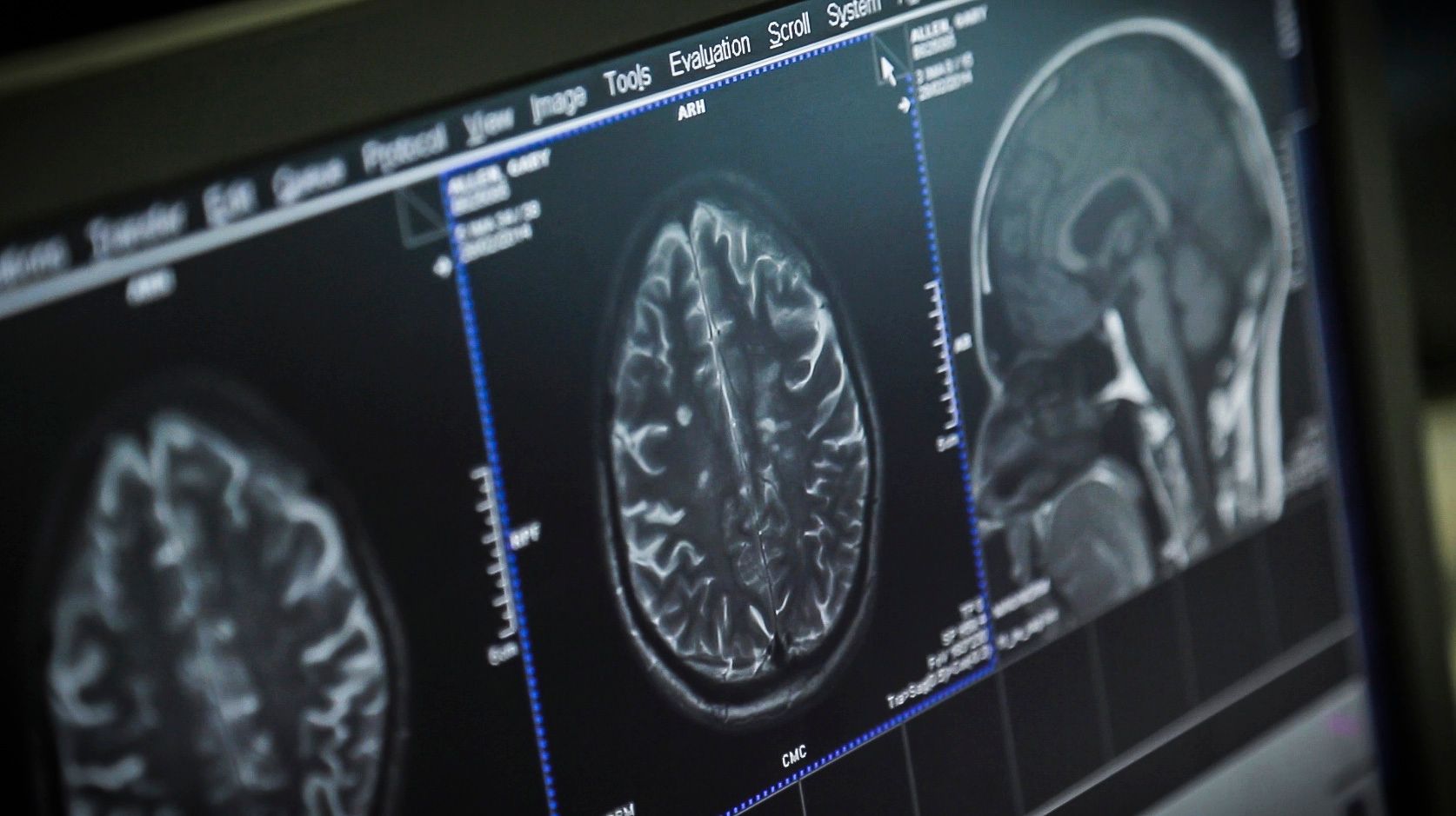
Uses of MRIs in MS
Mris for detecting demyelination and diagnosing ms.
- You may not always see a direct correlation between the MRI scan and your symptoms. Lesions seen on MRI may be small or have caused little damage. Your brain may have developed a workaround.
- Lesions in smaller areas, such as the brainstem, the spinal cord or the optic nerve are more likely to produce signs and symptoms of MS.
- People over age 50 may have small areas on their MRIs that resemble those seen in MS but are related to the aging process. A similar thing happens with people who experience migraine headaches.
MRIs for Assessing Risk After Clinically Isolated Syndrome (CIS) Diagnosis
- The number of lesions on an initial MRI of the brain (or spinal cord) can help assess your risk of developing a second attack in the future and being diagnosed with MS.
- The MRI can also identify a second neurological event in a person who has no additional symptoms. This helps confirm an MS diagnosis as early as possible.
MRIs for Tracking Disease Progress
Updates to MRI Recommendations, Part I
Updates to MRI Recommendations, Part II
Types and Purposes of MRI Scans
- T-1 weighted without gadolinium — may show dark areas (hypointensities) thought to indicate areas of permanent nerve damage
- T-1 weighted with gadolinium — may show bright areas (enhancing lesions) that indicate areas of active inflammation
- T-2 weighted — shows overall disease burden or lesion load (meaning the total number of lesions, both old and new)
- Fluid attenuated inversion recovery (FLAIR) — shows MS activity by reducing interference from the spinal fluid
Possible Risks Associated With Gadolinium-Based Contrast Agents (GBCAs)
- People with poor kidney function have increased risk of developing nephrogenic systemic fibrosis (NSF) if they are given GBCAs. This rare and serious disease causes thickening of the skin and damage to internal organs.
- Small amounts of the GBCAs can stay in your body for several months to years. It is not yet known if this retention of GBCA is harmful.
- Pregnant people should only receive a GBCAMRI during their pregnancy if it is considered essential. If possible, delay imaging if you are pregnant. The FDA has updated their recommendations .
- The FDA issued a safety communication on the use of GBCAs and recommended types of gadolinium less likely to be retained in the body.
- The FDA also required that the makers of gadolinium contrast agents do research to determine if GBCA deposits are harmful.
- Ask your doctor if you need to receive GBCA for your next MRI scan. It is not needed for all MRIs.
- Have your doctor check your kidney function with a blood test prior to receiving GBCA. This will reduce the risk of developing NSF. Ask the center doing your MRI what type of GBCA they use. Current macrocyclic agents are Dotarem (gadoterate meglumine), Gadavist (gadobutrol), ProHance (gadoteridol) andElucirem/Vueway (gadopiclenol). Macrocyclic agents are less likely to be retained in the body.
Different Magnets Provide Different Information
- Most conventional MRI machines are 1.5T or 3T.
- Open MRIs are usually less than 1.5T and do not provide the best images for detecting MS activity. But they may be used when someone has difficulty tolerating a closed MRI machine.
Thank you for visiting nature.com. You are using a browser version with limited support for CSS. To obtain the best experience, we recommend you use a more up to date browser (or turn off compatibility mode in Internet Explorer). In the meantime, to ensure continued support, we are displaying the site without styles and JavaScript.
- View all journals
Medical imaging articles from across Nature Portfolio
Medical imaging comprises different imaging modalities and processes to image human body for diagnostic and treatment purposes. It is also used to follow the course of a disease already diagnosed and/or treated.
Adapting vision–language AI models to cardiology tasks
Vision–language models can be trained to read cardiac ultrasound images with implications for improving clinical workflows, but additional development and validation will be required before such models can replace humans.
- Rima Arnaout
Related Subjects
- Bone imaging
- Brain imaging
- Magnetic resonance imaging
- Molecular imaging
- Radiography
- Radionuclide imaging
- Three-dimensional imaging
- Ultrasonography
- Whole body imaging
Latest Research and Reviews
Personalized coronary and myocardial blood flow models incorporating CT perfusion imaging and synthetic vascular trees
- Karthik Menon
- Muhammed Owais Khan
- Alison L. Marsden

Single-shot frequency offset measurement with HASTE using the selective parity approach
- Irina de Alba Alvarez
- Aidin Arbabi
- David G. Norris
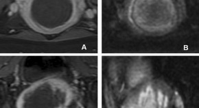
Diffusion-weighted imaging as a potential non-gadolinium alternative for immediate assessing the hyperacute outcome of MRgFUS ablation for uterine fibroids
- Yaoqu Huang
- Shouguo Zhou
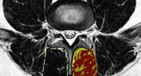
Spinal degeneration and lumbar multifidus muscle quality may independently affect clinical outcomes in patients conservatively managed for low back or leg pain
- Jeffrey R. Cooley
- Tue S. Jensen
- Jeffrey J. Hebert
Application of ultrasound to monitor in vivo residual bone movement within transtibial prosthetic sockets
- Niels Jonkergouw
- Maarten R. Prins
- Han Houdijk
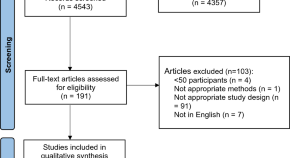
Optical coherence tomography angiography analysis methods: a systematic review and meta-analysis
- Ella Courtie
- James Robert Moore Kirkpatrick
- Richard J. Blanch
News and Comment
Artificial intelligence chatbot interpretation of ophthalmic multimodal imaging cases.
- Andrew Mihalache
- Ryan S. Huang
- Rajeev H. Muni
Blood glucose concentration measurement without finger pricking
A new sensor that detects optoacoustic signals generated by mid-infrared light enables measurement of glucose concentration from intracutaneous tissue rich in blood. This technology does not rely on glucose measurements in interstitial fluid or blood sampling and might yield the next generation of non-invasive glucose-sensing devices for improved diabetes management.
Enhancing diagnostic precision in liver lesion analysis using a deep learning-based system: opportunities and challenges
A recent study reported the development and validation of the Liver Artificial Intelligence Diagnosis System (LiAIDS), a fully automated system that integrates deep learning for the diagnosis of liver lesions on the basis of contrast-enhanced CT scans and clinical information. This tool improved diagnostic precision, surpassed the accuracy of junior radiologists (and equalled that of senior radiologists) and streamlined patient triage. These advances underscore the potential of artificial intelligence to enhance hepatology care, although challenges to widespread clinical implementation remain.
- Jeong Min Lee
- Jae Seok Bae
Population imaging cerebellar growth for personalized neuroscience
Growth chart studies of the human cerebellum, which is increasingly recognized as pivotal for cognitive development, are rare. Gaiser and colleagues utilized population-level neuroimaging to unveil cerebellar growth charts from childhood to adolescence, offering insights into brain development.
- Zi-Xuan Zhou
- Xi-Nian Zuo
The lucent yet opaque challenge of regulating artificial intelligence in radiology
- James M. Hillis
- Jacob J. Visser
- Katherine P. Andriole
Quick links
- Explore articles by subject
- Guide to authors
- Editorial policies
CT-ing is believing: Beckman’s new micro-CT scanner supports materials, life sciences research
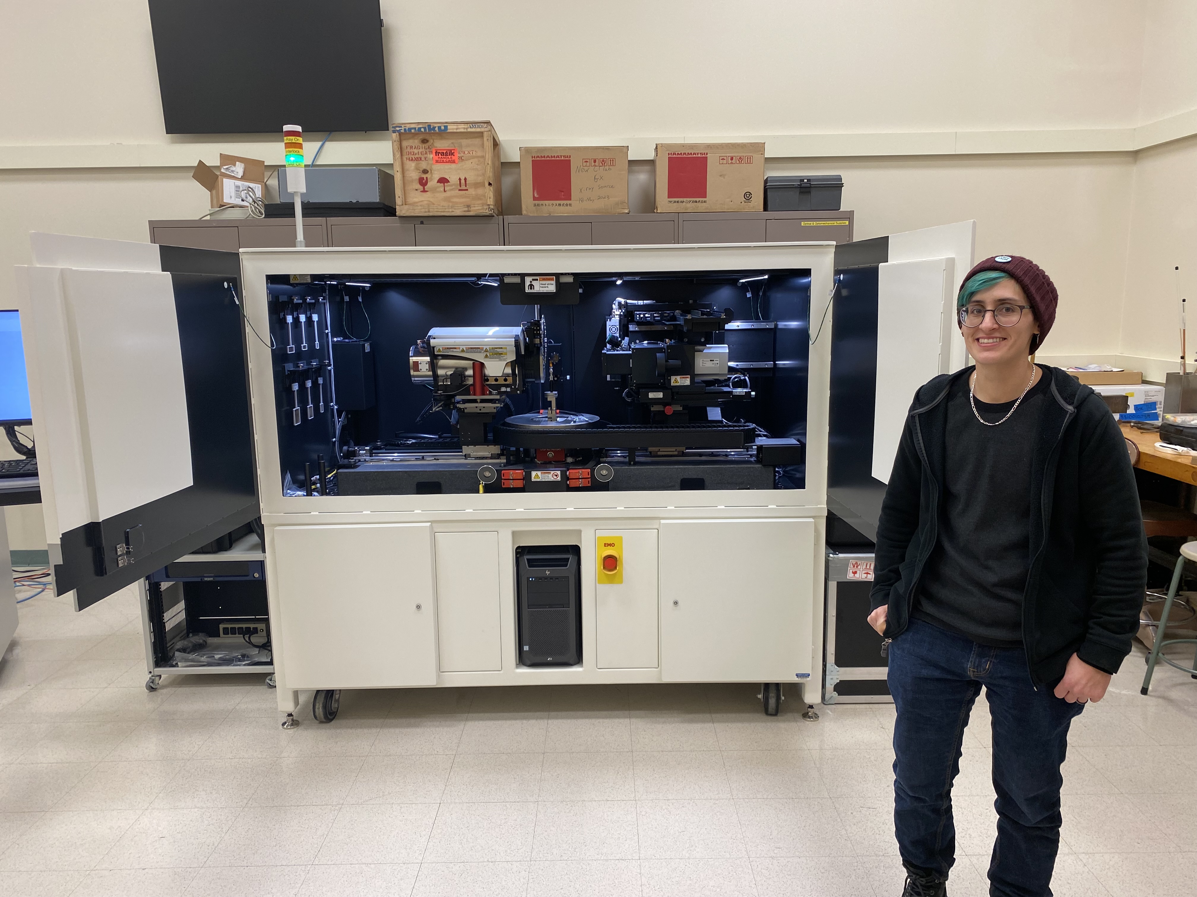
In conversation, a scintillating idea excites your discussion partners and transforms them into more inspired, illuminated versions of themselves. In physics, a scintillating material excites X-rays, transforming them into visible light. Micro-CT scanners, better known as X-ray microscopes, make use of both meanings.
Want a slice?
Computed tomography, abbreviated to CT, is an imaging technique not unlike a sandwich. Tomography, or “imaging with slices,” means capturing a series of cross-sectional X-ray scans (the slices) of an object — be it a vegetable, a mineral or even a person. Piled on top of one another like lettuce, turkey and Swiss, the images reconstruct the subject in 3D from the inside out — without ever making a cut.
According to Mayo Clinic, CT is useful for imaging most areas in the body but is best for “ bones, blood vessels and soft tissues ” like muscles. Doctors use it to diagnose tumors, spot blood clots and guide biopsies. At the Beckman Institute, interdisciplinary researchers use CT scans to study how our bodies move and heal and work toward preventing injuries like broken bones , torn ligaments and arthritis.
Good energy only
With microscopic computed tomography, also known as micro-CT or X-ray microscopy, researchers apply CT techniques to reconstruct samples as tiny and delicate as collagen and insect antennae . Most micro-CT scanners use only X-rays, which are invisible to the human eye, but Beckman’s new model also incorporates the kind of light we can see.

Converting high-energy photons like X-rays into visible light is called scintillation, Gibson said, warning that sometimes, lower-energy photons sneak through and muddle the results.
Handily, the Versa is equipped with a Ferris wheel of filters to block unnecessary rays and keep the energy high.
All about angles
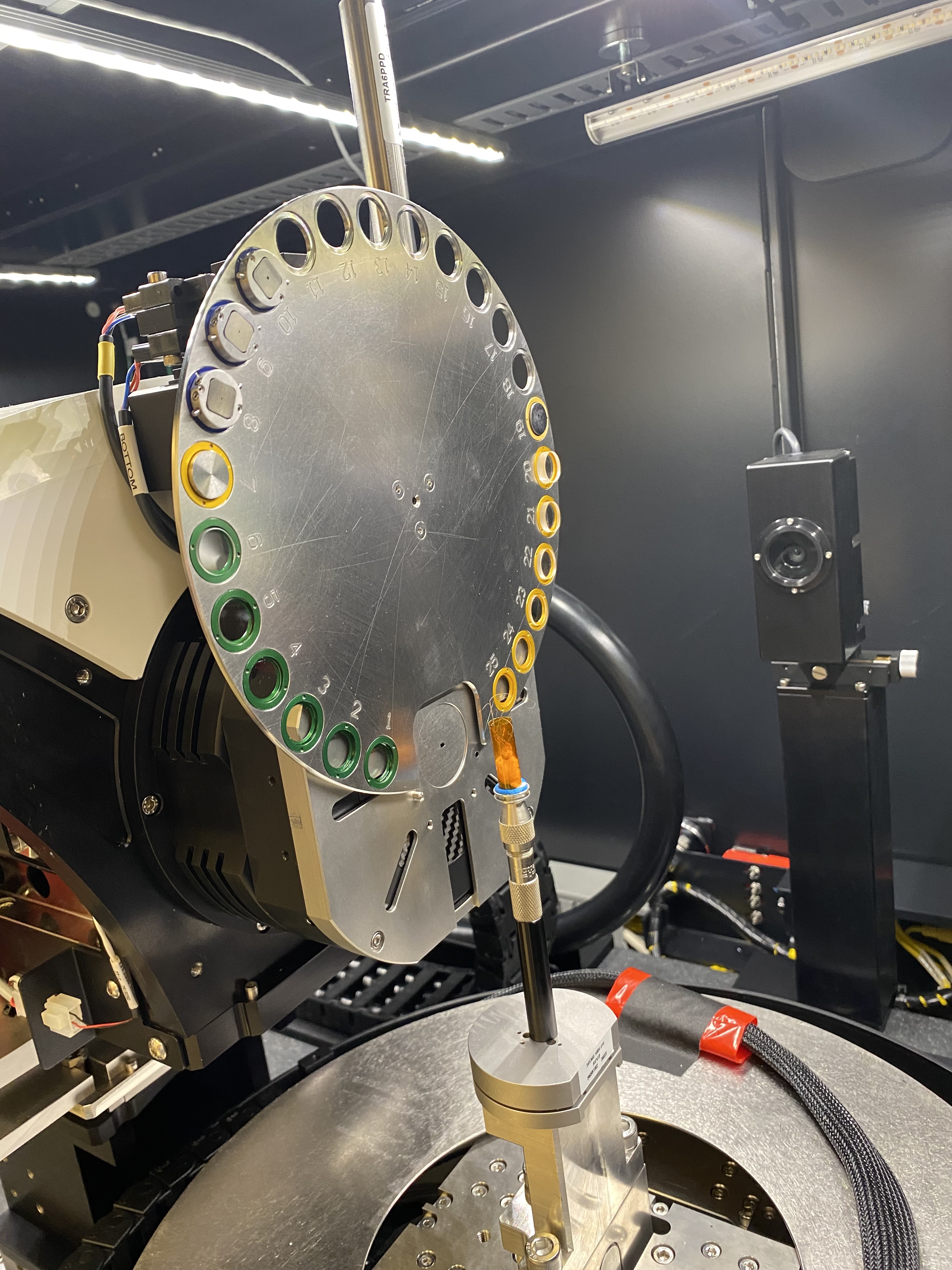
Capturing X-rays from all angles contributes to a complete, spatially accurate 3D reconstruction of each sample — what Gibson gratefully calls “non-destructive virtual dissection,” since it spares such delicate specimens as leafhopper insects and spiders.
Larger samples, though, need more elbow room. That’s why most micro-CT scanners require samples to squeeze into the allotted scanning window — or inch farther away until they fit.
“Imagine using a micro-CT to look at a slab of concrete. With most scanners, you’d need to move the block of concrete far away from the detector to keep the whole thing within that window,” said T. Josek , a Beckman microscopist.
Not so for the Versa 630, which “can take a high-quality scan of just a small subsection — like a crack inside the concrete — instead of scanning the whole thing and zooming in on the cracked area later,” Josek said.
Figuratively, it’s the difference between taking a professional portrait for your resume or zooming in on a grainy group photo. Literally, it may be the difference between a pothole-laden road and a highway built to last.
“There has been a push toward developing more sustainable materials that will last longer. We already know how to make something like concrete cheaply, and easily, but is it the best?” Josek said. “With building materials, hairline cracks are the start of where things go catastrophically wrong, and the micro-CT scanner at the Beckman Institute can see those cracks.”
“We’re providing the opportunity for researchers at the University of Illinois and beyond to start here and start small — see how a material behaves and what it looks like, and then scale up,” Josek said.
Speaking of small: the 630 Versa’s maximum imaging resolution is 40 nanometers, 2,500 times narrower than the width of a human hair .
Bop it, stretch it, scan it
The Versa 630 isn’t just for looking — two standout features allow researchers to get physical with their specimens, too.
Researchers across disciplines can use the compression/tensile stage to stretch, bend and compress their samples while scrutinizing them up close — a technique called in situ mechanical testing.

Mikayla Hoyle, a researcher in the Tissue Biomechanics Lab , will use this function to study how movement and exercise impact bone tissue. By adding weight to a tissue sample — to simulate the pounding of feet on pavement during a jog, for example — she “can analyze deformation to the bone by comparing a before image, when the sample is unloaded, with an after image, when load is applied,” she said, adding that the Versa 630 helps the researchers move beyond the surface of the bone to see what’s happening inside it.

The Versa 630 can also do diffraction contrast tomography, a technique usually found adjacent to a particle accelerator wherein researchers can determine grain orientation of a given material, like a metal, by tracking how X-rays bend as they pass through it.
Mixing materials (and people)
Interdisciplinary researchers are already lining up to scan their materials and specimens, Josek said.
“Sometimes, when you get new equipment, the hardest part is getting people to start using it. It’s exciting for us to have a new piece of equipment that just shows up, and immediately there’s a high demand,” they said.
The high demand is due in part to the collaboration that made acquiring the 630 Versa possible.
The scanner was installed in January 2024 and replaces the Microscopy Suite’s Xradia microXCT-400 CT scanner, an older model of the same line that became inoperable in 2023. Seeking a suitable replacement, Beckman microscopists worked with Paul Braun , a Beckman researcher and the director of the Materials Research Laboratory, to consider both institutes’ needs.

In addition to the Beckman Institute and the Materials Research Laboratory, The Grainger College of Engineering and Office for the Vice Chancellor of Research and Innovation committed funds to support purchasing the Versa 630.
The Beckman Microscopy Suite team is focused on finding projects on campus and beyond that might benefit from the Versa 630 and are providing comprehensive trainings for those new to the technology. Carl Zeiss Microscopy , the scanner’s manufacturer, is integral to these efforts.
"We at ZEISS are thrilled to be partnering with the Beckman Institute Microscopy Suite to deliver this flexible and intuitive system for X-ray microscopy," said Aubrey Funke, Carl Zeiss Microscopy Product Marketing Manager for Life Science EM/XRM. "The ability to image your sample, be it organic or material, becomes a less intimidating process on the ZEISS Xradia Versa 630. With high powered-resolution and expanded ease-of-use for all skill levels, the Versa 630 is ideal for diverse research capabilities across a spectrum of applications, including life science, electronics, materials research, and many more. We look forward to working with the Beckman Institute Microscopy Suite to support many diverse research projects.”
Beckman’s is the first 630 model for life science applications in the U.S.
First and foremost an entomologist, microscopist Gibson anticipates scanning more insect specimens.
“I am working with the paleontology lab at the Illinois Natural History Survey to CT-scan fossilized insects in amber — think the mosquitos from Jurassic Park,” he said.
As far as what the future holds for the Versa, Gibson said: “I personally do not know, but whatever it has in store I am excited for it!”
Excited, or scintillated?
Media contact: Jenna Kurtzweil, [email protected]
Share this story
In this article.

More stories by topic
- Microscopy Suite
- Frontiers in Oncology
- Radiation Oncology
- Research Topics
Magnetic Resonance Imaging for Radiation Therapy
Total Downloads
Total Views and Downloads
About this Research Topic
Since the introduction of magnetic resonance imaging (MRI) to radiation therapy (RT), it has been increasingly adopted in RT treatment planning for target and organ-at-risk (OAR) definition due to superior soft tissue contrasts. Recently, roles of MRI in the field of radiation oncology have been extended to ...
Keywords : MRI, MR-Linac, MR simulator, Functional Imaging, Radiotherapy
Important Note : All contributions to this Research Topic must be within the scope of the section and journal to which they are submitted, as defined in their mission statements. Frontiers reserves the right to guide an out-of-scope manuscript to a more suitable section or journal at any stage of peer review.
Topic Editors
Topic coordinators, recent articles, submission deadlines.
Submission closed.
Participating Journals
Total views.
- Demographics
No records found
total views article views downloads topic views
Top countries
Top referring sites, about frontiers research topics.
With their unique mixes of varied contributions from Original Research to Review Articles, Research Topics unify the most influential researchers, the latest key findings and historical advances in a hot research area! Find out more on how to host your own Frontiers Research Topic or contribute to one as an author.
- SUGGESTED TOPICS
- The Magazine
- Newsletters
- Managing Yourself
- Managing Teams
- Work-life Balance
- The Big Idea
- Data & Visuals
- Reading Lists
- Case Selections
- HBR Learning
- Topic Feeds
- Account Settings
- Email Preferences
Make Decisions with a VC Mindset
- Ilya A. Strebulaev

Venture capitalists’ unique approach to investment and innovation has played a pivotal role in launching one-fifth of the largest U.S. public companies. And three-quarters of the largest U.S. companies founded in the past 50 years would not have existed or achieved their current scale without VC support.
The question is, Why? What makes venture firms so good at finding start-ups that go on to achieve tremendous success? What skills do they have that experienced, networked, and powerful large corporations lack?
The authors’ research reveals that the venture mindset is characterized by several principles: the individual over the group, disagreement over consensus, exceptions over dogma, and agility over bureaucracy. This article offers guidance to traditional firms in using the VC mindset to spur innovation.
The key is to embrace risk, disagreement, and agility.
Idea in Brief
The opportunity.
Venture capitalists’ unique approach to investment and innovation has played a pivotal role in launching one-fifth of the largest U.S. public companies, demonstrating the power of the venture mindset.
The Challenge
Traditional companies often struggle to replicate the success of venture firms because of their aversion to risk and failure and their preference for consensus and stability.
The Solution
When faced with market changes or disruptive technology, big companies should adopt the venture mindset, prioritizing the individual over the group, disagreement over consensus, exceptions over dogma, and agility over bureaucracy.
Venture investors are the hidden hand behind the most innovative companies surrounding us. According to research conducted by one of us (Ilya), venture capitalists were causally responsible for the launch of one-fifth of the 300 largest U.S. public companies in existence today. They have played an essential role in unlocking the power of the internet, the mobile revolution, and now artificial intelligence in all its forms. Apple, Google, Moderna, Netflix, Airbnb, OpenAI, Salesforce, Tesla, Uber, and Zoom—these firms disrupted entire industries despite initially having fewer resources and less support and experience than their mature, successful, cash-rich competitors. All these businesses could theoretically have emerged from within an established company—but they didn’t. Instead, they were financed and shaped by VCs. Indeed, we estimate that three-quarters of the largest U.S. companies founded in the past 50 years would not have existed or achieved their current scale without VC support.
- IS Ilya A. Strebulaev is the David S. Lobel Professor of Private Equity and a professor of finance at the Stanford Graduate School of Business. He is also the founder of the Stanford GSB Venture Capital Initiative and a research associate at the National Bureau of Economic Research.
- AD Alex Dang is a venture builder and a digital strategy adviser. He was a partner at McKinsey and EY and launched numerous businesses at Amazon.
Partner Center
Numbers, Facts and Trends Shaping Your World
Read our research on:
Full Topic List
Regions & Countries
- Publications
- Our Methods
- Short Reads
- Tools & Resources
Read Our Research On:
What Are Americans’ Top Foreign Policy Priorities?
Protecting the u.s. from terrorism and reducing the flow of illegal drugs are top issues overall, but democrats and republicans have very different priorities, table of contents.
- Differences by partisanship
- Differences by age
- Acknowledgments
- The American Trends Panel survey methodology

Pew Research Center conducted this analysis to better understand Americans’ long-range foreign policy priorities. For this analysis, we surveyed 3,600 U.S. adults from April 1 to April 7, 2024. Everyone who took part in this survey is a member of the Center’s American Trends Panel (ATP), an online survey panel that is recruited through national, random sampling of residential addresses. This way nearly all U.S. adults have a chance of selection. The survey is weighted to be representative of the U.S. adult population by gender, race, ethnicity, partisan affiliation, education and other categories. Read more about the ATP’s methodology .
Here are the questions used for this analysis, along with responses, and its methodology .
Americans have a lot on their plates in 2024, including an important election to determine who will remain or become again president. But the world does not stop for a U.S. election, and multiple conflicts around the world as well as other issues of global prominence continue to concern Americans.
When asked to prioritize the long-range foreign policy goals of the United States, the majority of Americans say preventing terrorist attacks (73%), keeping illegal drugs out of the country (64%) and preventing the spread of weapons of mass destruction (63%) are top priorities. Over half of Americans also see maintaining the U.S. military advantage over other countries (53%) and preventing the spread of infectious diseases (52%) as primary foreign policy responsibilities.
About half of Americans say limiting the power and influence of Russia and China are top priorities. A recent annual threat assessment from the U.S. intelligence community focused heavily on those countries’ strengthening military relationship and their ability to shape the global narrative against U.S. interests.
Fewer than half of Americans say dealing with global climate change (44%) and getting other countries to assume more of the costs of maintaining world order (42%) are top priorities. The partisan gaps on these two issues are quite large:
- 70% of Democrats and Democratic-leaning independents say climate change should be a top priority, while 15% of Republicans and Republican leaners say this.
- 54% of Republicans say getting other countries to assume more of the costs of maintaining world order should be a top priority, compared with 33% of Democrats.
About four-in-ten Americans see limiting the power and influence of North Korea and Iran as top priorities. (The survey was conducted before Iran’s large-scale missile attack on Israel on April 13.) And about a third say the same about the U.S. being a leader in artificial intelligence, a technology that governments around the world are increasingly concerned about .
When it comes to goals that focus on international engagement, like strengthening the United Nations and NATO or finding a solution to the Israeli-Palestinian conflict, fewer than a third of Americans mark these as top foreign policy priorities.
Related: Fewer Americans view the United Nations favorably than in 2023
Only about a quarter of Americans prioritize promoting human rights in other countries, leading other countries in space exploration and reducing military commitments overseas. And similar shares say supporting Ukraine (23%) and Israel (22%) are top issues.
At the bottom of this list of foreign policy priorities are promoting global democracy ( a major policy push from the Biden administration ) and aiding refugees fleeing violence around the world – about two-in-ten Americans describe these as top concerns. These assessments come amid a recent global surge in asylum claims . Still, in Center surveys, democracy promotion has typically been at the bottom of Americans’ list of foreign policy priorities, even dating back to George W. Bush’s and Barack Obama’s administrations .
Overall, a majority of Americans say that all 22 long-range foreign policy goals we asked about should be given at least some priority. Still, about three-in-ten Americans say supporting Israel (31%), promoting democracy (28%) and supporting Ukraine (27%) should be given no priority.
The long-range foreign policy priority questions were also asked in 2018 and 2021, and since then there have been some significant shifts in responses:
- Since 2018, the public has become significantly more likely to say limiting the power and influence of China (+17 percentage points) and finding a solution to the Israeli-Palestinian conflict (+11) are top foreign policy priorities.
- Americans have also increased the emphasis they place on limiting the power and influence of Russia, particularly in the wake of the Russian invasion of Ukraine (+8 points since 2021).
- On the decline since 2018 are strengthening the UN and aiding refugees (-8 points each), reducing foreign military commitments (-6), and promoting and defending human rights in other countries (-5).
- Preventing the spread of infectious diseases is down 19 percentage points since 2021 – during the height of the COVID-19 pandemic – and about back to where it was in 2018.
These are among the findings from a Pew Research Center survey conducted April 1-7, 2024.
The survey of 3,600 U.S. adults shows that foreign policy remains a partisan issue. Republicans prioritize the prevention of terrorism, reducing the flow of illegal drugs into the country, and maintaining a military advantage over other nations. Meanwhile, Democrats prioritize dealing with climate change and preventing the spread of weapons of mass destruction (WMDs), but also preventing terrorist attacks.

There are also stark age differences on many of the policy goals mentioned, but for the most part, young adults are less likely than older Americans to say the issues we asked about are top priorities. The exceptions are dealing with climate change, reducing military commitments overseas, and promoting and defending human rights abroad – on these issues, 18- to 29-year-olds are significantly more likely than older Americans to assign top priority.
Even with these priorities, foreign policy generally takes the backset to domestic policy for most Americans: 83% say it is more important for President Joe Biden to focus on domestic policy, compared with 14% who say he should focus on foreign policy.
Americans are even less likely to prioritize international affairs than they were in 2019, when 74% wanted then-President Donald Trump to focus on domestic policy and 23% said he should focus on foreign policy.
Americans’ foreign policy priorities differ greatly by party. The largest divide, by a significant margin, is the 55 percentage point gap between Democrats and Republicans on dealing with global climate change (70% vs. 15%, respectively, see it as a top priority).
Supporting Ukraine, aiding refugees, reducing the spread of diseases, protecting human rights, and strengthening the UN are also issues on which Democrats are at least 20 points more likely than Republicans to prioritize. For example, 63% of Democrats say reducing the spread of infectious diseases is a top priority, compared with 41% of Republicans.
Republicans prioritize supporting Israel, reducing the flow of illegal drugs and maintaining a military advantage over other countries – among other security and hard power issues – significantly more than Democrats do. For example, more than half of Republicans (54%) say getting other countries to assume more of the costs of maintaining world order should be a top focus in foreign policy. Only a third of Democrats say the same.
The priority assigned to several issues is divided even further by ideology within parties. Take support for Israel and Ukraine as examples. Supporting Israel is generally a higher priority for Republicans than Democrats, but within the Republican Party, 48% of conservatives say it’s a top concern, while 18% of moderates and liberals agree. Previous Center research shows that conservative Republicans are especially likely to favor military aid to Israel .
Supporting Ukraine, something Democrats emphasize more than Republicans, is a top priority particularly for liberal Democrats (47%), while about three-in-ten moderate and conservative Democrats agree (29%). Democrats have also shown more willingness than Republicans to provide aid to Ukraine in its conflict with Russia.
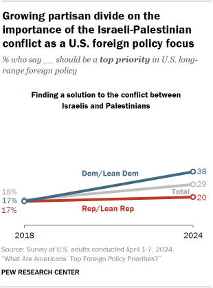
Generally, the partisan differences on the importance of several foreign policy issues have gotten smaller since 2021 , when most of these questions were last fielded. This is especially true for items related to the relative power of major countries, like the U.S. maintaining a military advantage and limiting the power and influence of both Russia and China.
However, finding a solution to the conflict between Israelis and Palestinians – a priority that saw no partisan difference at all when it was last asked about in 2018 – has an emerging partisan gap today. The share of Democrats who call this a top priority has more than doubled, while the share of Republicans has changed little.
Age differences persist on foreign policy issues. Older Americans prioritize most of the issues we asked about at higher rates than those ages 18 t0 29.
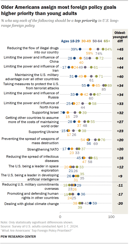
On four issues, there is at least a 40 percentage point gap between Americans ages 65 and older and young adults ages 18 to 29. The oldest Americans are more likely to prioritize reducing the flow of illegal drugs, limiting the power and influence of China and Iran, and maintaining a U.S. military advantage.
Those in the oldest age group are also more concerned than their younger counterparts on an additional 11 issues, ranging from support for Israel to U.S. leadership in space exploration.
For their part, young adults are more likely to say dealing with global climate change, reducing U.S. military commitments overseas, and promoting and defending human rights in other countries should be top foreign policy priorities.
Even starker patterns appear when looking at partisanship within two age groups – adults ages 18 to 49 and those 50 and older.
Among Democrats, older adults place particularly high priority on supporting Ukraine, strengthening NATO, and limiting the power and influence of Russia amid its war with Ukraine. Older Democrats are also more likely than younger ones to prioritize preventing the development of WMDs, curbing the spread of diseases, strengthening the UN and promoting democracy around the world, among other issues.
Among Republicans, those ages 50 and older are more likely than those ages 18 to 49 to prioritize supporting Israel, limiting the power and influence of Iran and China, getting other countries to assume more foreign policy costs, reducing the amount of illegal drugs entering the U.S., preventing terrorism, and maintaining a military advantage.
Sign up for our weekly newsletter
Fresh data delivery Saturday mornings
Sign up for The Briefing
Weekly updates on the world of news & information
- Environment & Climate
- Global Health
- Human Rights
- International Affairs
- United Nations
- War & International Conflict
Americans Remain Critical of China
As biden and trump seek reelection, who are the oldest – and youngest – current world leaders, a growing share of americans have little or no confidence in netanyahu, fewer americans view the united nations favorably than in 2023, rising numbers of americans say jews and muslims face a lot of discrimination, most popular, report materials.
1615 L St. NW, Suite 800 Washington, DC 20036 USA (+1) 202-419-4300 | Main (+1) 202-857-8562 | Fax (+1) 202-419-4372 | Media Inquiries
Research Topics
- Age & Generations
- Coronavirus (COVID-19)
- Economy & Work
- Family & Relationships
- Gender & LGBTQ
- Immigration & Migration
- Internet & Technology
- Methodological Research
- News Habits & Media
- Non-U.S. Governments
- Other Topics
- Politics & Policy
- Race & Ethnicity
- Email Newsletters
ABOUT PEW RESEARCH CENTER Pew Research Center is a nonpartisan fact tank that informs the public about the issues, attitudes and trends shaping the world. It conducts public opinion polling, demographic research, media content analysis and other empirical social science research. Pew Research Center does not take policy positions. It is a subsidiary of The Pew Charitable Trusts .
Copyright 2024 Pew Research Center
Terms & Conditions
Privacy Policy
Cookie Settings
Reprints, Permissions & Use Policy

IMAGES
VIDEO
COMMENTS
Magnetic resonance imaging (MRI) is a non-invasive biomedical imaging technique that uses a strong oscillating magnetic field to induce endogenous atoms such as hydrogen, or exogenously added ...
Introduction. With its ability to provide exceptional soft tissue contrast, combined with non‐ionising radiation exposure, magnetic resonance imaging (MRI) has become widely used in medical imaging globally. 1 However, the three electromagnetic fields used in MR imaging (the static magnetic field, the radiofrequency field and the time‐varying gradient magnetic fields) 2, 3 each create ...
A list of content based on research topics. New MRI technology, developed by Siemens in collaboration with researchers at The Ohio State University College of Medicine and College of Engineering, will expand imaging access for patients with implanted medical devices, severe obesity or claustrophobia.
Magnetic Resonance Imaging (MRI) is a non-invasive imaging technology that produces three dimensional detailed anatomical images. It is often used for disease detection, diagnosis, and treatment monitoring. It is based on sophisticated technology that excites and detects the change in the direction of the rotational axis of protons found in the water that makes up living tissues.
Topics in MRI: An Open Access Journal is an international, fully open access journal devoted to the publication of basic and clinical research, educational and review articles, and case reports related to the development and use of diagnostic applications of magnetic resonance in medicine. The goal of this journal is to provide a forum for research and clinical applications of MRI for ...
Topics in Magnetic Resonance Imaging is a leading information resource for professionals in the MRI community. This publication supplies authoritative, up-to-the-minute coverage of technical advances in this evolving field as well as practical, hands-on guidance from leading experts. Six times a year, TMRI focuses on a single timely topic of interest to radiologists.
Current diagnostic MRI scanners use cryogenic superconducting magnets in the range of 0.5 Tesla (T) to 1.5 T. By comparison, the Earth's magnetic field is 0.5 Gauss (G), which is equivalent to 0.00005 T. Cooling the magnet to a temperature close to absolute zero (0 K) allows such huge currents to be conducted; this is most commonly performed ...
Magnetic Resonance Imaging (MRI) is the first international multidisciplinary journal encompassing physical, life, and clinical science investigations as they relate to the development and use of magnetic resonance imaging. MRI is dedicated to both basic research, technological innovation and applications, providing a single forum for ...
This research topic is to focus on the technical advancements in the intellect and portable MRI, covering a broad range of applications including food science, medicine, and industry. The topics shall include the most recent progress in the following areas but not limited to: • Novel imaging methods, pulse sequences suitable for low-field MRI ...
Despite the potential importance of research using highly portable MRI in remote and resource-limited settings, there is little analysis of the attendant ethical, legal, and social issues (ELSI). To begin addressing this gap, this paper presents findings from the first phase of an envisioned multi-staged and iterative approach for creating ...
1. Introduction. In 1991, when functional MRI first emerged, the most pressing topics of discussion among researchers were about whether or not the observed signal changes were robustly and repeatedly localized to the "true" regions of activation, and whether or not "draining vein effects" occurring downstream from the true site of activation were a major confound in the interpretation ...
This Research Topic focuses on MR imaging only, may include adult and pediatric studies, and welcomes Reviews and Original Research articles on the following topics: Clinical applications of AI using MR images. Machine learning applications for prediction, detection, classification, registration, and segmentation.
Small-bore NMR/MRI systems -The Division of MR Research manages 2 other smaller bore NMR/MRI Service Centers suitable for studies of animal models, cell systems, and in vitro studies. The NMR Service Center - has 400 MHz and 500 MHz Bruker NMR spectrometers, and a Bruker 4.7T 40 cm-horizontal bore spectrometer/imager equipped with rapid ...
A thesis or dissertation, as some people would like to call it, is an integral part of the Radiology curriculum, be it MD, DNB, or DMRD. We have tried to aggregate radiology thesis topics from various sources for reference. Not everyone is interested in research, and writing a Radiology thesis can be daunting.
Keywords: MRI, fMRI, PET/MRI, MRgFUS, MPI, MRE . Important Note: All contributions to this Research Topic must be within the scope of the section and journal to which they are submitted, as defined in their mission statements.Frontiers reserves the right to guide an out-of-scope manuscript to a more suitable section or journal at any stage of peer review.
Quantitative PET-CT and SPECT-CT. Topics range from absolute quantitation with novel scatter, attenuation, and randoms corrections in model-based iterative reconstructions both in PET-CT and PET-MR to quantitative imaging in non-traditional radioisotopes such as Y-86, and absolute quantitation of neurotransmitter densities in dynamic PET imaging.
Research topics in this area can cover advancements in mammography, tomosynthesis, breast MRI, and molecular imaging. Students can explore topics related to breast density, imaging-guided biopsies, breast cancer screening, and the impact of artificial intelligence in breast imaging.
In MRI, ideal image reconstruction uses a simple Fourier transform. However, there are a number of cases where this approach does not work well. For example, when magnetic field variations distort the simple Fourier relationship leading to image distortion, and in parallel imaging, where multiple independent channels are acquired using reduced ...
In a paper titled, "Multimodal MRI reveals brainstem connections that sustain wakefulness in human consciousness," published in Science Translational Medicine, a group of researchers at ...
Magnetic resonance imaging (MRI) is currently the most sensitive, noninvasive way of imaging the brain, spinal cord or other areas of the body. It is the preferred imaging method to help establish an MS diagnosis and to monitor your disease course. MRI has made it possible to visualize the effects of MS and understand more about it. How MRI Works
Topics. Health and Medicine. Date April 25, 2024 2024-04-25. Media Contact. Corrie Pikul [email protected] 401-863-1862 All News. Share. ... undergraduates embrace the rare opportunity to conduct experiments and engage in research with state-of-the-art MRI technology.
Head Topics. United Kingdom. Latest ... DPABINet: A turn-key brain network and graph theory analysis platform based on MRI data 4/25/2024 8:21 PM medical_xpress; ⏱ Reading Time: 46 sec. here; 7 min. at publisher Quality Score: News: 39%; Publisher: 51%; Medicine Research News News. Medicine Research,Health Research News,Health Research ...
Research Open Access 27 Apr 2024 Scientific Reports Volume: 14, P: 9725 Optical coherence tomography angiography analysis methods: a systematic review and meta-analysis
The Beckman Institute for Advanced Science and Technology recently added a new X-ray microscope to its Microscopy Suite.The Zeiss Xradia 630 Versa micro-CT scanner is available to University of Illinois Urbana-Champaign investigators and may be used to support research projects around the world.
Keywords: MRI, MR-Linac, MR simulator, Functional Imaging, Radiotherapy . Important Note: All contributions to this Research Topic must be within the scope of the section and journal to which they are submitted, as defined in their mission statements.Frontiers reserves the right to guide an out-of-scope manuscript to a more suitable section or journal at any stage of peer review.
Research on the following topics could be especially valuable: Factors affecting the use of AOMs, such as take-up rates and patients' adherence to drugs currently on the market; and Expectations about the prices and effectiveness of AOMs that are being developed.
Venture capitalists' unique approach to investment and innovation has played a pivotal role in launching one-fifth of the largest U.S. public companies. And three-quarters of the largest U.S ...
Americans have a lot on their plates in 2024, including an important election to determine who will remain or become again president. But the world does not stop for a U.S. election, and multiple conflicts around the world as well as other issues of global prominence continue to concern Americans.. When asked to prioritize the long-range foreign policy goals of the United States, the majority ...