
- Upload Ppt Presentation
- Upload Pdf Presentation
- Upload Infographics
- User Presentation
- Related Presentations


Mitochondria
By: JenniferDwayne Views: 2213

Basic Human Needs - Nutrition
By: JenniferDwayne Views: 1184

Leading a Healthy Lifestyle
By: JenniferDwayne Views: 1235

Psychoactive Drugs
By: JenniferDwayne Views: 1277
Physiotherapy in Occupational Health
By: JenniferDwayne Views: 1126

Facial Nerve Paralysis
By: FrankMarco Views: 466
Approach To The Ent Patient
By: medhelp Views: 1925

Individuals With Hearing Impairments
By: medhelp Views: 356

Sense of Hearing and Equilibrium
By: medhelp Views: 342

Ear Infections
By: FrankMarco Views: 438

- About : Professor, College of Nursing and Health Sciences
- Occupation : Medical Professional
- Specialty : MD
- Country : United States of America
HEALTH A TO Z
- Eye Disease
- Heart Attack
- Medications
Pete’s PowerPoint Station
- Science Index
- Math/Maths Index
- Language Arts/Literature Index
- Social Studies Index
- Holidays Index
- Art, Music, and Many More, A-Z
- Meteorology
- Four Seasons
- Pre-Algebra
- Trigonometry
- Pre-Calculus & Calculus
- Language Arts
- Punctuation
- Social Studies
- World Religions
- US Government
- Criminal Justice
- Famous People
- American History
- World History
- Ancient History
- The Middle Ages
- Architecture
- All Topics, A–Z
- Privacy & Cookie Policy
- Presentations
Human Respiratory System
Free presentations in powerpoint format.
Understanding the Respiratory System
Respiratory System Anatomy
Respiratory System Diagrammed in Humans
Respiratory System and It’s Physical Assessment in Humans
Respiratory System and Its Study in Biology
Respiratory System: How It Works
Respiratory System: Step-by-Step Instructions
Respiratory System: History of Its Study
What Is the Purpose of the Respiratory System?
Respiratory System Infections
Respiratory System – Middle School Science
The Respiratory System
Respiration
See Also: Human Body Systems , The Human Body
For Teachers
Lots of Lessons – Human Respiratory System, Lungs
Free Video Clips
Free Clipart
Home Collections Medical Lungs
Lungs Presentation Templates
Dive in presenters these free lungs powerpoint templates and google slides themes show what it's like to find the perfect place through compelling visualizations. you can save infographics of your choice and add them to your slides for a better look choose and forget doubts about editing.

Create captivating medical presentations with our user-friendly, Free Lungs PowerPoint Templates and Google Slides Themes!
- Informative graphics: We use simple, colorful pictures to explain lung anatomy, different lung issues, and more. These visuals make things easier to grasp and add interest to your presentation.
- Ready-to-go slides: Each slide comes with spaces for text and pre-filled info, so you can add your content easily and customize the presentation as needed.
- Simple editing: You can change anything on the slides, like text, images, and layouts. Make them your own by picking your favorite colors and fonts, or add your pics and videos.
- Lung anatomy: Learn about different lung parts and what they do, with detailed explanations and visuals.
- Lung infections: Check out different lung infections, their symptoms, and treatments.
- Lung diseases: Understand conditions like asthma, COPD, and lung cancer.
- Human lungs: Discover cool facts about how our lungs work.
- Medical presentations: Find professional slides tailored for med pros and students.
- Different formats: Pick from 4:3 or 16:9 aspect ratios, depending on what you need.
- Portrait or landscape: Choose how your slides look, depending on your style.
- Free or premium: We've got options for every budget.
- Royalty-free: Utilize our slides without copyright concerns.
We're here to help you!
What are lungs powerpoint templates.
Lungs PowerPoint Templates are specially made slides that we can use to make presentations on lung themes, human lungs, lung diseases, the anatomy of lungs, lung infection, etc.
Where can we use these Lungs PPT Slides?
We can use these Lungs PPT Slides in hospitals, medical conferences, seminars, schools, medical institutes, and wherever we need lungs theme presentations.
How can I make Lungs PPT Slides in a presentation?
We can make Lungs PPT Slides in presentations with clipart images or infographics to depict lung-related concepts. Pre-designed slides available online provide perfect layouts to reduce our stress in preparing presentations.
Who can use Lungs PowerPoint Templates?
Medical professionals, teachers, students, and lab technicians can use these Lungs PowerPoint Templates to make presentations or posters.
Why do we need Lungs PowerPoint Slides?
Lung PowerPoint Slides will help us instantly make impactful presentations to create awareness of lung infection and disease. It can also be used as an excellent teaching aid to discuss the anatomy of the lungs.
Where can I find Free Lungs PPT Templates?
There are countless service providers available online. Slide Egg is one of the best platforms among them, where you can find a range of Free Lungs PPT Templates.
Got any suggestions?
We want to hear from you! Send us a message and help improve Slidesgo
Top searches
Trending searches

memorial day
12 templates

holy spirit
36 templates

environmental science

21 templates

american history
74 templates

13 templates
Respiratory System Workshop for Medical Students
It seems that you like this template, respiratory system workshop for medical students presentation, free google slides theme, powerpoint template, and canva presentation template.
It's as simple as this: we need oxygen to live. Our respiratory system, thus, is quite important, so not only do we need to take care of it, we also must know as much about it as possible. If you are a medical student, you'll be glad to attend to workshops that are useful for your education. Teachers will also find this template a nice resource, as it allows them to explain the respiratory system in a visual way. The design focuses on the use of gradients (ranging from white to light blue, the color of safety), simple layouts and some health-themed icons and illustrations. Editing the contents is up to you!
Features of this template
- 100% editable and easy to modify
- 24 different slides to impress your audience
- Contains easy-to-edit graphics such as graphs, maps, tables, timelines and mockups
- Includes 500+ icons and Flaticon’s extension for customizing your slides
- Designed to be used in Google Slides, Canva, and Microsoft PowerPoint
- 16:9 widescreen format suitable for all types of screens
- Includes information about fonts, colors, and credits of the resources used
How can I use the template?
Am I free to use the templates?
How to attribute?
Combines with:
This template can be combined with this other one to create the perfect presentation:

Attribution required If you are a free user, you must attribute Slidesgo by keeping the slide where the credits appear. How to attribute?
Related posts on our blog.

How to Add, Duplicate, Move, Delete or Hide Slides in Google Slides

How to Change Layouts in PowerPoint

How to Change the Slide Size in Google Slides
Related presentations.

Premium template
Unlock this template and gain unlimited access

Register for free and start editing online

- Respiratory System Template
The respiratory system is one of the most crucial for the existence of living beings, since its main function is to provide the oxygen we use. For living beings and mammalian animals, the lungs are the most important organs within this apparatus. Of equal importance is the human respiratory system template for all our users, as it fits perfectly in presentations on academic, educational and preventive topics.
The modern design of the Respiratory System template for PowerPoint and Google Slides brings you many 100% editable health-inspired images, charts and vectors. All these elements can be perfectly used in an individual presentation, but you can also apply its compatibility with Canva for team editing online. If you want to be on par with the experts, you have to download this free ppt resource and start experimenting on all its professional and addictive slides.
Free Respiratory System Template for PowerPoint and Google Slides
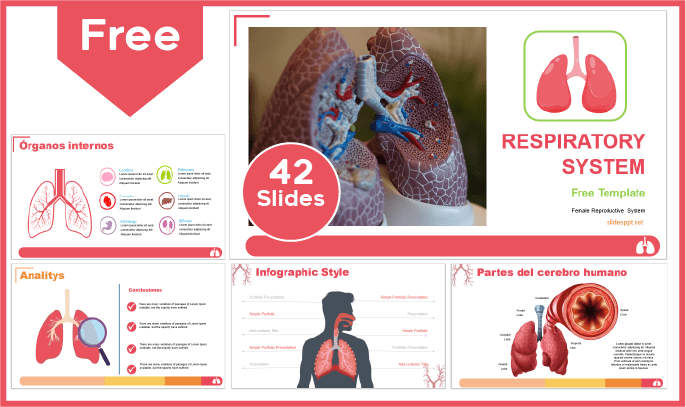
Main features
- 42 slides 100% editable
- 16:9 widescreen format suitable for all screens
- High quality royalty-free images
- Included resources: charts, graphs, timelines and diagrams
- More than 100 icons customizable in color and size
- Main font: Arial
- Predominant color: Red
Download this template
If you are impressed with this great ppt design, wait till you meet our newest category of biology PowerPoint templates and Google Slides themes . In this list you will have a number of customizable templates according to the selected theme, so just find the one you need and start working on its 100% editable and completely free content.
We use cookies to improve the experience of everyone who browses our website. Cookies Policy
Accept Cookies
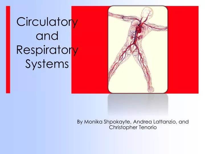
Circulatory and Respiratory Systems
Jul 24, 2014
1.23k likes | 2.19k Views
Circulatory and Respiratory Systems. By Monika Shpokayte , Andrea Lattanzio, and Christopher Tenorio. The Circulatory System. Transports: blood and oxygen from the lungs to the various tissues of the body O 2 from the lungs to the cells; CO 2 (a waste) from the cells to the lungs.
Share Presentation
- cardiac muscle
- red bone marrow
- circulatory system flash cards
- org courses liquid
- htm circulatory system quiz

Presentation Transcript
Circulatory and Respiratory Systems By Monika Shpokayte, Andrea Lattanzio, and Christopher Tenorio
The Circulatory System Transports: blood and oxygen from the lungs to the various tissues of the body O2 from the lungs to the cells; CO2 (a waste) from the cells to the lungs. other nutrients to cells (ex: glucose throughout the body) other wastes from cells (ex: ammonia to the liver) Hormones Also: Contains cells that fight infection (the lymphatic system) Stabilizes the pH and ionic concentration of the body fluids
The components of the human circulatory system include the heart, blood, red and white blood cells, platelets, blood vessels, and the lymphatic system.
How The Circulatory System Maintains Homeostasis All of the organ systems in the body contribute to homeostasis, but the circulatory system--the heart and blood vessels--is especially important. The heart pumps blood through the body to each of the other organs. Blood delivers the oxygen and nutrients these organs require. Without the cardiovascular system, none of the other systems in the body can function. In the Circulatory System, the heart, lungs, and blood vessels have to work together to maintain homeostasis.
The Heart: pumps oxygen rich blood, nutrients, hormones, and other things the body needs to maintain health, to organs and tissues.
Parts of the Heart The heart is made up of 4 different blood-filled areas. There are 2 chambers on each side of the heart. One chamber is on the top and the other is on the bottom. The 2 chambers on top are called the atria. The atria are the chambers that fill with the blood returning to the heart from the body and lungs. The heart has a left atrium and a right atrium. The two chambers on the bottom are called the ventricles. The heart has a left ventricle and a right ventricle. Their job is to squirt out the blood to the body and lungs. Running down the middle of the heart is a thick wall of muscle called the septum. The septum's job is to separate the left side and the right side of the heart.
Parts of the Heart There are 4 valves inside the heart. Mitral Valve & Tricuspid Valve - let blood flow from the atria to the ventricles Aortic Valve & Pulmonary Valve – control the flow of blood as it leaves the heart These valves all work to keep the blood flowing forward. They open up to let the blood move ahead, then they close quickly to keep the blood from flowing backward.
Blood Vessels Blood moves around many tubes called blood vessels. These blood vessels are attached to the heart. There are 3 types of blood vessels:
Blood Vessels
Blood Flow Through The Heart Watch the animation: Click Here
Types of Circulation 1. Pulmonary circulation -this is the path of blood between the heart and the lungs -blood from the body has little oxygen in it and carries carbon dioxide, a waste product from cells -it enters the right atrium and is pumped to the right ventricle -the right ventricle pumps blood through the pulmonary artery to the lungs -in the lungs, the blood releases carbon dioxide and picks up oxygen -the blood returns to the heart through the pulmonary veins and enters the left atrium 2. Systemic circulation - this is the path of blood between the heart and the rest of the body - from the left atrium blood is pumped to the left ventricle - the left ventricle is the strongest of the four chambers because it must pump blood throughout the entire body - from the left ventricle blood enters the aorta, the largest artery in the body - the aorta branches into smaller and smaller arteries to reach all parts of the body - blood returning from the body enters the right atrium
Composition of Blood 45% Blood Cells Includes: Red Blood Cells or Erthryocytes: (99%) carry oxygen and carbon dioxide; no nucleus; red from hemoglobin (iron-containing protein) and O2, produced in red bone marrow The other 1% B) White Blood Cells: help fight disease; many different types; major part of immune system C) Platelets: help blood clot when there is an injury 55% Plasma -Mainly 90-92 % water. -The straw-colored fluid that contains blood cells -Consists of: -Dissolved substances including electrolytes such as sodium, chlorine, potassiun, manganese, and calcium ions -Blood plasma proteins (albumin, globulin, fibrinogen) -Hormones
Composition of Blood
Cardiac Output/Pulse The human heart is composed mostly of cardiac muscle. The atria have relatively thin muscular walls, whereas the ventricles have thicker walls. The cardiac cycle consists of systole, during which the cardiac muscle contracts and the chambers pump blood, and diastole, when the heart chambers are relaxed and filling with blood. The cardiac output, or volume of blood pumped per minute into the systemic circuit, depends on heart rate and stroke volume, or quantity of blood pumped by each contraction of the left ventricle. Heart rate can be measured by taking the pulse, which is caused by the rhythmic stretching of arteries as the left ventricle contracts and pumps blood through them.
Blood Pressure -Is the hydrostatic force exerted against the wall of a blood vessel -Drives blood from the heart to the capillary beds -Much greater in arteries than in veins and is greatest during systole. -Results from combination of cardiac output and peripheral resistance -Measured with sphygmomanometer -When the heart relaxes between beats (diastole) the arterial pressure drops to about 80 mm Hg. This is called diastolic pressure. The pressure does not drop to 0 because the arterial walls are elastic and squeeze the blood. The 80 mm Hg diastolic pressure keeps blood flowing between beats. -When the ventricles contract (systole) the pressure in the arteries leaving the heart rises to about 120 millimeters of mercury (mm Hg). This is called systolic pressure. -Normal values: systolic/diastolic = 120/80 mm Hg.
Heart disease is a major leading cause of death in the United States. Diseases of the Circulatory System • Hypertension – aka high blood pressure -damages the endothelium and initiates plaque formation -promotes atherosclerosis; increases risk of heart attack or stroke -caused by stress, poor diet, genetics, and smoking -easily treated by drugs, diet, and exercise • Atherosclerosis -a diet high in cholesterol can result in the deposit of fatty material in the wall of the artery. -the diameter narrows which increases pressure and decreases flow. -if the artery is blocked completely it can cause a heart attack or stroke. -can be improved by decreasing fat and cholesterol in the diet and with exercise. Blockage of coronary arteries leads to the death of cardiac muscle in a heart attack; blockage of arteries in the head leads to a stroke, the death of nervous tissue in the brain.
Feedback Mechanism An example of a feedback mechanism in the human circulatory system would be the increase in heart rate and respiratory rate which occurs in response to increased exercise or other increased muscle cell activity. Heart rate is controlled via a bio-feedback loop in which special receptors located in the brain known as chemo-receptors monitor blood oxygen levels. As oxygen levels fall, the chemo-receptors sense falling oxygen levels and the brain sends electrical signals with increasing frequency to the heart. This causes the heart to beat faster as well and produce more cardiac contractions. As oxygen-rich blood reaches the brain, the chemo-receptors sense this restored oxygen level and the rate at which the heart beats slows. In this way, oxygen levels to the brain remain relatively constant and homeostasis is achieved.
Fun Facts about the Circulatory System -One drop of blood contains a half a drop of plasma, 5 million Red Blood Cells, 10 Thousand White Blood Cells and 250 Thousand Platelets. -The heart beats about 3 billion times during a lifetime. -You have thousands of miles of blood vessels in your body - you could wrap your blood vessels around the equator TWICE! -10 million blood cells die in the human body every second, the same quantity is produced at the same time. -Blood circulates the entire body in 20 seconds. -An average heart pumps about 450 gallons of blood everyday. -An average adult's body has about 5 liters of blood in it and a baby's body has about 1 liter of blood in it. -A human heart is a muscle which is the size of a clenched fist.
Source Page Animation of the heart: http://www.innerbody.com/anim/heart.html Heart and Circulation Game: http://www.e-learningforkids.org/Courses/Liquid_Animation/Body_Parts/Heart_and_Circulation/index.htm Circulatory System Quiz: http://www.quia.com/rr/30450.html Complete Review of Circulatory System: http://www.biology-questions-and-answers.com/the-circulatory-system.html Circulatory System Flash Cards: http://quizlet.com/798593/circulatory-system-flash-cards/
References • http://kvhs.nbed.nb.ca/gallant/biology/biology.html • http://kidshealth.org/kid/htbw/heart.html • http://regentsprep.org/regents/biology/units/homeostasis/feedback.cfm • http://www.ivy-rose.co.uk/HumanBody/Blood/Blood_StructureandFunctions.php • http://www.fi.edu/learn/heart/systems/circulation.html • http://www.nlm.nih.gov/medlineplus/ency/article/003398.htm • http://www.buzzle.com/articles/circulatory-system-facts.html Next up is the RESPIRATORY SYSTEM……
What Is The Respiratory System? It functions in the exchange of oxygen and carbon dioxide between the cells of the body and the external environment. In humans, this system includes the lungs and the passageways that carry air to the lungs.
Anatomy Of The Respiratory System The respiratory system is composed of numerous organs used to carry air into and out of the lungs. The system includes the nose and its nasal cavities, pharynx, larynx, trachea, bronchi, bronchioles, and the lungs.
The Nose and Nasal Cavities The nose is considered the normal route by which air enters the respiratory system. The external position of the nose is composed of cartilage and skin. The internal portion is called the nasal cavities, which is lined with a mucous membrane. The openings of the nasal cavities to the external environment are called external nares or nostrils. The nasal cavity is divided medially by the nasal septum. The cavity is then further divided into passageways by bony extensions called superior, middle, and inferior nasal conchae. The nose has three main functions: (1) an intake for oxygen and outlet of carbon dioxide; (2) a dust and germ filter of air, using the cilia and mucus of the nasal passages; and (3) adding water vapor (moisture) to the air so that the lungs are not dried out.
Pharynx The pharynx is also known as the throat. This region extends from the nasal cavities to the larynx. The pharynx serves as a passageway for both the digestive system and respiratory system. At its distal end, the pharynx branches into two tubes: the esophagus, which leads to the stomach; and the larynx, which leads to the lungs. The pharynx, or throat, is an air passage leading from the mouth and nose to the lungs. Mucus from the nasal passages drains down the throat to the stomach, where trapped germs are killed by stomach acid. The throat is also a passageway for food, leading to the esophagus.
Larynx The larynx is a cartilaginous structure connecting the pharynx and trachea at the level of the cervical vertebrae. It is composed of connective tissue containing nine pieces of cartilage in boxlike formation. The largest piece of cartilage in the larynx is called the “Adam’s apple”, or thyroid cartilage. A third cartilage is called the epiglottis. It is a leaf-shaped “lid” at the entry to the larynx. The function of it is to seal of the respiratory tract when food or liquids pass into the esophagus. The epiglottis closes off the trachea when we swallow. Below the epiglottis is the larynx or voice box. This contains 2 vocal cords, which vibrate when air passes by them. With our tongue and lips we convert these vibrations into speech. The area at the top of the trachea, which contains the larynx, is called the glottis.
Trachea The trachea furnishes an open passageway for incoming and outgoing air. Its ciliated cells also filter the air before it enters the bronchi, brushing mucus-entrapped particles to the pharynx to be swallowed. The trachea is about four to five inches long in the middle of the neck and is supported by a series of C shaped rings of cartilage stacked one upon another and open at the dorsal aspect. The trachea branches into two primary bronchi, which both have the same structure as the trachea. The trachea, or windpipe, is the air passage through the neck that connects the nose and throat with the lungs, and allows the free flow of air between the lungs and outside. When entering air leaves the throat, it first passes through the epiglottis, the flap that prevents food from entering the windpipe and lungs. It then passes through the larynx, or voice box, which contains the vocal chords when sound vibrations are formed. Only then does the air pass through the windpipe.
Bronchi They become smaller and smaller as they as they extend into the lungs, and eventually their diameter is reduced to about one millimeter. The bronchi that are at about one millimeter are called the bronchiole, which are composed of entirely smooth muscle that is supported by connective tissue. The function of this part of the respiratory system is to bring air into the lungs. The bronchi are the main distribution air passages that branch off the windpipe to carry air to all parts of the lungs.
Bronchioles Bronchioles are small airways of the respiratory system extending from the bronchi into the lobes of the lung. There are two divisions of bronchioles: The terminal bronchioles that passively conduct inspired air from the bronchi to the respiratory bronchioles and expired air from the respiratory bronchioles to the bronchi. The respiratory bronchioles function similarly, allowing the exchange of air and waste gases between the alveolar ducts and the terminal bronchioles. Each bronchiole ends in a grape-like cluster of tiny air sacs called alveoli.
The lungs’ function is to exchange oxygen (O2) from the air into the blood stream, and to take carbon dioxide (CO2) out of the blood stream and back into the air. Lungs The lungs are paired organs occupying most of the space of the thoracic cavity. They consist of millions of small cup-shaped out-pockets or sacs called alveoli. Each of the pair of organs are situated within the rib cage, consisting of elastic sacs with branching passages into which air is drawn, so that oxygen can pass into the blood and carbon dioxide be removed.
The Breathing Process • Breathing is the mechanism by which mammals ventilate their lungs (bring air in and out). • Breathing brings oxygen to the respiratory surface (lung) and rids the body of waste (CO2) by expelling it to the outside • In the process of breathing, air moves into and out of the alveoli. Breathing takes advantage of the principle that air flows from a region of higher pressure to a region of lower pressure. • Breathing occurs in two stages: exhalation and inhalation.
Inhalation • Inhalation is the process of bringing air INTO the lungs. • During inhalation, the following events occur: • 1) The ribs move up and out • 2) The diaphragm moves down. • 3) The intercostal muscles contract • When the above happens it increases the volume of the chest cavity. This creates a low pressure inside the chest. The pressure inside the chest is less than the pressure outside the body. • Air “rushes” into the lungs from the outside causing the lungs to inflate.
Exhalation • The process of removing air FROM the lung. • During Exhalation, the following events occur: • 1) The ribs move down and in. • 2) The diaphragm moves up • 3) The intercostal muscles relax. • When this happens, it decreases the volume of the chest cavity. This creates a high pressure inside the chest. The pressure inside the chest is greater than the pressure outside the body. Air is forced out of the lung. The lungs deflate.
Under resting conditions and during a normal breath, about 500 milliliters of air enter and leave the lungs. This air volume is called resting tidal volume. • The largest volume of air that can be exchanged in the lungs is the vital capacity of the lungs. Because this requires maximum inspiration and expiration producing exhaustive effort on the effort on the part of the muscles, the vital capacity of the lungs is hardly ever reached for long. Volume for the Lungs
Breathing is controlled by contractions of the respiratory muscles , which are controlled by nerve stimulations. • The main area for respiratory muscle control is a portion of the brain called the respiratory control center. Its located in the brain stem and includes parts of the medulla oblongata and pons. • Deep and rapid breathing is called hyperventilation. Control of Breathing
Gas Exchange Oxygen and carbon dioxide are transported to and from the lungs by slightly different mechanisms. At the alveoli, oxygen in the air is exchanged for carbon dioxide in the blood. The driving force behind this exchange is a passive process called diffusion. At the alveoli, red blood cells pass through a microscopic capillary at the surface of the alveolar sac. Oxygen from the alveolar sac diffuses across the respiratory membrane into the plasma and then into the interior surface of the red blood cells.
Respiratory Disorders The five major respiratory disorders: LUNG CANCER PEUMONIA ASTHMA BRONCHITIS EMPHASYEMA
Lung Cancer • The uncontrolled and invasive growth of abnormal cells within the lungs. • The leading killer of men and women in North America, mostly due to smoking. • The abnormal cells become a malignant tumor (group of cells) known as a carcinoma. • The carcinoma eventually takes over healthy cells, killing them.
Pneumonia • A disease of the lungs causing the alveoli to inflame (swell) and fill with liquids. • This interferes with the alveoli’s normal ability to take in oxygen causing the body’s cells to starve for oxygen. • There are TWO (2) main types of Pneumonia • Lobar Pneumonia ▪ This is pneumonia that affects a lobe of a lung. • Bronchial Pneumonia ▪ This is pneumonia that affects patches throughout both lungs
Asthma A disease where the airways and lungs of a person can become obstructed because they narrow and cut off air flow. Bronchioles can constrict (narrow) because of muscle spasms. Can occur at any age. Persons normally suffer from “Asthma Attacks”
Bronchitis A condition where the bronchioles become inflamed and filled with mucus resulting in a reduction of air flow into the lungs It can be caused by smoking or infected from sickness, such as the cold or flu. It is treated by antibiotics.
Emphysema Activity: Can You Breath? You will need a straw to perform this activity. Run in place for 30 seconds. Put the straw in your mouth and breathe ONLY through the straw, not your nose. Resume normal breathing without the straw. The effort needed to breathe through the straw resembles the characteristic shortness of breath caused by emphysema. • The swelling and scarring of alveoli in the lung resulting in loss of elasticity (cannot inflate and deflate) of the alveoli. • This causes some of them to burst resulting in a decrease of surface area for gas exchange. • Difficulty breathing is a result. • Smoking is usually the main cause for emphysema. • It can be cured if the person stops smoking and develops a healthier life-style.
How The Respiratory System Maintains Homeostasis • Homeostasis is maintained by the respiratory system in two ways: gas exchange and regulation of blood pH. Gas exchange is performed by the lungs by eliminating carbon dioxide (CO2), a waste product given off by cellular respiration. As CO2 exits the body, oxygen needed for cellular respiration enters the body through the lungs. • The body needs oxygen (O2) to provide energy released in the cellular respiration chemical reaction. Every cell in the body must have a continuous supply of oxygen. That oxygen is delivered to each cell by red blood cells in the circulatory system. Homeostasis is maintained by keeping a constant level of oxygen in the blood, supplied by the lungs. • The principle functions of the respiratory system are: Ventilate the lungs Extract oxygen from the air and transfer it to the bloodstream Excrete carbon dioxide and water vapor Maintain the acid base of the blood
Breathing/Feedback Mechanism • Breathing is clearly an involuntary process (you don't have to think about it), and like many involuntary processes (such as heart rate) it is controlled by a region of the brain called the medulla. The medulla and its nerves are part of the autonomic nervous system (i.e. involuntary). • The region of the medulla that controls breathing is called the respiratory center. The respiratory center transmits regular nerve impulses to the diaphragm and intercostal muscles to cause inhalation. Stretch receptors in the alveoli and bronchioles detect inhalation and send inhibitory signals to the respiratory centre to cause exhalation. This negative feedback system is continuous and prevents damage to the lungs. • Ventilation is also under voluntary control from the cortex, the voluntary part of the brain. This allows you to hold your breath or blow out candles, but it can be overruled by the autonomic system in the event of danger. For example, if you hold your breath for a long time, the carbon dioxide concentration in the blood increases so much that the respiratory center forces you to gasp and take a breath.
Activity: Crossword Puzzle
Fun Facts About The Respiratory System • When you are sleepy or drowsy the lungs do not take enough oxygen from the air. This causes a shortage of oxygen in our bodies. The brain senses this shortage of oxygen and sends a message that causes you to take a deep long breath---a YAWN. • Sneezing is like a cough in the upper breathing passages. It is the body's way of removing an irritant from the sensitive mucous membranes of the nose. Many things can irritate the mucous membranes. Dust, pollen, pepper or even a cold blast of air are just some of the many things that may cause you to sneeze. • Hiccups are the sudden movements of the diaphragm. It is involuntary --- you have no control over hiccups. There are many causes of hiccups. The diaphragm may get irritated, you may have eaten to fast, or maybe some substance in the blood could even have brought on the hiccups. • Human lungs have approximately 1500 miles of airways. • The right lung in humans is larger than the left lung in order to accommodate the heart. • The saying that ‘Laughter is the best medicine’ may have some truth to it. It is said that laughing helps boost the immune system.
Source Page • Respiratory Basics Animation: http://www.wisc-online.com/objects/ViewObject.aspx?ID=AP15104 • Diagram of Respiratory System:http://www.smm.org/heart/lungs/vascular.htm • Respiratory System Game:http://www.e-learningforkids.org/Courses/Liquid_Animation/Body_Parts/Respiratory_System/index.html • Lung Animation: http://www.innerbody.com/anim/lungs.html • Printable Notes About Circulatory/Respiratory Systems: http://kvhs.nbed.nb.ca/gallant/biology/circulation_respiration_notes.html
- More by User

Chapter 37: Circulatory and Respiratory Systems
Circulation: Structure and Function. TRANSPORTATIONCells need to get nutrients and oxygen and get rid of wastesConsists of heart, blood vessels, and blood. Heart: parts. Hollow muscular organ that pumps bloodEnclosed by a protective layer called the pericardiumThe thick layer of muscle is calle
368 views • 19 slides
Respiratory and Circulatory Systems
Respiratory and Circulatory Systems. Other Animal’s Respiration: . Sponge, hydra, planaria – diffusion from water Earthworm – skin Insect – spiracles and trachea lead to air sacs Fish – gills . Other Animal’s Circulation:. Sponge, hydra, planaria – diffusion Earthworm – aortic arches
417 views • 16 slides
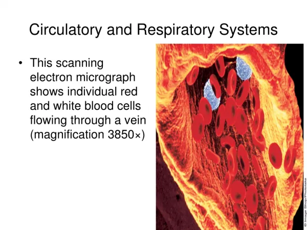
Circulatory and Respiratory Systems. This scanning electron micrograph shows individual red and white blood cells flowing through a vein (magnification 3850×). Circulatory and Respiratory Systems. The Circulatory System. Your heartbeat is a sign of life itself
1.91k views • 179 slides
The Circulatory and Respiratory Systems
The Circulatory and Respiratory Systems. By Hannah and Cathie. The Circulatory System. Overview body has about 5 liters of blood continually traveling through it
552 views • 36 slides
Chapter 20: Respiratory and Circulatory Systems
Chapter 20: Respiratory and Circulatory Systems. 20.1 What is Respiration?. Respiration: The process that moves oxygen and carbon dioxide between the atmosphere and the cells inside the body. Oxygen moves into the body in three stages:.
980 views • 66 slides

How are the two systems controlled?. The Circulatory and Respiratory Systems. Autonomic nervous system. What is the FUNCTION of the circulatory system?. To bring oxygen to tissues/organs throughout the body To remove carbon dioxide from tissues/organs throughout the body.
208 views • 9 slides
The Respiratory And Circulatory Systems
The Respiratory And Circulatory Systems. By Cody Sugihara. The circulatory system. This system moves your blood throughout your body Blood vessels are the roads that carry blood. http://en.wikipedia.org/wiki/File:Human_Heart_and_Circulatory_System.png. Types of blood vessels.
274 views • 11 slides

Circulatory and Respiratory Systems. Breathing, Blood, Keeping your Circulatory and Respiratory systems healthy!. A Breathtaking Feat!. Page 743 Amy Van Dyken’s story of success. Measuring your Breathing. Copy this Chart onto your paper. Hold your breath class averages.
364 views • 21 slides

The Circulatory and Respiratory Systems. Learning Goals. By the end of this unit you should be able to: Identify and describe the main functions of the circulatory and respiratory system Identify and describe the different types of blood cells and give examples of their function
293 views • 15 slides

Circulatory and Respiratory Systems. Ch 33.1 & 33.3. Types of Circulatory System. Open Circulatory System. Closed Circulatory System. Types of Circulation. Single loop Circulation. Double loop Circulation. Mammalian Heart-Chamber Evolution . Double Pump
495 views • 15 slides

Circulatory and Respiratory Systems. EQ: How does our body provide cells with O 2 and nutrients and removes waste and CO 2 ?. How Circulatory System Maintains Homeostasis. The respiratory and circulatory systems work together to maintain homeostasis
422 views • 12 slides

Circulatory and Respiratory Systems. Circulatory System (section 1) . Connects the muscles and organs of the body through an extensive system of vessels that transport blood, a mixture of specialized cells, and fluid. Circulatory System (section 1) .
1.39k views • 47 slides
Circulatory and Respiratory
Circulatory and Respiratory. Mrs. Geib Lincoln Elementary 4 th grade. The Respiratory System. The Cardiovascular/Circulatory System. The Components of Blood. The Heart. Blood Vessels, Pulmonary Circulation. Maintaining Cardiovascular Health.
230 views • 7 slides

Chapter 47: Circulatory and Respiratory Systems
Chapter 47: Circulatory and Respiratory Systems. 47-1 The Circulatory System. 47-2 Blood. 47-3 The Respiratory System. 47-1 The Circulatory System. I. The Heart (THORACIC cavity, behind STERNUM). Moves blood PULMONARY and SYSTEMIC loops.
1.43k views • 122 slides

Circulatory and Respiratory systems
Circulatory and Respiratory systems. Shade in low oxygen blood as blue and highly oxygenated blood as red. from head. to lungs. from trunk. to head/arms. from lungs. from lungs. to trunk. a. Artery Carries blood away from heart. B. Right atria Receives blood from body. C.
495 views • 31 slides

The Circulatory and Respiratory Systems. Chapter 49. Invertebrate Circulatory Systems. Sponges, Cnidarians, and nematodes lack a separate circulatory system - Sponges circulate water using many incurrent pores and one excurrent pore
815 views • 53 slides

Circulatory and Respiratory Systems. Open and Closed Circulatory Systems. How do open and closed circulatory systems compare? In an open circulatory system, blood is only partially contained within a system of blood vessels as it travels through the body.
539 views • 38 slides

1.82k views • 179 slides

Respiratory and Circulatory Systems. Chapter 30: Respiratory and Circulatory Systems I. Respiratory and Circulatory Functions (30.1). A. The respiratory and circulatory systems work together to maintain homeostasis. 1. Every cell in body needs nutrients and oxygen to function.
526 views • 41 slides
Unit 6 Respiratory and Circulatory Systems
Unit 6 Respiratory and Circulatory Systems. UNIT 6: PHYSIOLOGY Chapter 30: Respiratory and Circulatory Systems I. Respiratory and Circulatory Functions (30.1). A. The respiratory and circulatory systems work together to maintain homeostasis.
518 views • 42 slides

IMAGES
VIDEO
COMMENTS
Nose or Mouth - moistens and heats the air before going into the trachea. Cilia and mucus trap dirt in the air. Larynx - part of the trachea where our vocal cords are located.; Trachea - the tube that leads from the nose and mouth to the lungs. The walls have rings of cartilage to protect the trachea and prevent it from collapsing.
Presentation Transcript. Major Organs and Functions • Nose:The only Externally visible part of the respiratory system. • During the process of breathing, air passes through the external nares (nostrils) • The interior of the nose is called the nasal cavity, which is divided by the midline/nasal septum. • The Respiratory Mucosa lining ...
The respiratory system allows for oxygen to enter the body and carbon dioxide to exit through a series of major organs. Air enters through the nose or mouth and passes through the pharynx, larynx, trachea, bronchi and into the lungs where gas exchange occurs in the alveoli. Oxygen then passes into the bloodstream and carbon dioxide passes out ...
Download our Google Slides and PPT templates about Lungs and breathe life with your slides! Free Easy to edit Professional. ... Freepik Free vectors, ... Download the "Lungs and Respiratory System" presentation for PowerPoint or Google Slides. The education sector constantly demands dynamic and effective ways to present information.
Free Download The Respiratory System PowerPoint Presentation. Major Functions of the Respiratory System To supply the body with oxygen and dispose of carbon dioxide Respiration - four distinct processes must happen Pulmonary ventilation - moving air into and out of the lungs External respiration - gas exchange between the lungs and the blood Transport - transport of oxygen and carbon ...
For Teachers. Lots of Lessons - Human Respiratory System, Lungs. Free Video Clips. Free Clipart. Pete's PowerPoint Station is your destination for free PowerPoint presentations for kids and teachers about Human Respiratory System, and so much more.
Free Google Slides theme, PowerPoint template, and Canva presentation template. Download the "Lungs and Respiratory System" presentation for PowerPoint or Google Slides. The education sector constantly demands dynamic and effective ways to present information. This template is created with that very purpose in mind.
DISORDERS OF RESPIRATORY. EMPHYSEMA Emphysema is a lung. PNEUMONIA Pneumonia is an inflammatory condition. LUNG ABSCESS Lung abscess. LUNG COLLAPSE A collapsed lung. APNEA Apnea or apnoea. LUNG TUMOURS Lung cancer, Anatomy and physiology of the respiratory system - Download as a PDF or view online for free.
Sep 6, 2016 • Download as PPSX, PDF •. 347 likes • 149,476 views. Dr. Binu Babu Nursing Lectures Incredibly Easy. Respiratory System - Physiology. Health & Medicine. Slideshow view. Download now. Respiratory System - Physiology - Download as a PDF or view online for free.
Tags. Professional Blue Illustration Medical Health Minimalist Breakthrough Research Lungs Anatomy. Download this blue colored medical template for Google Slides and PPT to share you research about respiratory system.
SlidesCarnival templates have all the elements you need to effectively communicate your message and impress your audience. Download your presentation as a PowerPoint template or use it online as a Google Slides theme. 100% free, no registration or download limits. Create engaging presentations about the respiratory system with these lung templates.
Presentation Transcript. Functions of upper respiratory tract: nasal/oral cavities and trachea • Nose/Mouth: • Filters air, Warms air, Moistens air. Also Provides resonance in speech (so you don't sound funny) • Larynx: "voice box" Holds our vocal chords. • Trachea: • Commonly called your "wind pipe.".
Presentation Transcript. The Respiratory System Chapter 21. Introduction • The trillions of cells making up the body require a continuous supply of oxygen to carry out vita functions • We can survive only a few minutes without oxygen • As cells use oxygen, they give off carbon dioxide a waste product of cellular respiration which the body ...
The respiratory system aids in breathing, also called pulmonary ventilation. In pulmonary ventilation, air is inhaled through the nasal and oral cavities (the nose and mouth). It moves through the pharynx, larynx, and trachea into the lungs. Then air is exhaled, flowing back through the same pathway. Changes to the volume and air pressure in ...
The Free Respiratory PowerPoint Templates Slide includes the x-ray image of a human body, in which lungs are highlighted. It has six text holders with well-research information about the lungs and respiratory system. This content-ready slide is the best tool to teach the students about the respiratory system.
Lungs Presentation Templates. Dive in presenters! These free lungs PowerPoint templates and Google Slides Themes show what it's like to find the perfect place through compelling visualizations. You can save infographics of your choice and add them to your slides for a better look! Choose and forget doubts about editing!
Free Google Slides theme, PowerPoint template, and Canva presentation template. Do you need to share some details and info about diseases related to the respiratory system? Use this template with gradients in blue to explain the illnesses, diagnoses and recommendations. The infographics that we have included will be quite useful to you as well!
The respiratory system includes: Breathing in, also known as inhalation, causes our lungs to become larger. The muscles between the ribs, called the intercostal muscles, help the chest to expand (get bigger), which helps air flow into the lungs. Breathing out, or exhalation, is when air is released from the lungs.
Free Google Slides theme, PowerPoint template, and Canva presentation template ... Teachers will also find this template a nice resource, as it allows them to explain the respiratory system in a visual way. The design focuses on the use of gradients (ranging from white to light blue, the color of safety), simple layouts and some health-themed ...
ANATOMY OF RESPIRATORY SYSTEM DR SADIA FARHAN. Anatomy of the Respiratory System • Consists of all structures in the body that make up the airway and help us breathe • Diaphragm • Intercostal muscles • Accessory muscles of breathing • Nerves from the brain and spinal cord to those muscles. Anatomy of the Respiratory System.
Free Respiratory System Template for PowerPoint and Google Slides. Main features. 42 slides 100% editable. 16:9 widescreen format suitable for all screens. High quality royalty-free images. Included resources: charts, graphs, timelines and diagrams. More than 100 icons customizable in color and size. Main font: Arial.
The Human Respiratory System. The Human Respiratory System. Biology 314 Mr. Doron. Game Plan . Introduction to the respiratory system Pathway the air takes Role of the nasal cavity Role of the pharynx Role of the epiglottis Role of the trachea Role of the bronchi & lungs Fun FAQ's (yawning, sneezing, hiccups). 308 views • 18 slides
In the Circulatory System, the heart, lungs, and blood vessels have to work together to maintain homeostasis. The Heart: pumps oxygen rich blood, nutrients, hormones, and other things the body needs to maintain health, to organs and tissues. Parts of the Heart The heart is made up of 4 different blood-filled areas.