Thank you for visiting nature.com. You are using a browser version with limited support for CSS. To obtain the best experience, we recommend you use a more up to date browser (or turn off compatibility mode in Internet Explorer). In the meantime, to ensure continued support, we are displaying the site without styles and JavaScript.
- View all journals
- Explore content
- About the journal
- Publish with us
- Sign up for alerts
- Review Article
- Published: 04 December 2019

Neuroplasticity in cognitive and psychological mechanisms of depression: an integrative model
- Rebecca B. Price 1 &
- Ronald Duman 2
Molecular Psychiatry volume 25 , pages 530–543 ( 2020 ) Cite this article
269 Citations
86 Altmetric
Metrics details
- Neuroscience
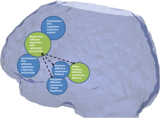
Chronic stress and depressive-like behaviors in basic neuroscience research have been associated with impairments of neuroplasticity, such as neuronal atrophy and synaptic loss in the medial prefrontal cortex (mPFC) and hippocampus. The current review presents a novel integrative model of neuroplasticity as a multi-domain neurobiological, cognitive, and psychological construct relevant in depression and other related disorders of negative affect (e.g., anxiety). We delineate a working conceptual model in which synaptic plasticity deficits described in animal models are integrated and conceptually linked with human patient findings from cognitive science and clinical psychology. We review relevant reports including neuroimaging findings (e.g., decreased functional connectivity in prefrontal-limbic circuits), cognitive deficits (e.g., executive function and memory impairments), affective information processing patterns (e.g., rigid, negative biases in attention, memory, interpretations, and self-associations), and patient-reported symptoms (perseverative, inflexible thought patterns; inflexible and maladaptive behaviors). Finally, we incorporate discussion of integrative research methods capable of building additional direct empirical support, including using rapid-acting treatments (e.g., ketamine) as a means to test this integrative model by attempting to simultaneously reverse these deficits across levels of analysis.
This is a preview of subscription content, access via your institution
Access options
Subscribe to this journal
Receive 12 print issues and online access
251,40 € per year
only 20,95 € per issue
Buy this article
- Purchase on Springer Link
- Instant access to full article PDF
Prices may be subject to local taxes which are calculated during checkout
Distinct µ-opioid ensembles trigger positive and negative fentanyl reinforcement

Cortico-cortical transfer of socially derived information gates emotion recognition

Emotions and brain function are altered up to one month after a single high dose of psilocybin
Chisholm D, Sweeny K, Sheehan P, Rasmussen B, Smit F, Cuijpers P, et al. Scaling-up treatment of depression and anxiety: a global return on investment analysis. Lancet Psychiatry. 2016;3:415–24.
PubMed Google Scholar
Wang PS, Lane M, Olfson M, Pincus HA, Wells KB, Kessler RC. Twelve-month use of mental health services in the United States: results from the National Comorbidity Survey Replication. Arch Gen Psychiatry. 2005;62:629–40.
Abdallah CG, Sanacora G, Duman RS, Krystal JH. Ketamine and rapid-acting antidepressants: a window into a new neurobiology for mood disorder therapeutics. Annu Rev Med. 2015;66:509–23.
CAS PubMed Google Scholar
Duman RS, Aghajanian GK, Sanacora G, Krystal JH. Synaptic plasticity and depression: new insights from stress and rapid-acting antidepressants. Nat Med. 2016;22:238–49.
CAS PubMed PubMed Central Google Scholar
Duman RS, Aghajanian GK. Synaptic dysfunction in depression: potential therapeutic targets. Science. 2012;338:68–72.
Kashdan TB, Rottenberg J. Psychological flexibility as a fundamental aspect of health. Clin Psychol Rev. 2010;30:865–78.
PubMed PubMed Central Google Scholar
Joormann J. Cognitive inhibition and emotion regulation in depression. Curr Dir Psychol Sci. 2010;19:161–6.
Google Scholar
Disner S, Beevers C, Haigh EAP, Beck AT. Neural mechanisms of the cognitive model of depression. Nat Rev Neurosci. 2011;12:467–77.
Beck AT, Bredemeier K. A unified model of depression: integrating clinical, cognitive, biological, and evolutionary perspectives. Clin Psychol Sci. 2016;4:596–619.
Moda-Sava RN, Murdock MH, Parekh PK, Fetcho RN, Huang BS, Huynh TN, et al. Sustained rescue of prefrontal circuit dysfunction by antidepressant-induced spine formation. Science. 2019;364:eaat8078.
Murrough JW, Iosifescu DV, Chang LC, Al Jurdi RK, Green CM, Perez AM, et al. Antidepressant efficacy of ketamine in treatment-resistant major depression: a two-site randomized controlled trial. Am J Psychiatry. 2013;170:1134–42.
Xu Y, Hackett M, Carter G, Loo C, Galvez V, Glozier N, et al. Effects of low-dose and very low-dose dose ketamine among patients with major depression: a systematic review and meta-analysis. Int J Neuropsychopharmacol. 2015;19:pyv124.
PubMed Central Google Scholar
Beurel E, Nemeroff CB. Interaction of stress, corticotropin-releasing factor, arginine vasopressin and behaviour. Curr Top Behav Neurosci. 2014;18:67–80.
Luscher B, Fuchs T. GABAergic control of depression-related brain states. Adv Pharmacol. 2015;73:97–144.
Waters RP, Rivalan M, Bangasser DA, Deussing JM, Ising M, Wood SK, et al. Evidence for the role of corticotropin-releasing factor in major depressive disorder. Neurosci Biobehav Rev. 2015;58:63–78.
Fee C, Banasr M, Sibille E. Somatostatin-positive gamma-aminobutyric acid interneuron deficits in depression: cortical microcircuit and therapeutic perspectives. Biol Psychiatry. 2017;82:549–59.
Wohleb ES, Franklin T, Iwata M, Duman RS. Integrating neuroimmune systems in the neurobiology of depression. Nat Rev Neurosci. 2016;17:497–511.
McEwen BS, Nasca C, Gray JD. Stress effects on neuronal structure: hippocampus, amygdala, and prefrontal cortex. Neuropsychopharmacology. 2016;41:3–23.
Anacker C, Hen R. Adult hippocampal neurogenesis and cognitive flexibility—linking memory and mood. Nat Rev Neurosci. 2017;18:335–46.
Koo JW, Chaudhury D, Han MH, Nestler EJ. Role of mesolimbic brain-derived neurotrophic factor in depression. Biol Psychiatry. 2019;86:738–48.
Cathomas F, Murrough JW, Nestler EJ, Han MH, Russo SJ. Neurobiology of Resilience: interface between mind and body. Biol Psychiatry. 2019;86:410–20.
Duman RS, Sanacora G, Krystal JH. Altered connectivity in depression: GABA and glutamate neurotransmitter deficits and reversal by novel treatments. Neuron. 2019;102:75–90.
Arnone D. Functional MRI findings, pharmacological treatment in major depression and clinical response. Prog Neuropsychopharmacol Biol Psychiatry. 2019;91:28–37.
Castrén E. Is mood Chem? Nat Rev Neurosci. 2005;6:241–6.
Castren E, Hen R. Neuronal plasticity and antidepressant actions. Trends Neurosci. 2013;36:259–67.
Changeaux JP, Danchin A. Selective stabilisation of developing synapses as a mechanism for the specification of neuronal networks. Nature. 1976;264:705–12.
Wang Q, Timberlake MA 2nd, Prall K, Dwivedi Y. The recent progress in animal models of depression. Prog Neuropsychopharmacol Biol Psychiatry. 2017;77:99–109.
Ota K, Liu R, Voleti B, Maldonado-Aviles J, Duric V, Iwata M, et al. REDD1 is essential for stress-induced synaptic loss and depressive behavior. Nat Med. 2014;20:531–5.
Wohleb ES, Terwilliger R, Duman CH, Duman RS. Stress-induced neuronal colony stimulating factor 1 provokes microglia-mediated neuronal remodeling and depressive-like behavior. Biol Psychiatry. 2018;83:38–49.
Roozendaal B, McEwen BS, Chattarji S. Stress, memory and the amygdala. Nat Rev Neurosci. 2009;10:423–33.
Patel D, Anilkumar S, Chattarji S, Buwalda B. Repeated social stress leads to contrasting patterns of structural plasticity in the amygdala and hippocampus. Behav Brain Res. 2018;347:314–24.
Caddy C, Amit BH, McCloud TL, Rendell JM, Furukawa TA, McShane R, et al. Ketamine and other glutamate receptor modulators for depression in adults. Cochrane Database Syst Rev. 2015:CD011612.
McCloud TL, Caddy C, Jochim J, Rendell JM, Diamond PR, Shuttleworth C, et al. Ketamine and other glutamate receptor modulators for depression in bipolar disorder in adults. Cochrane Database Syst Rev. 2015:CD011611.
Zanos P, Moaddel R, Morris PJ, Georgiou P, Fischell J, Elmer GI, et al. NMDAR inhibition-independent antidepressant actions of ketamine metabolites. Nature. 2016;533:481–6.
Li N, Lee B, Liu RJ, Banasr M, Dwyer JM, Iwata M, et al. mTOR-dependent synapse formation underlies the rapid antidepressant effects of NMDA antagonists. Science. 2010;329:959–64.
Liu RJ, Duman C, Kato T, Hare B, Lopresto D, Bang E, et al. GLYX-13 produces rapid antidepressant responses with key synaptic and behavioral effects distinct from ketamine. Neuropsychopharmacology. 2017;42:1231–42.
Burgdorf J, Zhang XL, Nicholson KL, Balster RL, Leander JD, Stanton PK, et al. GLYX-13, a NMDA receptor glycine-site functional partial agonist, induces antidepressant-like effects without ketamine-like side effects. Neuropsychopharmacology. 2013;38:729–42.
Newport DJ, Carpenter LL, McDonald WM, Potash JB, Tohen M, Nemeroff CB, et al. Ketamine and other NMDA antagonists: early clinical trials and possible mechanisms in depression. Am J Psychiatry. 2015;172:950–66.
Abdallah C, Averill L, Gueorguieva R, Goktas S, Purohit P, Ranganathan M, et al. Rapamycin, an immunosuppressant and mTORC1 inhibitor, triples the antidepressant response rate to ketamine at two weeks following treatment. 2018. https://doi.org/10.1101/500959 .
Sen S, Duman R, Sanacora G. Serum brain-derived neurotrophic factor, depression, and antidepressant medications: meta-analyses and implications. Biol Psychiatry. 2008;64:527–32.
Kang HJ, Voleti B, Hajszan T, Rajkowska G, Stockmeier CA, Licznerski P, et al. Decreased expression of synapse-related genes and loss of synapses in major depressive disorder. Nat Med. 2012;18:1413–7.
Price RB, Eldreth DA, Mohlman J. Deficient prefrontal attentional control in late-life generalized anxiety disorder: an fMRI investigation. Transl Psych. 2011;1:e46.
CAS Google Scholar
Price RB, Allen KB, Silk JS, Ladouceur CD, Ryan ND, Dahl RE, et al. Vigilance in the laboratory predicts avoidance in the real world: a dimensional analysis of neural, behavioral, and ecological momentary data in anxious youth. Dev Cogn Neurosci. 2016;19:128–36.
Siegle GJ, Thompson W, Carter CS, Steinhauer SR, Thase ME. Increased amygdala and decreased dorsolateral prefrontal BOLD responses in unipolar depression: related and independent features. Biol Psychiatry. 2007;61:198–209.
Gotlib IH, Joormann J. Cognition and depression: current status and future directions. Annu Rev Clin Psychol. 2010;6:285–312.
Dozois D, Beck A. Cognitive schemas, beliefs and assumptions. In: Dobson K, Dozois D, editors. Risk factors in depression. Oxford, England: Elsevier/Academic Press; 2008, p. 121–43.
de Raedt R, Koster EHW. Understanding vulnerability for depression from a cognitive neuroscience perspective: a reappraisal of attentional factors and a new conceptual framework. Cogn Affect Behav Neurosci. 2010;10:50–70.
Stange JP, Connolly SL, Burke TA, Hamilton JL, Hamlat EJ, Abramson LY, et al. Inflexible cognition predicts first onset of major depressive episodes in adolescence. Depress Anxiety. 2016;33:1005–12.
Schmaal L, Veltman DJ, van Erp TG, Samann PG, Frodl T, Jahanshad N, et al. Subcortical brain alterations in major depressive disorder: findings from the ENIGMA Major Depressive Disorder working group. Mol Psychiatry. 2016;21:806–12.
Schmaal L, Hibar DP, Samann PG, Hall GB, Baune BT, Jahanshad N, et al. Cortical abnormalities in adults and adolescents with major depression based on brain scans from 20 cohorts worldwide in the ENIGMA Major Depressive Disorder Working Group. Mol Psychiatry. 2017;22:900–9.
Chen G, Guo Y, Zhu H, Kuang W, Bi F, Ai H, et al. Intrinsic disruption of white matter microarchitecture in first-episode, drug-naive major depressive disorder: a voxel-based meta-analysis of diffusion tensor imaging. Prog Neuropsychopharmacol Biol Psychiatry. 2017;76:179–87.
Park HJ, Friston K. Structural and functional brain networks: from connectionsto cognition. Science. 2013;342:1238411.
Sporns O, Chialvo DR, Kaiser M, Hilgetag CC. Organization, development and function of complex brain networks. Trends Cogn Sci. 2004;8:418–25.
Kaiser RH, Andrews-Hanna JR, Wager TD, Pizzagalli DA. Large-Scale network dysfunction in major depressive disorder: a meta-analysis of resting-state functional connectivity. JAMA Psychiatry. 2015;72:603–11.
Abdallah CG, Averill LA, Collins KA, Geha P, Schwartz J, Averill C, et al. Ketamine treatment and global brain connectivity in major depression. Neuropsychopharmacology. 2017;42:1210–9.
Hirshfeld-Becker DR, Gabrieli JDE, Shapero BG, Biederman J, Whitfield-Gabrieli S, Chai XJ. Intrinsic functional brain connectivity predicts onset of major depression disorder in adolescence: a pilot study. Brain Connect. 2019;9:388–98.
Jiang X, Shen Y, Yao J, Zhang L, Xu L, Feng R, et al. Connectome analysis of functional and structural hemispheric brain networks in major depressive disorder. Transl Psychiatry. 2019;9:136.
Greicius MD, Flores BH, Menon V, Glover GH, Solvason HB, Kenna H, et al. Resting-state functional connectivity in major depression: abnormally increased contributions from subgenual cingulate cortex and thalamus. Biol Psychiatry. 2007;62:429–37.
Rive MM, van Rooijen G, Veltman DJ, Phillips ML, Schene AH, Ruhe HG. Neural correlates of dysfunctional emotion regulation in major depressive disorder. A systematic review of neuroimaging studies. Neurosci Biobehav Rev. 2013;37:2529–53.
Carballedo A, Scheuerecker J, Meisenzahl E, Schoepf V, Bokde A, Moller HJ, et al. Functional connectivity of emotional processing in depression. J Affect Disord. 2011;134:272–9.
Anand A, Li Y, Wang Y, Wu J, Gao S, Bukhari L, et al. Activity and connectivity of brain mood regulating circuit in depression: a functional magnetic resonance study. Biol Psychiatry. 2005;57:1079–88.
Ochsner KN, Ray RD, Cooper JC, Robertson ER, Chopra S, Gabrieli JD, et al. For better or for worse: neural systems supporting the cognitive down- and up-regulation of negative emotion. Neuroimage. 2004;23:483–99.
Zilverstand A, Parvaz MA, Goldstein RZ. Neuroimaging cognitive reappraisal in clinical populations to define neural targets for enhancing emotion regulation. A systematic review. Neuroimage. 2017;151:105–16.
Pico-Perez M, Radua J, Steward T, Menchon JM, Soriano-Mas C. Emotion regulation in mood and anxiety disorders: a meta-analysis of fMRI cognitive reappraisal studies. Prog Neuropsychopharmacol Biol Psychiatry. 2017;79:96–104.
Johnstone T, van Reekum CM, Urry HL, Kalin NH, Davidson RJ. Failure to regulate: counterproductive recruitment of top-down prefrontal-subcortical circuitry in major depression. J Neurosci. 2007;27:8877–84.
Thompson SM, Kallarackal AJ, Kvarta MD, Van Dyke AM, LeGates TA, Cai X. An excitatory synapse hypothesis of depression. Trends Neurosci. 2015;38:279–94.
Koechlin E. Prefrontal executive function and adaptive behavior in complex environments. Curr Opin Neurobiol. 2016;37:1–6.
Wagner S, Muller C, Helmreich I, Huss M, Tadic A. A meta-analysis of cognitive functions in children and adolescents with major depressive disorder. Eur Child Adolesc Psychiatry. 2015;24:5–19.
Wagner S, Doering B, Helmreich I, Lieb K, Tadic A. A meta-analysis of executive dysfunctions in unipolar major depressive disorder without psychotic symptoms and their changes during antidepressant treatment. Acta Psychiatr Scandinavica. 2012;125:281–92.
McClintock SM, Husain MM, Greer TL, Cullum CM. Association between depression severity and neurocognitive function in major depressive disorder: a review and synthesis. Neuropsychology. 2010;24:9–34.
Roca M, Vives M, Lopez-Navarro E, Garcia-Campayo J, Gili M. Cognitive impairments and depression: a critical review. Actas Esp Psiquiatr. 2015;43:187–93.
Smith EE, Jonides J. Storage and executive processes in the frontal lobes. Science. 1999;283:1657–61.
Fuster JM. The prefrontal cortex. 3rd edn. Philadelphia, PA: Lippincott-Raven; 1997.
Murrough JW, Iacoviello B, Neumeister A, Charney DS, Iosifescu DV. Cognitive dysfunction in depression: neurocircuitry and new therapeutic strategies. Neurobiol Learn Mem. 2011;96:553–63.
Motter JN, Pimontel MA, Rindskopf D, Devanand DP, Doraiswamy PM, Sneed JR. Computerized cognitive training and functional recovery in major depressive disorder: a meta-analysis. J Affect Disord. 2016;189:184–91.
Siegle GJ, Price RB, Jones N, Ghinassi F, Painter T, Thase ME. You gotta work at it: pupillary indices of task focus are prognostic for response to a neurocognitive intervention for depression. Clin Psychol Sci. 2014;2:455–71.
Zhou FC, Wang YY, Zheng W, Zhang Q, Ungvari GS, Ng CH,et al. Prospective memory deficits in patients with depression: a meta-analysis. J Affect Disord. 2017;220:79–85.
Van Vreeswijk MF, De Wilde EJ. Autobiographical memory specificity, psychopathology, depressed mood and the use of the autobiographical memory test: a meta analysis. Behav Res Ther. 2004;42:731–43.
Pollock LR, Williams JM. Effective problem solving in suicide attempters depends on specific autobiographical recall. Suicide Life Threat Behav. 2001;31:386–96.
Williams JM, Ellis NC, Tyers C, Healy H, Rose G, MacLeod AK. The specificity of autobiographical memory and imageability of the future. Mem Cogn. 1996;24:116–25.
McEwen BS, Sapolsky RM. Stress and cognitive function. Curr Opin Neurobiol. 1995;5:205–16.
Mathews A, MacLeod C. Cognitive vulnerability to emotional disorders. Annu Rev Clin Psychol. 2005;1:167–95.
Peckham AD, McHugh RK, Otto MW. A meta-analysis of the magnitude of biased attention in depression. Depress Anxiety. 2010;27:1135–42.
Gilboa-Schechtman E, Erhard-Weiss D, Jeczemien P. Interpersonal deficits meet cognitive biases: memory for facial expressions in depressed and anxious men and women. Psychiatry Res. 2002;113:279–93.
Ridout N, Astell AJ, Reid IC, Glen T, O’Carroll RE. Memory bias for emotional facial expressions in major depression. Cogn Emot. 2003;17:101–22.
Hirsch CR, Meeten F, Krahe C, Reeder C. Resolving ambiguity in emotional disorders: the nature and role of interpretation biases. Annu Rev Clin Psychol. 2016;12:281–305.
Milders M, Bell S, Platt J, Serrano R, Runcie O. Stable expression recognition abnormalities in unipolar depression. Psychiatry Res. 2010;179:38–42.
Persad SM, Polivy J. Differences between depressed and nondepressed individuals in the recognition of and response to facial emotional cues. J Abnorm Psychol. 1993;102:358–68.
van Randenborgh A, Pawelzik M, Quirin M, Kuhl J. Bad roots to grow: deficient implicit self-evaluations in chronic depression with an early onset. J Clin Psychol. 2016;72:580–90.
Franck E, De Raedt R, De Houwer J. Implicit but not explicit self-esteem predicts future depressive symptomatology. Behav Res Ther. 2007;45:2448–55.
Phillips WJ, Hine DW, Thorsteinsson EB. Implicit cognition and depression: a meta-analysis. Clin Psychol Rev. 2010;30:691–709.
Nock MK, Park JM, Finn CT, Deliberto TL, Dour HJ, Banaji MR. Measuring the suicidal mind: implicit cognition predicts suicidal behavior. Psychol Sci. 2010;21:511–7.
Cha CB, Najmi S, Park JM, Finn CT, Nock MK. Attentional bias toward suicide-related stimuli predicts suicidal behavior. J Abnorm Psychol. 2010;119:616–22.
Barch DM, Pagliaccio D, Luking K. Mechanisms underlying motivational deficits in psychopathology: similarities and differences in depression and schizophrenia. Curr Top Behav Neurosci. 2016;27:411–49.
Chen C, Takahashi T, Nakagawa S, Inoue T, Kusumi I. Reinforcement learning in depression: a review of computational research. Neurosci Biobehav Rev. 2015;55:247–67.
Dombrovski AY, Szanto K, Clark L, Aizenstein HJ, Chase HW, Reynolds CF 3rd, et al. Corticostriatothalamic reward prediction error signals and executive control in late-life depression. Psychol Med. 2015;45:1413–24.
Dombrovski AY, Szanto K, Clark L, Reynolds CF, Siegle GJ. Reward signals, attempted suicide, and impulsivity in late-life depression. JAMA Psychiatry. 2013;70:1020–30.
Gradin VB, Kumar P, Waiter G, Ahearn T, Stickle C, Milders M, et al. Expected value and prediction error abnormalities in depression and schizophrenia. Brain. 2011;134:1751–64.
Kumar P, Waiter G, Ahearn T, Milders M, Reid I, Steele JD. Abnormal temporal difference reward-learning signals in major depression. Brain. 2008;131:2084–93.
Dombrovski AY, Hallquist MN. The decision neuroscience perspective on suicidal behavior: evidence and hypotheses. Curr Opin Psychiatry. 2017;30:7–14.
Berridge KC. Motivation concepts in behavioral neuroscience. Physiol Behav. 2004;81:179–209.
McGaugh JL. The amygdala modulates the consolidation of memories of emotionally arousing experiences. Annu Rev Neurosci. 2004;27:1–28.
Cahill L, Alkire MT. Epinephrine enhancement of human memory consolidation: interaction with arousal at encoding. Neurobiol Learn Mem. 2003;79:194–8.
Beevers CG, Clasen PC, Enock PM, Schnyer DM. Attention bias modification for major depressive disorder: effects on attention bias, resting state connectivity, and symptom change. J Abnorm Psychol. 2015;124:463–75.
White LK, Sequeira S, Britton JC, Brotman MA, Gold AL, Berman E, et al. Complementary features of attention bias modification therapy and cognitive-behavioral therapy in pediatric anxiety disorders. Am J Psychiatry. 2017;174:775–84.
Mathews A, MacLeod C. Cognitive approaches to emotion and emotional disorders. Annu Rev Psychol. 1994;45:25–50.
MacLeod C. Cognitive bias modification procedures in the management of mental disorders. Curr Opin Psychiatry. 2012;25:114–20.
Jones EB, Sharpe L. Cognitive bias modification: a review of meta-analyses. J Affect Disord. 2017;223:175–83.
Harmer CJ, Goodwin GM, Cowen PJ. Why do antidepressants take so long to work? A cognitive neuropsychological model of antidepressant drug action. Br J Psychiatry. 2009;195:102–8.
Association AP. Diagnostic and statistical manual of mental disorders. Arlington, VA: American Psychiatric Publishing; 2013.
Nolen-Hoeksema S, Morrow J, Fredrickson BL. Response styles and the duration of episodes of depressed mood. J Abnorm Psychol. 1993;102:20–8.
McEvoy PM, Mahoney AEJ, Moulds ML. Are worry, rumination, and post-event processing one and the same? Development of the repetitive thinking questionnaire. J Anxiety Disord. 2010;24:509–19.
Beck JG. Cognitive therapy: basics and beyond. New York, NY: The Guilford Press; 1995.
Beck AT, Dozois DJ. Cognitive therapy: current status and future directions. Annu Rev Med. 2011;62:397–409.
Teasdale JD, Segal ZV, Williams JMG, Ridgeway VA, Soulsby JM, Lau MA. Prevention of relapse/recurrence in major depression by mindfulness-based cognitive therapy. J Consult Clin Psychol. 2000;68:615–23.
Segal ZV, Walsh KM. Mindfulness-based cognitive therapy for residual depressive symptoms and relapse prophylaxis. Curr Opin Psychiatry. 2016;29:7–12.
Segal ZV, Teasdale JD, Williams JMG. Mindfulness-based cognitive therapy: theoretical rationale and empirical status. In: Folette SC, Linehan MM (eds.) Mindfulness and acceptance: Expanding the cognitive-behavioral tradition. New York, NY: Guilford Press; 2004.
Davidson J, Turnbull CD. Diagnostic significance of vegetative symptoms in depression. Br J Psychiatry. 1986;148:442–6.
Dimidjian S, Barrera M Jr., Martell C, Munoz RF, Lewinsohn PM. The origins and current status of behavioral activation treatments for depression. Annu Rev Clin Psychol. 2011;7:1–38.
Ratcliffe M. Experiences of Depression: a study in phenomenology. Oxford, UK: Oxford University Press; 2015.
Martell CR, Dimidjian S, Herman-Dunn R. Behavioral activation for depression: a clinician’s guide. New York: The Guilford Press; 2010.
Dimidjian S, Hollon SD, Dobson KS, Schmaling KB, Kohlenberg RJ, Addis ME, et al. Randomized trial of behavioral activation, cognitive therapy, and antidepressant medication in the acute treatment of adults with major depression. J Consult Clin Psychol. 2006;74:658–70.
Murrough JW, Collins KA, Fields J, DeWilde KE, Phillips ML, Mathew SJ, et al. Regulation of neural responses to emotion perception by ketamine in individuals with treatment-resistant major depressive disorder. Transl Psychiatry. 2015;5:e509.
Gartner M, Aust S, Bajbouj M, Fan Y, Wingenfeld K, Otte C, et al. Functional connectivity between prefrontal cortex and subgenual cingulate predicts antidepressant effects of ketamine. Eur Neuropsychopharmacol. 2019;29:501–8.
Li CT, Chen MH, Lin WC, Hong CJ, Yang BH, Liu RS, et al. The effects of low-dose ketamine on the prefrontal cortex and amygdala in treatment-resistant depression: A randomized controlled study. Hum Brain Mapp. 2016;37:1080–90.
Ionescu DF, Felicione JM, Gosai A, Cusin C, Shin P, Shapero BG, et al. Ketamine-Associated Brain Changes: a review of the neuroimaging literature. Harv Rev Psychiatry. 2018;26:320–39.
Downey D, Dutta A, McKie S, Dawson GR, Dourish CT, Craig K, et al. Comparing the actions of lanicemine and ketamine in depression: key role of the anterior cingulate. Eur Neuropsychopharmacol. 2016;26:994–1003.
Reed JL, Nugent AC, Furey ML, Szczepanik JE, Evans JW, Zarate CA, Jr. Ketamine normalizes brain activity during emotionally valenced attentional processing in depression. Neuroimage Clin. 2018;20:92–101.
Evans JW, Szczepanik J, Brutsche N, Park LT, Nugent AC, Zarate CA Jr. Default mode connectivity in major depressive disorder measured up to 10 days after ketamine administration. Biol Psychiatry. 2018;84:582–90.
Jett JD, Boley AM, Girotti M, Shah A, Lodge DJ, Morilak DA. Antidepressant-like cognitive and behavioral effects of acute ketamine administration associated with plasticity in the ventral hippocampus to medial prefrontal cortex pathway. Psychopharmacology. 2015;232:3123–33.
Nikiforuk A, Popik P. Ketamine prevents stress-induced cognitive inflexibility in rats. Psychoneuroendocrinology. 2014;40:119–22.
Shiroma PR, Albott CS, Johns B, Thuras P, Wels J, Lim KO. Neurocognitive performance and serial intravenous subanesthetic ketamine in treatment-resistant depression. Int J Neuropsychopharmacol. 2014;17:1805–13.
Permoda-Osip A, Kisielewski J, Bartkowska-Sniatkowska A, Rybakowski JK. Single ketamine infusion and neurocognitive performance in bipolar depression. Pharmacopsychiatry. 2015;48:78–9.
Price RB, Iosifescu DV, Murrough JW, Chang LC, Al Jurdi RK, Iqbal SZ, et al. Effects of ketamine on explicit and implicit suicidal cognition: a randomized controlled trial in treatment-resistant depression. Depress Anxiety. 2014;31:335–43.
Price RB, Nock MK, Charney DS, Mathew SJ. Effects of intravenous ketamine on explicit and implicit measures of suicidality in treatment-resistant depression. Biol Psychiatry. 2009;66:522–6.
Price RB, Kuckertz JM, Siegle GJ, Ladouceur CD, Silk JS, Ryan ND, et al. Empirical recommendations for improving the stability of the dot-probe task in clinical research. Psychol Assess. 2015;27:365–76.
Price RB, Brown V, Siegle GJ. Computational modeling applied to the dot-probe task yields improved reliability and mechanistic insights. Biol Psychiatry. 2019;85:606–12.
Rodebaugh TL, Scullin RB, Langer JK, Dixon DJ, Huppert JD, Bernstein A, et al. Unreliability as a threat to understanding psychopathology: the cautionary tale of attentional bias. J Abnorm Psychol. 2016;125:840–51.
Auxéméry Y. Post-traumatic psychiatric disorders: PTSD is not the only diagnosis. Presse Med. 2018;47:423–30.
Williams LM. Precision psychiatry: a neural circuit taxonomy for depression and anxiety. Lancet Psychiatry. 2016;3:472–80.
Feder A, Parides MK, Murrough JW, Perez AM, Morgan JE, Saxena S, et al. Efficacy of intravenous ketamine for treatment of chronic posttraumatic stress disorder: a randomized clinical trial. JAMA Psychiatry. 2014;71:681–8.
Glue P, Medlicott NJ, Harland S, Neehoff S, Anderson-Fahey B, Le Nedelec M, et al. Ketamine’s dose-related effects on anxiety symptoms in patients with treatment refractory anxiety disorders. J Psychopharmacol. 2017;31:1302–5.
Price RB, Gates K, Kraynak TE, Thase ME, Siegle GJ. Data-driven subgroups in depression derived from directed functional connectivity paths at rest. Neuropsychopharmacology. 2017;42:2623–32.
Price RB, Lane S, Gates K, Kraynak TE, Horner MS, Thase ME, et al. Parsing heterogeneity in the brain connectivity of depressed and healthy adults during positive mood. Biol Psychiatry. 2017;81:347–57.
Langenecker SA, Mickey BJ, Eichhammer P, Sen S, Elverman KH, Kennedy SE, et al. Cognitive control as a 5-HT1A-Based domain that is disrupted in major depressive disorder. Front Psychol. 2019;10:691.
Price RB, Rosen D, Siegle GJ, Ladouceur CD, Tang K, Allen KB, et al. From anxious youth to depressed adolescents: prospective prediction of 2-year depression symptoms via attentional bias measures. J Abnorm Psychol. 2015;125:267–78.
Fried EI, Nesse RM. Depression is not a consistent syndrome: an investigation of unique symptom patterns in the STAR*D study. J Affect Disord. 2014;172C:96–102.
Santarelli L, Saxe M, Gross C, Surget A, Battaglia F, Dulawa S, et al. Requirement of hippocampal neurogenesis for the behavioral effects of antidepressants. Science. 2003;301:805–9.
Hoogendam JM, Ramakers GM, Di Lazzaro V. Physiology of repetitive transcranial magnetic stimulation of the human brain. Brain Stimul. 2010;3:95–118.
Dukart J, Regen F, Kherif F, Colla M, Bajbouj M, Heuser I, et al. Electroconvulsive therapy-induced brain plasticity determines therapeutic outcome in mood disorders. Proc Natl Acad Sci USA. 2014;111:1156–61.
Cassilhas RC, Tufik S, de Mello MT. Physical exercise, neuroplasticity, spatial learning and memory. Cell Mol Life Sci. 2016;73:975–83.
Wilkinson S, Holtzheimer PE, Gao S, Kirwin D, Price R. Leveraging neuroplasticity to enhance adaptive learning: the potential for synergistic somatic-behavioral treatment combinations to improve clinical outcomes in depression. Biol Psychiatry. 2019;85:454–65.
Cullen KR, Westlund MK, Klimes-Dougan B, Mueller BA, Houri A, Eberly LE, et al. Abnormal amygdala resting-state functional connectivity in adolescent depression. JAMA Psychiatry. 2014;71:1138–47.
Young KD, Zotev V, Phillips R, Misaki M, Drevets WC, Bodurka J. Amygdala real-time functional magnetic resonance imaging neurofeedback for major depressive disorder: a review. Psychiatry Clin Neurosci. 2018;72:466–81.
Young KD, Siegle GJ, Misaki M, Zotev V, Phillips R, Drevets WC, et al. Altered task-based and resting-state amygdala functional connectivity following real-time fMRI amygdala neurofeedback training in major depressive disorder. Neuroimage Clin. 2018;17:691–703.
Dell’osso B, Camuri G, Castellano F, Vecchi V, Benedetti M, Bortolussi S, et al. Meta-review of metanalytic studies with repetitive transcranial magnetic stimulation (rTMS) for the treatment of major depression. Clin Pr Epidemiol Ment Health. 2011;7:167–77.
Derryberry D, Reed MA. Anxiety-related attentional biases and their regulation by attentional control. J Abnorm Psychol. 2002;111:225–36.
Hsu KJ, Beard C, Rifkin L, Dillon DG, Pizzagalli DA, Bjorgvinsson T. Transdiagnostic mechanisms in depression and anxiety: the role of rumination and attentional control. J Affect Disord. 2015;188:22–7.
Sumner JA. The mechanisms underlying overgeneral autobiographical memory: an evaluative review of evidence for the CaR-FA-X model. Clin Psychol Rev. 2012;32:34–48.
Hitchcock C, Werner-Seidler A, Blackwell SE, Dalgleish T. Autobiographical episodic memory-based training for the treatment of mood, anxiety and stress-related disorders: a systematic review and meta-analysis. Clin Psychol Rev. 2017;52:92–107.
Clarke PJ, Notebaert L, Macleod C. Absence of evidence or evidence of absence: reflecting on therapeutic implementations of attentional bias modification. BMC Psychiatry. 2014;14:8.
Franklin JC, Fox KR, Franklin CR, Kleiman EM, Ribeiro JD, Jaroszewski AC, et al. A brief mobile app reduces nonsuicidal and suicidal self-injury: evidence from three randomized controlled trials. J Consult Clin Psychol. 2016;84:544–57.
Download references
Acknowledgements
This project was supported in part by National Institute of Mental Health grant number R01MH113857 (RBP).
Author information
Authors and affiliations.
Departments of Psychiatry and Psychology, University of Pittsburgh, Pittsburgh, PA, USA
Rebecca B. Price
Department of Psychiatry, Yale University, New Haven, CT, USA
Ronald Duman
You can also search for this author in PubMed Google Scholar
Corresponding author
Correspondence to Rebecca B. Price .
Ethics declarations
Conflict of interest.
The authors declare that they have no conflict of interest.
Additional information
Publisher’s note Springer Nature remains neutral with regard to jurisdictional claims in published maps and institutional affiliations.
Rights and permissions
Reprints and permissions
About this article
Cite this article.
Price, R.B., Duman, R. Neuroplasticity in cognitive and psychological mechanisms of depression: an integrative model. Mol Psychiatry 25 , 530–543 (2020). https://doi.org/10.1038/s41380-019-0615-x
Download citation
Received : 07 June 2019
Revised : 18 November 2019
Accepted : 19 November 2019
Published : 04 December 2019
Issue Date : March 2020
DOI : https://doi.org/10.1038/s41380-019-0615-x
Share this article
Anyone you share the following link with will be able to read this content:
Sorry, a shareable link is not currently available for this article.
Provided by the Springer Nature SharedIt content-sharing initiative
This article is cited by
Towards a network-based operationalization of plasticity for predicting the transition from depression to mental health.
- Claudia Delli Colli
- Flavia Chiarotti
- Igor Branchi
Nature Mental Health (2024)
Glial-restricted precursors stimulate endogenous cytogenesis and effectively recover emotional deficits in a model of cytogenesis ablation
- Joana Martins-Macedo
- Bruna Araújo
- Luísa Pinto
Molecular Psychiatry (2024)
TNF-α/TNFR1 activated astrocytes exacerbate depression-like behavior in CUMS mice
- Mengjiao Gao
- Huihui Chai
Cell Death Discovery (2024)
Sex-Related and Brain Regional Differences of URB597 Effects on Modulation of MAPK/PI3K Signaling in Chronically Stressed Rats
- Milica Jankovic
- Natasa Spasojevic
- Sladjana Dronjak
Molecular Neurobiology (2024)
A whole transcriptome profiling analysis for antidepressant mechanism of Xiaoyaosan mediated synapse loss via BDNF/trkB/PI3K signal axis in CUMS rats
BMC Complementary Medicine and Therapies (2023)
Quick links
- Explore articles by subject
- Guide to authors
- Editorial policies

Harnessing neuroplasticity: modern approaches and clinical future
Affiliation.
- 1 a Division of Applied Biomedical Sciences and Biotechnology, School of Health Sciences , International Medical University , Kuala Lumpur , Malaysia.
- PMID: 29667473
- DOI: 10.1080/00207454.2018.1466781
Background and purpose : Neurological diseases and injuries to the nervous system may cause inadvertent damage to neuronal and synaptic structures. Such phenomenon would lead to the development of neurological and neurodegenerative disorders which might affect memory, cognition and motoric functions. The body has various negative feedback systems which can induce beneficial neuroplastic changes in mediating some neuronal damage; however, such efforts are often not enough to ameliorate the derogatory changes. Materials and methods: Articles discussing studies to induce beneficial neuroplastic changes were retrieved from the databases, National Center for Biotechnology Information (NCBI) and MEDLINE, and reviewed. Results: This review highlights the significance of neuroplasticity in restoring neuronal functions and current advances in research to employ this positive cellular event by inducing synaptogenesis, neurogenesis, clearance of toxic amyloid beta (Aβ) and tau protein aggregates, or by providing neuroprotection. Compounds ranging from natural products (e.g. bilobalides, curcumin) to novel vaccines (e.g. AADvac1, RG7345) have been reported to induce long-lasting neuroplasticity in vitro and in vitro . Activity-dependent neuroplasticity is also inducible by regimens of exercises and therapies with instances in human studies proving major successes. Lastly, mechanical stimulation of brain regions through therapeutic hypothermia or deep brain stimulation has given insight on the larger scale of neuroplasticity within the nervous system. Conclusion: Harnessing neuroplasticity may not only offer an arm in the vast arsenal of approaches being taken to tackle neurological disorders, such as neurodegenerative diseases, but from ample evidence, it also has major implications in neuropsychological disorders.
Keywords: Neuroplasticity; neurogenesis; neurorestoration; physical exercise; synaptogenesis.
Publication types
- Forecasting
- Nervous System Diseases / diagnosis
- Nervous System Diseases / metabolism*
- Nervous System Diseases / therapy*
- Neuronal Plasticity / drug effects
- Neuronal Plasticity / physiology*
- Neurons / drug effects
- Neurons / metabolism*
- Neurons / pathology
- Neuroprotective Agents / pharmacology
- Neuroprotective Agents / therapeutic use
- Neuroprotective Agents
Suggestions or feedback?
MIT News | Massachusetts Institute of Technology
- Machine learning
- Social justice
- Black holes
- Classes and programs
Departments
- Aeronautics and Astronautics
- Brain and Cognitive Sciences
- Architecture
- Political Science
- Mechanical Engineering
Centers, Labs, & Programs
- Abdul Latif Jameel Poverty Action Lab (J-PAL)
- Picower Institute for Learning and Memory
- Lincoln Laboratory
- School of Architecture + Planning
- School of Engineering
- School of Humanities, Arts, and Social Sciences
- Sloan School of Management
- School of Science
- MIT Schwarzman College of Computing
MIT scientists discover fundamental rule of brain plasticity
Press contact :.

Previous image Next image
Our brains are famously flexible, or “plastic,” because neurons can do new things by forging new or stronger connections with other neurons. But if some connections strengthen, neuroscientists have reasoned, neurons must compensate lest they become overwhelmed with input. In a new study in Science , researchers at the Picower Institute for Learning and Memory at MIT demonstrate for the first time how this balance is struck: when one connection, called a synapse, strengthens, immediately neighboring synapses weaken based on the action of a crucial protein called Arc.
Senior author Mriganka Sur said he was excited but not surprised that his team discovered a simple, fundamental rule at the core of such a complex system as the brain, where 100 billion neurons each have thousands of ever-changing synapses. He likens it to how a massive school of fish can suddenly change direction, en masse, so long as the lead fish turns and every other fish obeys the simple rule of following the fish right in front of it.
“Collective behaviors of complex systems always have simple rules,” says Sur, the Paul E. and Lilah Newton Professor of Neuroscience in the Picower Institute and the Department of Brain and Cognitive Sciences at MIT. “When one synapse goes up, within 50 micrometers there is a decrease in the strength of other synapses using a well-defined molecular mechanism.”
This finding, he said, provides an explanation of how synaptic strengthening and weakening combine in neurons to produce plasticity.
Multiple manipulations
Though the rule they found was simple, the experiments that revealed it were not. As they worked to activate plasticity in the visual cortex of mice and then track how synapses changed to make that happen, lead authors Sami El-Boustani and Jacque Pak Kan Ip, postdocs in Sur’s lab, accomplished several firsts.
In one key experiment, they invoked plasticity by changing a neuron’s “receptive field,” or the patch of the visual field it responds to. Neurons receive input through synapses on little spines of their branch-like dendrites. To change a neuron’s receptive field, the scientists pinpointed the exact spine on the relevant dendrite of the neuron, and then closely monitored changes in its synapses as they showed the mouse a target in a particular place on a screen that differed from the neuron’s original receptive field. Whenever the target was in the new receptive field position they wanted to induce, they reinforced the neuron’s response by flashing a blue light inside the mouse’s visual cortex, instigating extra activity just like another neuron might. The neuron had been genetically engineered to be activated by light flashes, a technique called “optogenetics.”
The researchers did this over and over. Because the light stimulation correlated with each appearance of the target in the new position in the mouse’s vision, this caused the neuron to strengthen a particular synapse on the spine, encoding the new receptive field.
“I think it’s quite amazing that we are able to reprogram single neurons in the intact brain and witness in the living tissue the diversity of molecular mechanisms that allows these cells to integrate new functions through synaptic plasticity,” El-Boustani says.
As the synapse for the new receptive field grew, the researchers could see under the two-photon microscope that nearby synapses also shrank. They did not observe these changes in experimental control neurons that lacked the optogenetic stimulation.
But then they went further to confirm their findings. Because synapses are so tiny, they are near the limit of the resolution of light microscopy. So after the experiments the team dissected the brain tissues containing the dendrites of manipulated and control neurons and shipped them to co-authors at the Ecole Polytechnique Federal de Lausanne in Switzerland. They performed a specialized, higher-resolution, 3-D electron microscope imaging, confirming that the structural differences seen under the two-photon microscope were valid.
“This is the longest length of dendrite ever reconstructed after being imaged in vivo,” said Sur, who also directs the Simons Center for the Social Brain at MIT.
Of course, reprogramming a mouse’s genetically engineered neuron with flashes of light is an unnatural manipulation, so the team did another more classic “monocular deprivation” experiment in which they temporarily closed one eye of a mouse. When that happens synapses in neurons related to the closed eye weaken and synapses related to the still open eye strengthen. Then when they reopened the previously closed eye, the synapses rearrange again. They tracked that action, too, and saw that as synapses strengthen, their immediate neighbors would weaken to compensate.
Solving the mystery of the Arc
Having seen the new rule in effect, the researchers were still eager to understand how neurons obey it. They used a chemical tag to watch how key “AMPA” receptors changed in the synapses and saw that synaptic enlargement and strengthening correlated with more AMPA receptor expression while shrinking and weakening correlated with less AMPA receptor expression.
The protein Arc regulates AMPA receptor expression, so the team realized they had to track Arc to fully understand what was going on. The problem, Sur said, is that no one had ever done that before in the brain of a live, behaving animal. So the team reached out to co-authors at the Kyoto University Graduate School of Medicine and the University of Tokyo, who invented a chemical tag that could do so.
Using the tag, the team could see that the strengthening synapses were surrounded with weakened synapses that had enriched Arc expression. Synapses with reduced amount of Arc were able to express more AMPA receptors whereas increased Arc in neighboring spines caused those synapses to express less AMPA receptors.
“We think Arc maintains a balance of synaptic resources,” Ip says. “If something goes up, something must go down. That’s the major role of Arc.”
Sur says the study therefore solves a mystery of Arc: No one before had understood why Arc seemed to be upregulated in dendrites undergoing synaptic plasticity, even though it acts to weaken synapses, but now the answer was clear. Strengthening synapses increase Arc to weaken their neighbors.
Sur added that the rule helps explain how learning and memory might work at the individual neuron level because it shows how a neuron adjusts to the repeated simulation of another.
Ania Majewska, associate professor of neuroscience in the Center for Visual Science at the University of Rochester, says the study’s advanced methods allowed the team to achieve and important set of new results.
“Because of the difficulty in monitoring and manipulating the tiny and numerous synapses that connect neurons, most studies have been carried out in reduced preparations with artificial stimuli making it unclear how the mechanisms identified are actually implemented in the complicated circuits that function inside a brain reacting to its environment,” Majewska says. “This new study from the Sur lab has great impact because it combines cutting edge imaging and genetic tools to beautifully monitor the function of individual synapses inside a brain that is responding to behaviorally-relevant stimuli that elicit changes in neuronal responses.
“Given the results from this tour de force approach, we can now say that, in the intact brain, synapses that lie in close proximity to one another interact during changes in circuit function through a mechanism that involves a molecular cascade in which arc plays a critical role,” she said. “This information allows us to understand not only how neuronal circuits develop and remodel in a physiological setting, but provides clues that will be important in identifying how these processes go awry in various neurological diseases.”
In addition to Sur, El-Boustani and Ip, the paper’s other authors are Vincent Breton-Provencher, Ghraham Knott, Hiroyuki Okuno and Haruhiko Bito.
Funding for the research came from the Picower Institute Innovation Fund, The Simons Center for the Social Brain, a Marie Curie Postdoctoral Fellowship, a Human Frontier Science Program Long-Term Fellowship, the National Institutes of Health, the National Science Foundation, and KAKENHI.
Share this news article on:
Related links.
- Paper: "Locally coordinated synaptic plasticity of visual cortex neurons in vivo"
- Mriganka Sur
- Department of Brain and Cognitive Sciences
Related Topics
- Neuroscience
- Brain and cognitive sciences
- Picower Institute
Related Articles

Calcium-based MRI sensor enables more sensitive brain imaging

MIT neuroscientists give "invisible" cells a new look

Picower neuroscientists reveal fundamental discovery about cortical neurons
Previous item Next item
More MIT News

Looking for a specific action in a video? This AI-based method can find it for you
Read full story →

Controlled diffusion model can change material properties in images

Sophia Chen: It’s our duty to make the world better through empathy, patience, and respect

Using art and science to depict the MIT family from 1861 to the present

Convening for cultural change

Q&A: The power of tiny gardens and their role in addressing climate change
- More news on MIT News homepage →
Massachusetts Institute of Technology 77 Massachusetts Avenue, Cambridge, MA, USA
- Map (opens in new window)
- Events (opens in new window)
- People (opens in new window)
- Careers (opens in new window)
- Accessibility
- Social Media Hub
- MIT on Facebook
- MIT on YouTube
- MIT on Instagram
Articles on Neuroplasticity
Displaying 1 - 20 of 33 articles.

Lifestyle changes can reduce dementia risk by maintaining brain plasticity — but the time to act is now
Saskia Sivananthan , McGill University and Laura Middleton , University of Waterloo
Medication can help you make the most of therapy − a psychologist and neuroscientist explains how
Rebecca Price , University of Pittsburgh
Memories may be stored in the membranes of your neurons
John Katsaras , University of Tennessee ; Charles Patrick Collier , University of Tennessee , and Dima Bolmatov, Ph.D. , University of Tennessee

Turning down the volume of pain – how to retrain your brain when you get sensitised
Joshua Pate , University of Technology Sydney

Shorter days affect the mood of millions of Americans – a nutritional neuroscientist offers tips on how to avoid the winter blues
Lina Begdache , Binghamton University, State University of New York

Fatherhood changes men’s brains, according to before-and -after MRI scans
Darby Saxbe , USC Dornsife College of Letters, Arts and Sciences and Magdalena Martínez García , Instituto de Investigación Sanitaria Gregorio Marañón IiSGM

Ketamine paired with looking at smiling faces to build positive associations holds promise for helping people with treatment-resistant depression

Game of Thrones star Emilia Clarke is missing ‘quite a bit’ of her brain. How can people survive and thrive after brain injury?
Anthony Hannan , Florey Institute of Neuroscience and Mental Health
The biological switch that could turn neuroplasticity on and off in the brain – podcast
Gemma Ware , The Conversation and Daniel Merino, The Conversation

Swimming gives your brain a boost – but scientists don’t know yet why it’s better than other aerobic activities
Seena Mathew , University of Mary Hardin-Baylor

Scottish independence: what’s at stake in May elections
Gemma Ware , The Conversation

Scotland: Why May election is crucial for independence movement, and the UK – podcast
Astrocyte cells in the fruit fly brain are an on-off switch that controls when neurons can change and grow
Sarah DeGenova Ackerman , University of Oregon

Brains manage neurons like air traffic controllers manage airplane movements
Jérémie Lefebvre , L’Université d’Ottawa/University of Ottawa

Your brain on sugar: What the science actually says
Amy Reichelt , Western University

An impaired sense of smell can signal cognitive decline, but ‘smell training’ could help
Anna Wolf , The University of Melbourne and Alex Bahar-Fuchs , The University of Melbourne

Does microdosing improve your mood and performance? Here’s what the research says
Vince Polito , Macquarie University

Play games with your kids this summer to boost their brains
Neha Shivhare , Simon Fraser University and David Kaufman , Simon Fraser University

Controversial brain study has scientists rethinking neuron research
Janice R. Naegele , Wesleyan University
A war made me realize: The world needs biomedical engineers
Zahra Moussavi , University of Manitoba
Related Topics
- Alzheimer's disease
- Brain plasticity
- Mental health
- Neurogenesis
- Neuroscience
Top contributors

Professor and Head of Epigenetics and Neural Plasticity, Florey Institute of Neuroscience and Mental Health

Postdoctoral Fellow, UO Institute of Neuroscience and Howard Hughes Medical Institute, University of Oregon

Senior Lecturer (Adjunct), Nutritional neuroscientist, University of Adelaide

Associate Professor of Psychiatry and Psychology, University of Pittsburgh

Associate Professor of Psychology, Brandeis University

Lecturer in Psychology and Cognitive Science, University of Sheffield

Head of Developmental Psychobiology Lab, Deakin University

Senior Lecturer in Bioethics, Australian National University

Emeritus Professor, Department of Cognitive Science, Macquarie University

Senior lecturer, The Open University

Emeritus Professor, The University of Melbourne

Scientia Professor of Ageing and Mental Health, Centre for Healthy Brain Ageing, UNSW Sydney

Senior Research Fellow in Cognitive Science, Macquarie University

Associate Professor of Health and Wellness Studies, Binghamton University, State University of New York

Lecturer in Rehabilitation, University of South Australia
- X (Twitter)
- Unfollow topic Follow topic

Neuroplasticity
Reviewed by Psychology Today Staff
Neuroplasticity is the brain’s capacity to continue growing and evolving in response to life experiences. Plasticity is the capacity to be shaped, molded, or altered; neuroplasticity, then, is the ability for the brain to adapt or change over time, by creating new neurons and building new networks.
Historically, scientists believed that the brain stopped growing after childhood . But current research shows that the brain is able to continue growing and changing throughout the lifespan, refining its architecture or shifting functions to different regions of the brain.
The importance of neuroplasticity can’t be overstated: It means that it is possible to change dysfunctional patterns of thinking and behaving and to develop new mindsets, new memories, new skills, and new abilities.
- The Science of Neuroplasticity
- Neuroplasticity in Everyday Life
- How to Stimulate Neuroplasticity

Neuroplasticity encompasses how nerve cells adapt to circumstances—to respond to stimulation by generating new tendrils of connection to other nerve cells, called synapses, and to respond to deprivation and excess stress by weakening connections.
Neuroplasticity underlies the capacity for learning and memory , and it enables mental and behavioral flexibility. Research has firmly established that the brain is a dynamic organ and can change its design throughout life, responding to experience by reorganizing connections—via so-called “wiring” and “rewiring.” Scientists sometimes refer to the process of neuroplasticity as structural remodeling of the brain.
The brain changes most rapidly in childhood , but it’s now clear that the brain continues to develop throughout life. At any time, day-to-day behaviors can have measurable effects on brain structure and function. For example, a well-known study of British taxi drivers found that memorizing the city streets led to changes in the memory center, the hippocampus, and that those who had driven for longer had more expansion in the hippocampus. These changes in middle age highlight the role of neuroplasticity in learning across the lifespan .
Neurogenesis refers to the creation of new brain cells. Scientists long believed that the brain was not capable of producing new neurons, but modern research has revealed that certain regions of the brain, particularly the hippocampus, are capable of generating new cells throughout adult life.
One of the core concepts of neuroplasticity is known as Hebb’s rule: Neurons that fire together, wire together. In other words, the more that neurons communicate with one another, the stronger their connection will be. Similarly, connections that are not used will be lost.
New research suggests how neurogenesis plays a role in that process. As new cells are created, they build robust connections and have a higher likelihood of “winning out” in the battle to connect than older neurons, demonstrating the interplay between neurogenesis and neuroplasticity .

The ability of the brain to change and grow in response to experience enables people to bounce back from setbacks and adversity—to be resilient. They can bend without breaking.
The disruption of neuroplasticity by severe stress or adversity is characteristic of such conditions as depression and post- traumatic stress disorder. There is quite literally a loss of synapses. In those disorders, people get stuck in neural ruts of negative thinking /feeling/behaving or fear -based memories.
All psychotherapy is intended to foster resilience ; the goal is to help people examine distressing feelings and experience and redirect them into more functional patterns, restoring cognitive and behavioral flexibility.
Aging is thought to decrease resilience through the cumulative detrimental effects of stress on neuroplasticity. The dynamic capacity of the brain to rewire itself in response to experience makes a case for lifelong stimulation as a way to maintain optimal brain health and to decrease the risk of dementia and degenerative disorders like Alzheimer’s disease.
People who have endured traumatic brain injuries have revealed the remarkable capacity for the brain to change and heal. The brain can move critical functions from a damaged area to a healthy one, or recreate connections that were lost.
One powerful example is former U.S. Representative Gabrielle Giffords, who was tragically shot in the head in 2011. She could not speak following the incident, but in the years since, music therapy helped Giffords to recover the ability to express herself.
After a limb is amputated or lost, most people continue to feel sensations in that body part. They often feel pain, but they may also experience sensations such as being touched or wearing clothing. This fascinating phenomenon is due to neurons that continue to transmit sensory information about the body part that they previously controlled.
Due to brain plasticity, the amount of neural "real estate" devoted to a particular body part can increase or decrease. Sometimes this happens quickly; for example, losing a middle finger can lead neighboring fingers to soon take over that territory. Other times this happens slowly or not at all, as in the case of people whose phantom limbs persist for decades.
The existence of neuroplasticity creates the foundation for mental health treatment through rigorous and intensive cognitive training. It means that shifting beliefs and habits through talk therapy can create biological changes that can help overcome conditions such as anxiety and depression. Brain imaging studies have borne this out, demonstrating that therapy can produce lasting changes in brain structure and connectivity.
Humans are creatures of habits, and we often develop routines from which we seldom deviate. But a few practices can help foster cognitive flexibility and overall adaptability: 1. Do something you know how to do, but do it differently. For example, take a different routine home from work, or cook something that you wouldn’t usually make. 2. Pursue new challenges and experiences, such as learning a new language or picking up painting or martial arts. 3. Meet new people, because a diversity of viewpoints can expand your thinking.

It is not only possible but necessary to use your mind and your body to reshape your brain. Enhancing synaptic connectivity through any of a variety of means actively promotes cognitive and mental health and blunts the impact of negative stimuli.
One of the most powerful ways to open up “windows of plasticity” in the brain is physical activity. Aerobic exercise helps the brain as much as the heart. In the brain, it stimulates the release of the substance known as brain-derived neurotropic factor (BDNF), which sets in motion the growth of new synaptic connections and bolsters the strength of signals transmitted from neuron to neuron.
BDNF helps pave networks of neuronal correction, promoting mental and behavioral flexibility. Stress is known to weaken expression of BDNF. Studies show that walking an hour a day, 5 out of 7 days a week, increases brain matter in the hippocampus, the seat of learning and memory.
All drugs known to alleviate depression stimulate the release of BDNF and other biological molecules that promote nerve cell growth and neuroplasticity. Many other nonpharmacologic methods have been shown to directly stimulate and maintain neuroplasticity. They include:
- Engaging in positive social interactions
- Participating in novel activities
- Engaging in play
- Being in enriched and stimulating environments
- Practicing and repeating positive activities—even mentally rehearsing them
- Engaging in mental training strategies such as mindfulness meditation
- Developing a sense of purpose in life.
The proteins responsible for regulating the processes of cell birth and cell death in the brain are known as neurotrophic factors, one of which is BDNF. When a neuron obtains an adequate amount of these proteins during development, it survives, while neurons that do not receive enough die. As these proteins are not abundant, neurons must compete for them during development and even into old age.
Consequently, decreased levels of BDNF have been associated with neurodegenerative disorders such as Parkinson’s disease, Alzheimer’s disease, multiple sclerosis, and Huntington’s disease. Higher levels are associated with improved cognitive functioning, mental health, and memory.
Rigorous exercise can be especially beneficial for neurogenesis and memory. One study found that three weeks of high-intensity cycling and five weeks of aerobic exercise improved cognitive functioning and increased levels of BDNF. Another found that BDNF levels increased with aerobic exercise and that this corresponded with a small increase in hippocampal volume as well.
Sometimes it’s difficult to develop new habits, thought patterns, or social skills—even if we want to. But a few concrete steps can help rewire responses that seem to be entrenched in the brain. First, label the response you want to change. Second, identify the new response that you want to develop. Third, explore what factors might reduce the unwanted response and boost the desired response. Lastly, repeatedly practice the new response so that it becomes ingrained.

Master the art of presence and single-tasking to combat overwhelm, reclaim focus, and rediscover the joy of being fully present in every moment.

In addition to the effects of exercise on adrenal gland functioning, research indicates that exercise has the power to reduce PTSD symptoms by rewiring the brain.

Studies show that experiencing marital conflict as a child makes people more vulnerable to conflict in their adult romantic relationships. Here's how to stop the cycle.

Get out of your comfort zone and learn to rewire your ADHD brain to improve your cognitive skills.

Left-brained? Right-brained? No, just...brained.

How is neuroscientific research affirming that the pleasure of sound and rhythm is linked to the internal rhythms of our heartbeat, our breathing, and our pulse?

It may require very little daily cannabis consumption to produce long-term neuroprotection in the older brain.

Neuropsychopharmacology is another emerging discipline in the "neuro realm," focusing on drug effects and neural constructs on brain and human behavior.

Once viewed only as a general anesthetic or a drug of abuse, ketamine is now being explored as a treatment for a wide range of psychiatric disorders.

Nobel Prize-winning scientists, professional athletes, and tech innovators have utilized psychedelic medicines to enhance their creativity and performance. Read on to learn how.
- Find a Therapist
- Find a Treatment Center
- Find a Psychiatrist
- Find a Support Group
- Find Online Therapy
- United States
- Brooklyn, NY
- Chicago, IL
- Houston, TX
- Los Angeles, CA
- New York, NY
- Portland, OR
- San Diego, CA
- San Francisco, CA
- Seattle, WA
- Washington, DC
- Asperger's
- Bipolar Disorder
- Chronic Pain
- Eating Disorders
- Passive Aggression
- Personality
- Goal Setting
- Positive Psychology
- Stopping Smoking
- Low Sexual Desire
- Relationships
- Child Development
- Self Tests NEW
- Therapy Center
- Diagnosis Dictionary
- Types of Therapy

At any moment, someone’s aggravating behavior or our own bad luck can set us off on an emotional spiral that threatens to derail our entire day. Here’s how we can face our triggers with less reactivity so that we can get on with our lives.
- Emotional Intelligence
- Gaslighting
- Affective Forecasting
- Neuroscience

Personalize Your Experience
Log in or create an account for a personalized experience based on your selected interests.
Already have an account? Log In
Free standard shipping is valid on orders of $45 or more (after promotions and discounts are applied, regular shipping rates do not qualify as part of the $45 or more) shipped to US addresses only. Not valid on previous purchases or combined with any other promotional offers.
Register for an enhanced, personalized experience.
Receive free access to exclusive content, a personalized homepage based on your interests, and a weekly newsletter with topics of your choice.
Home / Healthy Aging / The power of neuroplasticity: How your brain adapts and grows as you age
The power of neuroplasticity: How your brain adapts and grows as you age
Please login to bookmark.

Over the summer, I spent an evening with my wife’s family, many of whom had recently flown in from France for their annual visit. All crammed on the back patio, we were quite the crowd: my wife and I, her mother, her sister, her nephew, her aunt, and family friends all catching up while sharing drinks. By convenience and habit, the majority of the conversation was in French, which I don’t speak. However, I’d known the visit was coming and had done my best to prepare. I enlisted my wife for tutoring sessions, changed all my favorite shows to French subtitles and dubs, and practiced with a language learning app every day for months.
Of course, aside from courtesies and a few phrases here and there, the conversation was too fast and too complex for me to keep up with. Mostly, I just enjoyed the challenge and quick translations from my mother-in-law.
However, what really struck me was how effortlessly my 4-year-old nephew worked the crowd. As the two most recent additions to the family — through birth in my nephew’s case and marriage in mine — he and I were both still learning the language. Though he was often too shy to speak, it was clear he knew what was going on and was able to adapt to the language much more quickly.
Unlike me, my nephew is not poring over grammar books or language apps — he simply has the advantage of a younger brain with incredible neuroplasticity.
“The ability of the brain to change — to adapt based on the environment, stimuli or experiences — is termed broadly as neuroplasticity,” says Mayo Clinic expert Prashanthi Vemuri, Ph.D., who researches the brain and neurodegenerative disorders.
Though it’s true that people of any age can benefit from the power of neuroplasticity, the brain does change as you get older, meaning it’s important to understand how to care for your cognitive health.
Below, Dr. Vemuri discusses exactly what neuroplasticity is, why it matters and how to optimize your brain’s potential.
Understanding neuroplasticity, even as you age
To understand neuroplasticity, it’s important to get familiar with the basic functioning of the brain. The brain is composed of billions of neurons — nerve cells that collect, process and send information — as well as a complex network of electrical circuits that allow these neurons to “talk” with one another. These connections are crucial, as neurons in the brain also can send messages to other parts of the body through the nervous system. In short, neuroplasticity is the brain’s ability to form and adapt this vast network of neural connections.
When you’re younger, your brain has an abundance of young neurons, which helps your brain take in new information quickly and form new neural connections. And this greater plasticity is exactly why kids have a much easier time learning a new language than adults do, explains Dr. Vemuri.
“Your brain is still developing when you are young — the brain volume is increasing, the brain connectivity is still maturing and the brain development hasn’t yet peaked,” says Dr. Vemuri. “Your brain is still growing and because of that, you can learn new things and the brain adapts much more easily.”
Dr. Vemuri says brain development continues to mature into mid-to-late 20s. From there, the brain slowly shrinks, with the rate of shrinkage increasing after 60 years of age. This change can affect cognitive functions like memory, processing speed, decision-making and learning — all the areas that may leave you feeling a little less sharp as you get older.
However, the brain still has an incredible capacity for change, in large part due to neuroplasticity. Though the number of neurons may decline with age, emerging research has shown that neuroplasticity helps the brain retain its ability to adapt both structurally and functionally throughout life. In short, neuroplasticity means you can retrain your brain, tap into new skills and maybe even learn a new language, no matter your age.
How neuroplasticity can help heal the brain after damage
Interestingly, neuroplasticity can play a key role in helping people bounce back from serious conditions like stroke and even COVID-19.
During a stroke, adequate blood supply doesn’t reach a portion of the brain or bleeding occurs in the brain, typically due to a blocked or burst blood vessel. As a result, brain cells become damaged or die. However, the brain can sometimes recover from this damage, says Dr. Vemuri.
“Let’s say you experience motor or speech symptoms with the stroke — that is, difficulty with mobility or speech. You could, over time with a lot of practice, recover that function because the brain functionally reorganizes itself.”
Additionally, neuroplasticity is helping some people recover from COVID-19. An estimated 20% of those who acquire the illness experience a change in their sense of taste and smell, with another 20% experiencing prolonged changes lasting for weeks to months. But in an estimated 95% of people with these changes, neuroplasticity helps senses improve in less than a year — most effectively through olfactory retraining , which involves smelling scents like clove or lemon to train the nerves to heal and adapt.
How to maintain your neuroplasticity
There are a number of strategies to maintain, and potentially even improve, your brain health.
Dr. Vemuri says sleep is one of the most important — though often overlooked — strategies to maintain your brain health and reduce the risk of Alzheimer’s disease and other types of dementia. Researchers believe that sleep disruption is associated with beta-amyloid, a protein that can harden into plaque — an early sign of the Alzheimer’s cascade.
During sleep, the brain clears itself of toxins like the amyloid protein, Dr. Vemuri explains, potentially lowering the risk of Alzheimer’s. In fact, studies show that people who don’t sleep enough may be twice as likely to develop Alzheimer’s disease, in addition to having an increased risk of dementia.
Other lifestyle factors like regular exercise, managing stress and blood pressure, limiting alcohol consumption, not smoking, and maintaining a strong social network all play a role in maintaining brain health.
And research suggests that the phrase “use it or lose it” applies to your brain and cognitive abilities. To use neuroplasticity to your advantage, especially as you age, Dr. Vemuri recommends regularly stimulating your brain with puzzles and challenges like sudoku, Wordle, or family game night. The more you cultivate this habit, the better. Research suggests that the benefits of these activities accrue over your lifetime.
Likewise, research suggests that you can build up your cognitive reserve — or how your brain copes with certain changes or even cognitive decline — through moderately challenging activities like reading, playing an instrument or learning a new skill. In fact, people who spend more time learning tend to have neural networks better equipped to adapt to the changes brought on by brain disorders.
Retirement is an especially important time to focus on neuroplasticity, says Dr. Vemuri, as many people experience a significant shift in lifestyle at this time.
Often, “cognitive function can decline because you’re doing less complicated tasks and the demands on the brain are lower,” she says. “Retirement therefore presents an opportunity to continue using it to keep it.”

Relevant reading
Mayo Clinic on Incontinence
For those living with incontinence, an overactive bladder can make day-to-day life unmanageable. Mayo Clinic on Incontinence is a modern-day guide to the new medications, therapies, treatment plans, and surgical options available to those living with incontinence. If you’re suffering from unwanted symptoms of incontinence—like an uncontrollable bladder and bowel…

Discover more Healthy Aging content from articles, podcasts, to videos.
You May Also Enjoy

by Jamie M. Bogle, Au.D., Ph.D.

by Paul D. Pettit, M.D., Anita H. Chen, M.D.

by Derek J. Lomas, M.D., Paras H. Shah, M.D.

by Lynne S. Peterson, M.D.

Privacy Policy
We've made some updates to our Privacy Policy. Please take a moment to review.
- Frontiers in Human Neuroscience
- Brain Health and Clinical Neuroscience
- Research Topics
Neuroplasticity and Neurorehabilitation
Total Downloads
Total Views and Downloads
About this Research Topic
In the history of neuroscience it had long been a virtually axiomatic belief that the mature mammalian nervous system was hardwired and fixed. This view goes back to the work of Louis Broca in the 1850s and has been perhaps most famously articulated by Ramon y Cajal. The immature nervous system was thought to ...
Important Note : All contributions to this Research Topic must be within the scope of the section and journal to which they are submitted, as defined in their mission statements. Frontiers reserves the right to guide an out-of-scope manuscript to a more suitable section or journal at any stage of peer review.
Topic Editors
Topic coordinators, recent articles, submission deadlines.
Submission closed.
Participating Journals
Total views.
- Demographics
No records found
total views article views downloads topic views
Top countries
Top referring sites, about frontiers research topics.
With their unique mixes of varied contributions from Original Research to Review Articles, Research Topics unify the most influential researchers, the latest key findings and historical advances in a hot research area! Find out more on how to host your own Frontiers Research Topic or contribute to one as an author.

An official website of the United States government
The .gov means it’s official. Federal government websites often end in .gov or .mil. Before sharing sensitive information, make sure you’re on a federal government site.
The site is secure. The https:// ensures that you are connecting to the official website and that any information you provide is encrypted and transmitted securely.
- Publications
- Account settings
Preview improvements coming to the PMC website in October 2024. Learn More or Try it out now .
- Advanced Search
- Journal List
- Neural Plast
- v.2019; 2019
Finding the Intersection of Neuroplasticity, Stroke Recovery, and Learning: Scope and Contributions to Stroke Rehabilitation
Leeanne carey.
1 Occupational Therapy, School of Allied Health, Human Sciences and Sport, College of Science, Health and Engineering, La Trobe University, Bundoora, VIC 3086, Australia
2 Neurorehabilitation and Recovery, Stroke Division, Florey Institute of Neuroscience and Mental Health, Heidelberg VIC 3084, Australia
Alistair Walsh
Achini adikari.
3 Research Centre for Data Analytics and Cognition, La Trobe University, Bundoora, VIC 3086, Australia
Peter Goodin
4 Department of Medicine and Neurology, Melbourne Brain Centre, Royal Melbourne Hospital, Parkville, VIC 3050, Australia
Damminda Alahakoon
Daswin de silva, kok-leong ong, michael nilsson.
5 Faculty of Health and Medicine and Centre for Rehab Innovations, The University of Newcastle, Callaghan NSW 2308, Australia
6 LKC School of Medicine, Nanyang Technological University (NTU), 308232, Singapore
7 Djavad Mowafaghian Centre for Brain Health, Faculty of Medicine, University of British Columbia, Vancouver, BC, Canada V6T 1Z3
Neural plastic changes are experience and learning dependent, yet exploiting this knowledge to enhance clinical outcomes after stroke is in its infancy. Our aim was to search the available evidence for the core concepts of neuroplasticity, stroke recovery, and learning; identify links between these concepts; and identify and review the themes that best characterise the intersection of these three concepts.
We developed a novel approach to identify the common research topics among the three areas: neuroplasticity, stroke recovery, and learning. A concept map was created a priori , and separate searches were conducted for each concept. The methodology involved three main phases: data collection and filtering, development of a clinical vocabulary, and the development of an automatic clinical text processing engine to aid the process and identify the unique and common topics. The common themes from the intersection of the three concepts were identified. These were then reviewed, with particular reference to the top 30 articles identified as intersecting these concepts.
The search of the three concepts separately yielded 405,636 publications. Publications were filtered to include only human studies, generating 263,751 publications related to the concepts of neuroplasticity ( n = 6,498), stroke recovery ( n = 79,060), and learning ( n = 178,193). A cluster concept map (network graph) was generated from the results; indicating the concept nodes, strength of link between nodes, and the intersection between all three concepts. We identified 23 common themes (topics) and the top 30 articles that best represent the intersecting themes. A time-linked pattern emerged.
Discussion and Conclusions
Our novel approach developed for this review allowed the identification of the common themes/topics that intersect the concepts of neuroplasticity, stroke recovery, and learning. These may be synthesised to advance a neuroscience-informed approach to stroke rehabilitation. We also identified gaps in available literature using this approach. These may help guide future targeted research.
1. Introduction
Neuroplasticity can be defined as the ability of the nervous system to respond to intrinsic or extrinsic stimuli by reorganizing its structure, function, and connections [ 1 ]. Neural plastic changes are associated with development [ 2 ] and learning [ 3 , 4 ]. They occur throughout the lifespan [ 5 ] and may be enhanced following injury [ 6 ]. They are influenced by experience [ 7 ] and the context [ 8 , 9 ] in which that experience occurs. The major drivers of neuroplastic change are meaningful behavior [ 10 ]. Evidence of neural plastic changes can be observed at various levels, e.g., cellular/synaptic changes, changes in the structure and function of brain regions and networks, and changes in behavior such as improved skill and adaptability [ 11 , 12 ]. Strong scientific evidence demonstrates that the brain has remarkable capacity for plasticity and reorganisation, yet exploiting this knowledge to enhance clinical outcomes is in its infancy.
After a brain injury, such as stroke, the person is challenged to sense, move, communicate, and engage in daily activities with the brain and body that are impacted by the stroke. Immediate and long-term effects of stroke include impairment in sensation, movement, cognition, psychological and emotional functions, and reduced independence and quality of life. There may be evidence of improvement and some regaining of lost skill. A trajectory of spontaneous and supported recovery over the days, weeks, and months after stroke has been described [ 13 , 14 ]. Yet rehabilitation outcomes are currently suboptimal and variable [ 15 , 16 ], and evidence supporting novel or more effective treatments is limited.
Neural plastic changes occur following brain injury, such as stroke [ 17 ]. The changes may occur in the days, weeks, months, and years following stroke [ 11 , 13 ]. They may be adaptive or maladaptive [ 18 , 19 ]. For example, a person can learn nonuse of the limb or develop dystonic postures following sensory loss [ 20 ]. However, we have yet to harness this window of opportunity for ongoing recovery both short- and long-term after stroke. The continuum of recovery after stroke presents opportunities for targeted rehabilitation to harness and enhance these mechanisms of neural plasticity for improved outcomes.
Neural plastic changes are experience and learning dependent . Learning is the process of acquiring a relatively lasting change in knowledge and skills [ 21 ]. Learning cannot be measured directly, and assessment may address different criterion indicators of learning [ 21 ]. The potential exists for the phenomenon of neural plasticity to be shaped by the experiences that occur following stroke [ 8 , 9 , 19 ] and to be positively impacted by rehabilitation [ 9 , 19 , 22 ]. The question is how can we build on and shape this experience and drive positive plasticity to achieve better outcomes for stroke survivors?
Neurorehabilitation may be defined as “facilitation of adaptive learning” [ 23 ]. Stroke rehabilitation founded on neuroscience is now recognised for its capacity to achieve more restorative outcomes [ 1 , 19 ]. Experience and learning-dependent plasticity are core to this change [ 12 , 23 ]. There are different conditions under which that plasticity may be enhanced, facilitated, and/or consolidated. These different conditions likely impact the type of neuroplasticity facilitated and behavioral outcomes observed. An advanced understanding of these will help guide the development of neuroscience-based interventions.
The aim of our scoping review was (i) to search the evidence available in relation to the three core concepts of neural plasticity, stroke recovery, and learning; (ii) to identify how these concepts are linked to each other; and (iii) to identify and discuss the themes/topics that best characterise the intersection of these three concepts, in order to better inform the neuroscience basis of stroke rehabilitation and stroke recovery.
In relation to neural plasticity, we were interested in the identification of evidence of neuroplastic changes, e.g., at cellular and neural network levels. This included evidence such as synaptic changes, brain networks, and functional connectivity. We anticipated this literature would be primarily found in neuroscience and neuroimaging type journals. For the concept of stroke recovery, we were interested in outcomes related to impairment, performance, participation, and quality of life, at different times in the recovery trajectory and in relation to rehabilitation. The concept of learning focused on the process of change and included domains such as experience, different types of learning, attention and cognition, adaptation, environment, motivation, and goal. Investigation of the links and intersection between these concepts has the potential to reveal the following: (1) the type of learning experience that can enhance neural plasticity; (2) the evidence that links neural plasticity and improved outcomes for stroke survivors; and (3) how the different learning experiences linked with neural plasticity might influence/contribute to better stroke outcomes.
In achieving our aim, we sought to develop and use a methodology that would enable a broad and comprehensive scoping of the current literature. This included identification of key topics represented in the literature that relate to the three core concepts and an approach that permits searching and identification of related terms that may be used by authors. This was important to maximise the likelihood that a broad range of terms that are likely to have similar or overlapping meaning was able to be searched and accessed.
2. Methodology
A series of steps were conducted to identify the common research interests among the three research areas: neuroplasticity, stroke recovery, and learning. A concept map was first developed to guide the review in relation to our aim. Figure 1 depicts the concept map comprising (a) the three main concepts (neuroplasticity, stroke recovery, and learning); (b) example main keywords related to each of the concepts; (c) arrows depicting the associations among each of the main concepts; and (d) numbers to indicate our key foci/associations of interest. The target population was adult humans with stroke. Health outcomes included improved function, such as skill, performance, and quality of life.

Concept map depicting the three main concepts and the potential associations between them.
Following the initial creation of the concept map, our approach was to scope the literature available in relation to each of the three core concepts separately and then identify the relationship (link) between each other. Given the amount of literature for each of the concepts, we adopted a novel approach to searching and clustering the large number of papers and identifying the links and intersection. In particular, we employed an automatic text processing engine ( Section 2.3 ) to aid the process and identify the unique and common topics among these research concepts. In this way, we were able to map the identified topics to components 1, 2, and 3 in the proposed concept map. A narrative review was then conducted of the common themes and the top articles that were identified as intersecting the three concepts.
Our novel approach consisted of three main phases: data collection and filtering, development of a clinical vocabulary, and the development of an automatic clinical text processing engine. The methodology to build the vocabulary and text processing engine is comprised of three main technical approaches: text mining [ 24 ] — to extract relevant information from the research articles and structure data according to our analysis; natural language processing (NLP) [ 25 ]—to create word embeddings and topic ontology; and text analysis [ 26 ] — to derive insights on how the three concepts are linked together based on the identified topic associations. The details of the techniques are further described in Sections 2.2 and 2.3 .
2.1. Data Collection and Filtering
A comprehensive literature search was conducted using PubMed to assemble research studies addressing neuroplasticity, stroke recovery, and learning. First, we conducted three separate and broad searches. We used the tree of MeSH headings associated with each of these concepts to ensure broad and comprehensive inclusion of data. For example, under the heading of learning [F02.463.425], this included 25 subheadings and further 32 subheadings under these subheadings. As an inclusion criteria for the collected studies, we selected research where experiments were conducted on humans.
The PubMed database was accessed using the Entrez Programming Utilities (E-utilities), a set of eight server side programs that provide a programmatic interface to the National Center for Biotechnology Information (NCBI) database system [ 27 ]. A python helper library, used to interact with the E-utilities and perform other formatting and data managing tasks, is available at https://github.com/alistairwalsh/informatician .
The three separate requests with the query terms “neuroplasticity[MeSH],” “stroke[MeSH],” and “learning[MeSH]” returned associated PubMed ID numbers, which were then used to retrieve all the information available for those articles. The resulting XML documents were then searched for an English abstract along with their article title, abstract, and index terms (i.e., mesh terms and/or keyword lists) to produce a collection of studies that were searched for terms of interest.
Three sources of data were collected and analysed for each article retrieved: title, abstract, and index terms, as identified in the article by the authors. This data was not only selected for its availability but also based on the expectation that key topic words should be captured in these sources. Further, data collected across these data sources should be comparable as the type of information included in abstracts is relatively uniform, with clear expectations, and is usually word limited, thus minimising bias due to variance in article length.
2.2. Development of a Clinical Vocabulary
Following the filtering of the collected documents, text mining tasks were performed to gain insights on the associations between the three concepts. Text mining is the process of extracting useful information from unstructured data and customization according to the requirements. For this purpose, it was necessary to build a vocabulary/initial seed word list, which could be used as the guide for text mining to extract relevant information. Therefore, a clinical vocabulary comprising of prominent topics in all three research areas was required. The following steps were undertaken to develop the vocabulary.
2.2.1. Domain Knowledge from Experts
An initial vocabulary was formed using the domain knowledge from experts. These topic vocabulary terms are listed in Table 1 . This initial vocabulary included keywords as well as key phrases. Three knowledge experts (LC, MN, and LB) contributed to the list.
Domain knowledge from experts used for each of the three concept areas.
2.2.2. Incorporating Index Terms Provided by Authors in Articles Retrieved
Index terms (keywords provided by authors) and MESH terms used by the authors for each article were included to further enrich the vocabulary.
2.2.3. Word Embedding Technique to Expand the Vocabulary
Word embedding is a machine learning technique that intelligently captures the context of a word in a document, i.e., capturing semantic and syntactic similarity as well as identifying the relation with other words. This technique was used to extract synonyms for the original list of terms (i.e., as outlined in Table 1 ). The extracted model was applied to the three sources of data from each article (i.e. title, abstract, and index terms). A word2vec model was trained from the collection of publications that can identify terms that were being used in a similar context. For instance, the word “consolidation” generated a similarly used word list (“formation,” “reconsolidation,” “storage,” and “acquisition”). The generated similar words were manually reviewed for relevance before adding to the vocabulary.
2.3. Development of an Automatic Clinical Text Processing Engine
To analyse the associations between the concepts, we developed an automatic clinical text processing engine, which is capable of automatically extracting key terms from documents and generating a concept link map. A series of natural language processing (NLP) techniques and text analysis were used for this purpose. NLP is known as the application of computational techniques to analyse natural language which is unstructured textual data [ 28 ]. The developed text processing engine is comprised of an array of NLP techniques to extract topics, calculate similarity, and create a concept link map which was used for the analysis of topic associations. The primary tasks of the developed engine are explained below.
2.3.1. Automatic Term Extraction
Intelligent search algorithms [ 29 ] were used to automatically extract relevant terms from the abstracts, titles, and index terms provided by the authors of the publications. The developed vocabulary was used for this purpose. The process generated lists of topics being discussed for each publication.
2.3.2. Term Similarity Identification
Once the terms were extracted, it was essential to identify the common terms between the three groups. We used NLP techniques to automatically group publications that have similar topics and thereby identify unique and common clusters of topics.
2.3.3. Weight Concept Link Map
The results were then used to generate a weighted concept link map illustrating the topics that connect the concepts together. The output concept map represented an overview of the topics that link the three concepts together. Each connection was given a score based on the number of publications, therefore allowing filtering out only the important connections.
The high-level process of the text analysis engine is illustrated in Figure 2 .

The high-level process of the methodology.
2.4. Investigation of Time-Linked Patterns in Keywords Used for Each Concept
We conducted a post hoc analysis to explore if any time-related patterns emerged in relation to the emergence of topics for each of the three concepts over time. First, the three core concepts were analysed with the date of the publication and for each topic; a percentage was calculated for each year indicating the use of that topic in a particular year (i.e., based on sum of times, each keyword was mentioned each year, from 1975 to 2018). We then analysed how the three concepts have been linked together from 1975 to 2018 to explore the emergence of patterns in the linking of concepts over time.
Searching the three core concepts separately yielded 405,636 publications. Publications were filtered to include only studies of humans, generating 263,751 publications from the three groups. This included studies related to the concepts of neuroplasticity ( n = 6,498), stroke recovery ( n = 79,060), and learning ( n = 178,193).
Figure 3 illustrates the topical associations between the three main concepts generated from the automatic text processing engine following the concept map. The three main nodes in the generated concept map represent the focus areas: neuroplasticity, stroke recovery, and learning. Each line connected to the nodes represents topics discussed related to the respective research area. The strength of each line is an indication of the quantity of publications. The encircled components of the generated diagram are based on the proposed concept link map in the methodology. The numbers indicate the links between the concepts as follows:
- Common themes being discussed in neuroplasticity and learning
- Common themes being discussed in neuroplasticity and stroke recovery
- Common themes being discussed in learning and stroke recovery with common themes in neuroplasticity

Generated concept map using the automatic text processing engine—showing 3 main concepts (nodes), strength of link between nodes (number of publications), identification of common themes being discussed based on the proposed concept link map (encircled areas 1, 2, and 3), and topics (words) that help to characterise the concept and/or the links between them.
The common themes identified between the main concepts are listed in Table 2 , together with an indication of the number of publications and normalised score (weights) for each theme.
Common themes identified linking concepts of neuroplasticity, stroke recovery, and learning.
The top 30 articles identified that intersect all three main concepts: neuroplasticity, learning, and stroke recovery, are listed in Table 3 . These articles were selected according to their weighting and are ordered with the most recent at the top. It is noted that 15 articles are reviews and nine are controlled trials. The full text of these articles was downloaded and reviewed for the narrative review.
Top 30 filtered articles that address common themes being discussed in learning and stroke recovery with common themes in neuroplasticity.
3.1. A Time-Based Analysis of the Terminology and the Evolution of Topics over Time
A post hoc analysis of the use of keywords (topics) for each concept and the evolution of how the topics link together over time revealed two outcomes: (1) overall topic distribution over time—this indicated how frequently a given topic was addressed in research studies each year thereby demonstrating the patterns over time; (2) the emergence of topics—this indicated when certain topics first appeared and how they evolved over time. Based on the patterns identified by these outcomes, we further examined the time-based topical associations to observe how the link (intersection) between the three concepts (neuroplasticity, learning, and stroke) had emerged over time. For demonstration purposes, we created three sets of publications based on the patterns detected by the time-based topic distribution. Three time periods emerged: (1) Early era (1975-1990); (2) Emerging era (1997-2003); and (3) Recent era (2012-2018). These time periods emerged primarily from the topic flow graph of neuroplasticity. Using the publications in these three groups, we analysed the evolution of the link between the three concepts. This process was automated by the proposed text mining approach.
Figure 4 highlights the outcomes of this analysis showing the associations of the concepts according to the aforementioned time periods. The Early era (1975-1990) was characterised by only a few topics in neuroplasticity. Prominent topics were “Stimulation,” “Consolidation,” and “Synapses.” The links between neuroplasticity, stroke, and learning are established. This was followed by the Emerging era (1997-2003), a time where many new topics (keywords) first appeared, particularly in relation to neuroplasticity, and more new directions of research were formed. The Recent period (2012-2018) revealed the latest research topics. Many new topics appeared in relation to all three concepts during this period. The link, Neuroplasticity-Stroke, was expanded with “Neurostimulation” and “Cortical activation” other than “Brain”; the link Neuroplasticity-Learning became stronger, with many more research studies; and the link Learning-Stroke emerged, linking all three concepts together.

Generated comparison to demonstrate the evolution of topics over three selected time periods. The weight of the links is a representation of the quantity of publications.
4. Discussion
The aim of this review was to identify the literature that links neuroplasticity, stroke recovery, and learning in order to advance our understanding of and provide direction for a neuroscience-informed approach to stroke rehabilitation. The concept map generated by the text processing engine provides an efficient and rigorous approach to identify associations between different research areas as well as insights on important research themes and topics within a large pool of research publications. Moreover, the weighted link map provided a quantitative measure of the significance of the relationship between the themes; thus, the important topics could be identified. Finally, the intersection between all three concepts was defined and common topics identified. Time-linked patterns emerged from our analysis of the evolution of the link between the three concepts.
4.1. A Novel Methodology to Reveal the Presence and Absence of Topics and How They Are Linked
The methodology used to conduct this review is novel. Commonly, when commencing a literature review, a basic search term of interest will return a very large number of articles. Subsequently, more complex search terms are added until a manageable number of articles are returned. This often means there is little knowledge of the articles being excluded before the human reviewers' start to look at the final articles. The approach detailed here of conducting an extremely broad search of the literature databases and using natural language processing to understand what is present means the choice of articles to include and perhaps more importantly, knowing what is being discarded from review, has the advantage of being controlled and repeatable.
The intent of our approach was to identify key topics related to the core concepts in a systematic and comprehensive manner, thus scoping the currently available literature in the field. To achieve this, our approach employed a broad range of terms that represent the current literature and captured words that might have similar or overlapping meaning between studies and over time. The use of machine learning approaches involving text mining, word embedding, and natural language processing enhanced this feature of our review. However, there are two important considerations when conducting a literature search across different domains and across large spans of time. First, do the different domains use the same term to mean the same concept or are the same terms used to mean different things in their own domain? Second, has the meaning of a term changed over time or were concepts referred to by a different term in the past? Word embedding, which maps words to vectors of real numbers, can help with this, as it understands the context. The meaning of words and word relationships is derived from their use in the text rather than any dictionary definition. In line with this, it can describe what is in the current literature. It does not however attempt to define or evaluate the terminology used.
4.2. Themes and Topics Linking Neuroplasticity, Stroke Recovery, and Learning
The approach used allowed the existing literature to inform the themes and topics that link the three main concepts. In this way, it not only confirmed but also expanded the topics identified by domain experts. The topics identified that linked only two concepts were often quite specialised and limited. In comparison, 23 common themes/topics emerged from the intersection between all three concepts. This is reinforcing and provides direction to inform an integrated neuroscience and learning-based approach to rehabilitation.
Our major focus was on themes, or topics, at the intersection of all three concepts. Cognition was the major theme identified (see Table 3 ), highlighting the importance of this topic. The review of the top 30 articles identified that cognition was discussed both in the context of impairment of cognitive functions post-stroke (e.g., [ 35 ]) and in the context of cognitive and information processing perspectives involved in learning. The evolution of cognitive processing perspectives to a blended approach between neural science and social-cognitive psychological science was highlighted [ 44 ]. In addition, the importance of brain networks and systems that support cognition and its role in recovery and learning-based rehabilitation was evident. For example, a dissociation between disrupted memory modifications in the presence of normal consolidation was reported and may be related to differences in a lesioned brain structure linked with macrostructure network anatomy and microstructural white matter integrity [ 37 ]. Clearly, cognition is important, highlighting the need to recognise and assess cognitive profiles of stroke survivors, even those with reported mild neurological impairment. The issue of cognitive decline [ 60 , 61 ] also needs to be considered.
As expected, Brain was also a topic that was represented in a large number of publications. As well as being a focus in its own right, it was often linked with terms such as brain function, brain damage, brain injury, brain plasticity, brain stimulation, brain imaging, brain activation, and brain networks. Stimulation was primarily referred to in the context of brain stimulation and adjunct therapeutic stimulation techniques, such as functional electrical stimulation (FES) [ 41 ]. This theme highlights the search for and possible role of adjunctive stimulation techniques to enhance neural plastic changes and stroke recovery. It highlights an area of research focus and proof of concept exploration of new therapies to try to manipulate plasticity and recovery.
Different types of learning were identified in the context of neuroplasticity and stroke recovery, representing a clear intersection of all three concepts (Link 3). These included task-based learning and activity-based learning . The common focus on learning in the context of tasks and/or activities ( n = 3,970 publications) was identified using this approach. The topic of task-specific training, a term often used in clinical settings, was also aligned. These learning approaches are seen as potential enhancers of neural plasticity [ 49 ]. Task-based learning and activity-based learning map to concepts of learning-dependent plasticity. The role of learning that is task- and/or activity-based appears to have relevance in the context of stroke recovery and rehabilitation. For example, changes in central nervous system (CNS) structure and function may be modified by “activity,” together with motor learning principles [ 55 ]. In fact, both neuroscience and learning approaches that are integrated into rehabilitation included task-based training as a core element of therapy, consistent with recommendations [ 1 , 9 , 12 , 23 , 57 ].
Aligned with this focus on task- and activity-based learning is skill and skill learning , focusing on the outcomes of learning. Skill learning in the context of stroke recovery and neurorehabilitation links learning-dependent plasticity with restorative therapies. The goal of learning-dependent plasticity is often the learning of a skill, such as juggling and playing a musical instrument. In the context of stroke recovery, it may be learning a sensorimotor skill, such as learning to grasp a cup in a more normal manner following paresis. We have clear evidence from animal studies that training is a critical ingredient to this change [ 10 , 62 ]. In human studies, evidence suggests that skill learning, but not strength training, induces cortical reorganization and cortical changes may only occur with learning of new skills and not just with repetitive use [ 9 , 63 ]. For example, recent evidence highlights that motor skill learning of a repeated sequence altered cortical activation by inducing a more normal, contralateral pattern of brain activation, whereas increasing general arm use did not induce motor learning or alter brain activity [ 63 ].
A relatively large proportion of the publications (20.78%) were focused on motor learning , movement , and motor control . This finding highlights the current focus on movement outcomes, potentially at the expense of other functions or more complex outcomes. A relatively small proportion of articles focused on language and speech (9.2%). In comparison, focus on sensation (vision or touch) appeared to be missing as did more complex outcomes such as daily activities and or transfer to novel and/or complex activities. This likely reflects where the field currently is, i.e., in its infancy, in relation to applying knowledge that integrates neural plasticity with learning and valued stroke recovery outcomes. Nevertheless, the value of learning paradigms, in particular motor learning paradigms, is growing and a push to “infuse” motor learning research into neurorehabilitation practice is argued for in this literature [ 44 ]. An interesting observation was that the capacity for functional restitution after brain damage was different in sensory and motor systems [ 34 ]. The authors identified the role of adaptation and perceptual learning and their linkages with plasticity, as potentially important. Such findings further highlight the importance of systematic investigation across different functions.
Interestingly, experience-dependent learning was identified as a topic linking only neuroplasticity and learning (not the 3-way intersection) in our review (Link 1). Experience-dependent learning is closely aligned with experience-dependent plasticity [ 12 ]. Experience-dependent plasticity refers to the brain's capacity to change in response to environmental stimuli (and learning). It has been a major focus of preclinical studies and has culminated in the evidence of “enriched environments” to enhance recovery. Key features of this type of plasticity include exposure to environments that have multiple sensory attributes, social context etc. [ 12 ]. The potential for enriched environments to impact neural plastic changes and stroke recovery has been identified [ 8 ]; however, it did not emerge from the current review that represents the collective focus of the field. Given the existing link between experience and neural plasticity, the potential to connect this link more strongly with stroke recovery through targeted research is highlighted.
A few topics highlighted outcomes and/or mechanisms of change at a neurobiological level. Those topics that spanned underlying mechanisms or biomarkers included connectivity , neuroimaging , BDNF , functional connectivity , and brain activation . The neurobiological mechanisms underlying recovery in patients with varying severity of impairment and in the longer term, are incompletely understood. New technologies are emerging and have a role in providing new insights [ 64 ] and in helping to predict recovery and ability to benefit from interventions [ 36 , 65 ]. For example, a predictive relationship was elucidated between the type of behavior, e.g., specific visual or distributed memory, and the brain lesion and network disruption [ 38 ]. This was possible using machine learning and multiple measures of the brain and behavior, i.e., resting functional connectivity (FC), lesion topography, and behavior in multiple domains (attention, visual memory, verbal memory, language, motor, and visual). A key role of distributed brain network disruption, beyond focal damage, was highlighted [ 38 ].
The process of and application of learning, including sequence learning to relearning and neurorehabilitation , were also identified as themes. Given the focus on learning and search terms used, it was interesting to note that the current literature often did not include topics that reflect a greater specificity in the nature of the learning, e.g., implicit and explicit learning. An exception was the identification of sequence learning as the approach to motor skill learning by Wadden et al. [ 36 ]. Again, this likely reflects the state of the science in the application these concepts to stroke rehabilitation. The issue of restitution of function, e.g., motor, versus adaptive motor learning strategies to compensate for motor impairments was identified but not resolved [ 39 ]. Nevertheless, we recommend this topic as an important avenue for future research on the basis that the process of learning is dynamic and could be disrupted following brain injury, and specific types of learning might be more beneficial following certain types of brain injury [ 23 ].
Of further interest is the fact that learning terms such as generalization and transfer (included in the MESH term for learning) did not emerge in any of the common themes. This is of potential concern given that outcomes associated with training and therapy need to be able to transfer to novel tasks and complex settings. The issue of sustainable and generalizable gains in motor skills and associated behaviors is highlighted in the rehabilitation literature [ 23 , 57 ]. It is known that transfer to tasks that have not been directly trained in therapy is often very limited [ 57 ]. Transfer of gains in skills to personally-important real-life activities is rarely spontaneous and relatively rarely reported. Improvement in personally important, real-life activities is critical [ 23 ]. However, sensorimotor rehabilitation is historically focused on impairment reduction, with limited focus given to transfer of gains to real-life activities. Greater attention to outcomes that demonstrate different gradients of transfer and generalisation is recommended.
Neuroplasticity, learning, and transfer to novel tasks may be promoted by task complexity [ 12 , 66 , 67 ]. Different neural networks are implicated for learning of sensorimotor skills and transfer [ 68 ] and the value of metacognition strategies suggested [ 69 ]. The need for specific strategies to enhance transfer is supported by evidence from motor learning and neuroscience [ 68 , 69 ]. Activity-dependent plasticity, defined as a form of neuroplasticity that arises from the use of cognitive functions and personal experience [ 67 ], would appear to be particularly relevant in this context. Interestingly, preliminary evidence suggests combined cognitive strategy and task-specific training improve transfer to untrained activities in subacute stroke [ 70 ].
Finally, learning modifiers was also identified as a topic. Factors that modify learning, its effectiveness, and impact at different times in the recovery trajectory are of interest. These factors ranged from factors such as BDNF [ 32 ] to adjunctive therapies, such as transcranial direct current stimulation [ 31 ] and robotics [ 42 , 51 ]. One of the top 30 articles addressed the time course of skill reacquisition after stroke [ 46 ]. Other factors that might be modifiers of learning such as stress, concentration, perception, emotion, mood, and fatigue were not identified as topics despite being included as search terms.
4.3. The Evolution of Themes and Topics over Time
Further analysis was carried out to explore the evolution and associations of topics over time. Our objective was to observe how the topics in neuroplasticity, stroke recovery, and learning had evolved over time (1975-2018) using the collected sample of research studies from 1975 to 2018. Only a few topics were identified in the early time period (1975-1990). The link between neuroplasticity and stroke was established via research focused on “Brain,” while the link between neuroplasticity and learning was established via studies on “Stimulation” and “Consolidation.” In contrast, the Emerging era (1997-2003) showed the appearance of many more topics in neuroplasticity and the links have more weight indicating the availability of more research studies. The analysis of research in the Recent era (2012-2018) disclosed the emergence of many new topics. The link between neuroplasticity and stroke recovery was further expanded by studies on “cortical activation” and “neurostimulation.” It was also observed that the link between stroke recovery and learning was established in this time period, thus linking all three concepts together.
As this analysis was automated by the text mining approach described, further analysis and comparison using different time periods will allow disclosing other interesting patterns and insights regarding the associations among the three concepts. We present this time-based topic analysis as further contribution to the proposed approach as it enables researchers to mine useful time-based patterns from many publications without manual processing.
4.4. Recommendations for Future Research
Some recommendations for future research emerge from our review. The development of computational models of salient neural processes [ 40 ], including plasticity and learning systems of the brain in the context of stroke rehabilitation, is recommended. While focus to date has been primarily on motor function, we should not lose sight of the need to target other functions, such as language and sensation. Further, systematic investigation of outcomes across a profile of outcomes, including impairment and performance, activities, and participation is recommended [ 71 ] to achieve the valued outcomes articulated by people living with stroke [ 72 ]. We should also give greater attention to the processes of learning and how they map to different types of neural plastic changes, i.e. experience-dependent, learning-dependent, and activity-dependent plasticity. This is important as the different types of plasticity are aligned with specific goals, experiences, and learning conditions and may be more able to be enhanced at different times in the recovery trajectory. It is unlikely that one type of learning or principle of training, such as intensity, is likely to meet the skill and activity outcomes valued.
The development of future interventions should match neuroscience and learning principles to specific outcomes. In particular, the need to systematically target the intersect between neural plasticity and learning to achieve better generalisation of training effects and transfer to novel tasks in the context of stroke rehabilitation is critical. With further understanding, the potential to individualise therapy emerges. This may include the recognition of underlying capacities that support a particular type of learning, through genetic variations and strategies that influence modifiers of learning, such as BDNF. Finally, future research should be directed at discovering drivers of the different types of plasticity, as well as when they might best be applied at different times in the recovery trajectory.
5. Conclusions
In summary, the novel approach taken in this review allowed us to identify and characterise not only the topics that are currently being investigated in the literature but also those that are not or are only infrequently mentioned. Identification of the common intersecting themes linked with the core concepts proposed now provides a foundation of literature that may be synthesised to advance a neuroscience-informed approach to stroke rehabilitation. Further, such an approach helps to identify gaps in the field that may be important, as researched and recommended in related fields. For example, the topics of transfer and generalisation have been extensively researched in the field of learning, but did not emerge as an intersection with neural plasticity and stroke recovery. The review of the concepts of neural plasticity, learning, and stroke recovery and the common themes and topics that link them has provided direction for future research, important in the development of new neuroscience and learning-based therapeutic approaches. Finally, the potential also exists to develop theoretical frameworks by which new interventions may be conceptualised, incorporating knowledge of the intersection between contributing fields of research.
Acknowledgments
We acknowledge the support for the analysis, write-up, and researchers from the James S. McDonnell Foundation 21st Century Science Initiative in Cognitive Rehabilitation-Collaborative Award (# 220020413). We also acknowledge the support from the National Health and Medical Research Council of Australia (NHMRC) Project grant (# 1022694), Career Development Award (# 307905), Centre of Research Excellence (# 1077898), and Partnership grant (# 1134495).
Conflicts of Interest
The authors have no conflict of interest to declare.
IMAGES
VIDEO
COMMENTS
neuron. neuroplasticity, capacity of neurons and neural networks in the brain to change their connections and behaviour in response to new information, sensory stimulation, development, damage, or dysfunction. Although some neural functions appear to be hard-wired in specific, localized regions of the brain, certain neural networks exhibit ...
The etymology of neuroplasticity breaks it down into two basic morphemes: neuro- plastic; with the "plastic" originally meaning "suitable for molding" as it comes first from the Greek term ...
Neuroplasticity is the ability of the nervous system to reorganize its structure and function in response to a subsequent variety of intrinsic or extrinsic stimuli, for instance, hippocampal neurogenesis, dendritic arborization, pruning, and synaptogenesis (Martinez-Morga & Martinez, 2017 ). Neuroplasticity is very well documented in rodents ...
Research in adult populations supports a wide range of temporal-related neuroplasticity from transient structural gray matter changes to more persistent changes which may be delayed and require several weeks to months to generate the necessary structural adaptations that might be revealed on imaging. 122,123 Integrating adequate time durations ...
3. Neuron Death. The research on neuroplasticity in adult brains was strongly stimulated by observations that brain neurons may die, for example, because of trauma or degenerative illnesses such as Parkinson's or Alzheimer's disease [].In the late 1990s, there were reports that even the stress that an individual experiences can kill neurons in the brain.
Introduction. Since 2011, our Director of NIMH has encouraged "the 21st century discipline of clinical neuroscience" to include brain-plasticity based strategies in providing clinical care (White, 2011).Neuroplasticity, the capacity of brain cells to change in response to intrinsic and extrinsic factors, can have negative or positive influence at any age across the entire lifespan.
Neuroplasticity plays a crucial role in developing and maintaining brain function, including learning and memory, as well as in recovery from brain injury and adaptation to environmental changes. In this review, we explore the vast potential of neuroplasticity in various aspects of brain function across the lifespan and in the context of disease.
Chronic stress and depressive-like behaviors in basic neuroscience research have been associated with impairments of neuroplasticity, such as neuronal atrophy and synaptic loss in the medial ...
Neuroplasticity, also known as neural plasticity or brain plasticity, is a process that involves adaptive structural and functional changes to the brain. A good definition is "the ability of the nervous system to change its activity in response to intrinsic or extrinsic stimuli by reorganizing its structure, functions, or connections." ...
Conclusion: Harnessing neuroplasticity may not only offer an arm in the vast arsenal of approaches being taken to tackle neurological disorders, such as neurodegenerative diseases, but from ample evidence, it also has major implications in neuropsychological disorders. Keywords: Neuroplasticity; neurogenesis; neurorestoration; physical exercise ...
Keywords: Imagery, motor control, neuroplasticity, learning, mental practice, mental skills, action representation.. Important Note: All contributions to this Research Topic must be within the scope of the section and journal to which they are submitted, as defined in their mission statements.Frontiers reserves the right to guide an out-of-scope manuscript to a more suitable section or journal ...
Topics View All →. Explore: Machine learning ... Funding for the research came from the Picower Institute Innovation Fund, The Simons Center for the Social Brain, a Marie Curie Postdoctoral Fellowship, a Human Frontier Science Program Long-Term Fellowship, the National Institutes of Health, the National Science Foundation, and KAKENHI. ...
Critical Periods for Experience-Dependent Plasticity. Age is a key determinant of experience-dependent cortical plasticity. Important structural and functional changes tend to predominantly occur early in life during time-limited epochs of stimulus-driven plasticity known as critical periods (Knudsen, 2004).A well-known example of this limited time-window was provided by the classic monocular ...
We are now entering the third decade of the 21st Century, and, especially in the last years, the achievements made by scientists have been exceptional, leading to major advancements in the fast-growing field of Mental Health. Frontiers have organized a series of Research Topics to highlight the latest advancements in science in order to be at the forefront of science in different fields of ...
Neuroplasticity refers to the ability of the brain to reorganize and modify its neural connections in response to environmental stimuli, experience, learning, injury, and disease processes. It encompasses a range of mechanisms, including changes in synaptic strength and connectivity, the formation of new synapses, alterations in the structure and function of neurons, and the generation of new ...
The biological switch that could turn neuroplasticity on and off in the brain - podcast. Gemma Ware, The Conversation and Daniel Merino, The Conversation. From the archive: new research helps ...
Keywords: Neuroplasticity, Neural Development, Editor's Pick . Important Note: All contributions to this Research Topic must be within the scope of the section and journal to which they are submitted, as defined in their mission statements.Frontiers reserves the right to guide an out-of-scope manuscript to a more suitable section or journal at any stage of peer review.
Neuroplasticity is the brain's capacity to continue growing and evolving in response to life experiences. ... Trending Topics. Coronavirus Disease 2019 ... But current research shows that the ...
Key topics that demand ongoing research, collaboration, and innovation include individualized treatments, long-term sustainability, modality combinations, translation into clinical practice, and ethical considerations [22-25]. Researchers and clinicians must address these issues to fully utilize neuroplasticity and open the door to more ...
Challenges and Perspectives in Plasticity Research. Numerous important findings on the mechanisms of LTP and LTD, such as the importance of NMDA receptors, their timing dependence, the locus of expression or the molecular mechanisms underlying LTP and LTD fueled intense debate by the end of the twentieth century (Madison et al., 1991; Malenka and Nicoll, 1993, 1999; Malenka and Bear, 2004).
Here we seek to highlight studies focusing on neuroplastic changes in response to rehabilitative interventions. The goal of this Research Topic is to highlight and promote studies looking at nervous system adaptations to rehabilitative interventions. While pathologies causing neurological deficits and techniques aimed at recovery can be quite ...
To use neuroplasticity to your advantage, especially as you age, Dr. Vemuri recommends regularly stimulating your brain with puzzles and challenges like sudoku, Wordle, or family game night. The more you cultivate this habit, the better. Research suggests that the benefits of these activities accrue over your lifetime.
Beginning 14 years ago, an efficacious form of neurorehabilitation termed Constraint-Induced Movement therapy or CI therapy was shown to produce marked neuroplastic changes in the brain. It has been proposed that CI therapy harnesses neuroplasticity in the service of restoring motor and language function lost as a result of such injuries to the ...
We developed a novel approach to identify the common research topics among the three areas: neuroplasticity, stroke recovery, and learning. A concept map was created a priori, and separate searches were conducted for each concept. The methodology involved three main phases: data collection and filtering, development of a clinical vocabulary ...