Reference management. Clean and simple.

Getting started with your research paper outline

Levels of organization for a research paper outline
First level of organization, second level of organization, third level of organization, fourth level of organization, tips for writing a research paper outline, research paper outline template, my research paper outline is complete: what are the next steps, frequently asked questions about a research paper outline, related articles.
The outline is the skeleton of your research paper. Simply start by writing down your thesis and the main ideas you wish to present. This will likely change as your research progresses; therefore, do not worry about being too specific in the early stages of writing your outline.
A research paper outline typically contains between two and four layers of organization. The first two layers are the most generalized. Each layer thereafter will contain the research you complete and presents more and more detailed information.
The levels are typically represented by a combination of Roman numerals, Arabic numerals, uppercase letters, lowercase letters but may include other symbols. Refer to the guidelines provided by your institution, as formatting is not universal and differs between universities, fields, and subjects. If you are writing the outline for yourself, you may choose any combination you prefer.
This is the most generalized level of information. Begin by numbering the introduction, each idea you will present, and the conclusion. The main ideas contain the bulk of your research paper 's information. Depending on your research, it may be chapters of a book for a literature review , a series of dates for a historical research paper, or the methods and results of a scientific paper.
I. Introduction
II. Main idea
III. Main idea
IV. Main idea
V. Conclusion
The second level consists of topics which support the introduction, main ideas, and the conclusion. Each main idea should have at least two supporting topics listed in the outline.
If your main idea does not have enough support, you should consider presenting another main idea in its place. This is where you should stop outlining if this is your first draft. Continue your research before adding to the next levels of organization.
- A. Background information
- B. Hypothesis or thesis
- A. Supporting topic
- B. Supporting topic
The third level of organization contains supporting information for the topics previously listed. By now, you should have completed enough research to add support for your ideas.
The Introduction and Main Ideas may contain information you discovered about the author, timeframe, or contents of a book for a literature review; the historical events leading up to the research topic for a historical research paper, or an explanation of the problem a scientific research paper intends to address.
- 1. Relevant history
- 2. Relevant history
- 1. The hypothesis or thesis clearly stated
- 1. A brief description of supporting information
- 2. A brief description of supporting information
The fourth level of organization contains the most detailed information such as quotes, references, observations, or specific data needed to support the main idea. It is not typical to have further levels of organization because the information contained here is the most specific.
- a) Quotes or references to another piece of literature
- b) Quotes or references to another piece of literature
Tip: The key to creating a useful outline is to be consistent in your headings, organization, and levels of specificity.
- Be Consistent : ensure every heading has a similar tone. State the topic or write short sentences for each heading but avoid doing both.
- Organize Information : Higher levels of organization are more generally stated and each supporting level becomes more specific. The introduction and conclusion will never be lower than the first level of organization.
- Build Support : Each main idea should have two or more supporting topics. If your research does not have enough information to support the main idea you are presenting, you should, in general, complete additional research or revise the outline.
By now, you should know the basic requirements to create an outline for your paper. With a content framework in place, you can now start writing your paper . To help you start right away, you can use one of our templates and adjust it to suit your needs.
After completing your outline, you should:
- Title your research paper . This is an iterative process and may change when you delve deeper into the topic.
- Begin writing your research paper draft . Continue researching to further build your outline and provide more information to support your hypothesis or thesis.
- Format your draft appropriately . MLA 8 and APA 7 formats have differences between their bibliography page, in-text citations, line spacing, and title.
- Finalize your citations and bibliography . Use a reference manager like Paperpile to organize and cite your research.
- Write the abstract, if required . An abstract will briefly state the information contained within the paper, results of the research, and the conclusion.
An outline is used to organize written ideas about a topic into a logical order. Outlines help us organize major topics, subtopics, and supporting details. Researchers benefit greatly from outlines while writing by addressing which topic to cover in what order.
The most basic outline format consists of: an introduction, a minimum of three topic paragraphs, and a conclusion.
You should make an outline before starting to write your research paper. This will help you organize the main ideas and arguments you want to present in your topic.
- Consistency: ensure every heading has a similar tone. State the topic or write short sentences for each heading but avoid doing both.
- Organization : Higher levels of organization are more generally stated and each supporting level becomes more specific. The introduction and conclusion will never be lower than the first level of organization.
- Support : Each main idea should have two or more supporting topics. If your research does not have enough information to support the main idea you are presenting, you should, in general, complete additional research or revise the outline.


- Departments
- For Students
- For Faculty
- Book Appointment

- Flow and Cohesion
- Reverse Outline
Paper Skeleton
- Creating a Research Space
- Personal Statements
- Literature Reviews
What Is a Skeleton?
A skeleton is the assemblage of a given paper’s first and last sentences of each paragraph.
Why Should I Use a Skeleton?
A skeleton can be used to address a bunch of different elements of a paper: precision of topic and concluding sentences, transitions, arrangement, repetition -- you name it. Mostly, it forces us to think of these sentences as joints to a skeleton, or moves being made in papers, and whether those moves are effective and accurate.
How Do I Perform a Skeleton?
First, copy and paste (or copy if working with a paper draft) the first and last sentences of each paragraph into a different document. Then, read them in the order they’re written and consider the moves these sentences are trying to make.
Example (the Following Skeleton Represents About One-Third of a Complete Draft):
P1: Topic: Jean Rhys' Good Morning, Midnight confines the reader to Sasha's declining mental state for the whole of the novel, robbing them of varied perspectives and enveloping them in her traumatic isolation. Conclusion: In doing so, Sasha creates a world within the world, one that exists behind the curtain of her mind, to remove herself from the pain of the present. P2: T: Terrance Hawkes argues that it is human nature to create worlds – stories, myths, and the like – to deal with the immediate world creatively, rather than directly. C: Deep within this well, Sasha finds herself mute during moments where she might defend herself, or dignify her actions. P3: T: Ewa Ziarek's writing in Female Bodies, Violence, and Form, help inform Sasha's silence as having resulted from (and be Rhys' response to) sexism and the abasement of females during the time of publication. C: However, Sasha's outward silence that is ventilated in her mind reveals a great deal about the nature of her isolation and her means of maintaining it. P4: T; Sasha's most telling method of isolation is what Ziarek refers to as 'petrified female tongue' (174), a silence that arises when a voice is needed most. C*: This is the present the novel takes place in. P5: T: Stuck in the now but desperately escaping to the safe place inside her head (which proves not much better), Sasha often reflects on the past to anesthetize the pain of the present. C: Sasha doesn't feel a connection with men like Mr. Blank but rather perceives herself as a damaged commodity, albeit one with a small measure of dignity *You’ll notice that this structure can and probably should be changed. Often we open and conclude in 1-2 sentences, and so paragraph 4’s last sentence is actually only half of the conclusion.
To What End?
Many observations may be made from the above skeleton, given a reading of the entire paper. Since it’s an old paper of my own, I see now that front-loading Hawkes and Ziarek into the paper might not be the most effective use of those readings. Moreover, I can see now the transition between such readings (P2C and P3T) is pretty loose.
[ Activity written by Luke Useted, May 2015. Image by Flickr user, Shaun Dunmall and used under Creative Commons license]
Click through the PLOS taxonomy to find articles in your field.
For more information about PLOS Subject Areas, click here .
Loading metrics
Open Access
Peer-reviewed
Research Article
A Unified Anatomy Ontology of the Vertebrate Skeletal System
* E-mail: [email protected]
Affiliations Department of Biology, University of South Dakota, Vermillion, South Dakota, United States of America, National Evolutionary Synthesis Center, Durham, North Carolina, United States of America
Affiliations National Evolutionary Synthesis Center, Durham, North Carolina, United States of America, Department of Biology, University of North Carolina, Chapel Hill, North Carolina, United States of America
Affiliation Department of Vertebrate Zoology and Anthropology, California Academy of Sciences, San Francisco, California, United States of America
Affiliation The Jacobs Neurological Institute, University at Buffalo, Buffalo, New York, United States of America
Affiliation Oregon Health and Science University, Portland, Oregon, United States of America
Affiliation Department of Biology, Dalhousie University, Halifax, Nova Scotia, Canada
Affiliation National Evolutionary Synthesis Center, Durham, North Carolina, United States of America
Affiliation Department of Ichthyology, The Academy of Natural Sciences, Philadelphia, Pennsylvania, United States of America
Affiliation Genomics Division, Lawrence Berkeley National Laboratory, Berkeley, California, United States of America
Affiliation The Jackson Laboratory, Bar Harbor, Maine, United States of America
Affiliation Zebrafish Information Network, University of Oregon, Eugene, Oregon, United States of America
Affiliation Department of Biomedical Sciences, Ontario Veterinary College, University of Guelph, Guelph, Ontario, Canada
Affiliations Zebrafish Information Network, University of Oregon, Eugene, Oregon, United States of America, Institute of Neuroscience, University of Oregon, Eugene, Oregon, United States of America
Affiliation Department of Biology, University of South Dakota, Vermillion, South Dakota, United States of America
- Wasila M. Dahdul,
- James P. Balhoff,
- David C. Blackburn,
- Alexander D. Diehl,
- Melissa A. Haendel,
- Brian K. Hall,
- Hilmar Lapp,
- John G. Lundberg,
- Christopher J. Mungall,

- Published: December 10, 2012
- https://doi.org/10.1371/journal.pone.0051070
- Reader Comments
The skeleton is of fundamental importance in research in comparative vertebrate morphology, paleontology, biomechanics, developmental biology, and systematics. Motivated by research questions that require computational access to and comparative reasoning across the diverse skeletal phenotypes of vertebrates, we developed a module of anatomical concepts for the skeletal system, the Vertebrate Skeletal Anatomy Ontology (VSAO), to accommodate and unify the existing skeletal terminologies for the species-specific (mouse, the frog Xenopus , zebrafish) and multispecies (teleost, amphibian) vertebrate anatomy ontologies. Previous differences between these terminologies prevented even simple queries across databases pertaining to vertebrate morphology. This module of upper-level and specific skeletal terms currently includes 223 defined terms and 179 synonyms that integrate skeletal cells, tissues, biological processes, organs (skeletal elements such as bones and cartilages), and subdivisions of the skeletal system. The VSAO is designed to integrate with other ontologies, including the Common Anatomy Reference Ontology (CARO), Gene Ontology (GO), Uberon, and Cell Ontology (CL), and it is freely available to the community to be updated with additional terms required for research. Its structure accommodates anatomical variation among vertebrate species in development, structure, and composition. Annotation of diverse vertebrate phenotypes with this ontology will enable novel inquiries across the full spectrum of phenotypic diversity.
Citation: Dahdul WM, Balhoff JP, Blackburn DC, Diehl AD, Haendel MA, Hall BK, et al. (2012) A Unified Anatomy Ontology of the Vertebrate Skeletal System. PLoS ONE 7(12): e51070. https://doi.org/10.1371/journal.pone.0051070
Editor: Marc Robinson-Rechavi, University of Lausanne, Switzerland
Received: May 23, 2012; Accepted: October 30, 2012; Published: December 10, 2012
Copyright: © 2012 Dahdul et al. This is an open-access article distributed under the terms of the Creative Commons Attribution License, which permits unrestricted use, distribution, and reproduction in any medium, provided the original author and source are credited.
Funding: This work was funded by grants from the National Science Foundation ( www.nsf.gov ) (DBI-0641025, DBI-1062404, DBI-1062542), National Institutes of Health ( www.nih.gov ) (HG002659), and the National Evolutionary Synthesis Center ( www.nescent.org ) (NSF EF-0423641, NSF #EF-0905606). The funders had no role in study design, data collection and analysis, decision to publish, or preparation of the manuscript.
Competing interests: The authors have declared that no competing interests exist.
Introduction
In the discipline of comparative morphology [1] , phenotypic diversity is described in free text in a variety of ways, including detailed anatomical studies, descriptions of new species, and characters used in phylogenetic analyses. However, it is often difficult to compare phenotypes across taxa because of the different terminologies used in these descriptions. Researchers studying different anatomical regions, different taxa, or working within different biological specialties often have dissimilar terminologies [2] . Furthermore, even when the same term is used, identifying publications that analyze the same structure is not trivial, and combining character matrices across studies is an even larger hurdle [3] . If phenotypic diversity were represented in a common and computable manner, one would be better able to explore the wealth of data available across a broad range of anatomy, development, and taxa and also to relate this information to different domains of biological knowledge such as genomics, comparative embryology, and functional morphology [4] , [5] . By grappling with phenotypic diversity in a structured and formal way, novel inquiries can be made across organismal phenotypic diversity, including evolved natural phenotypes and the mutant phenotypes of model systems.
This synthesis and discovery can be made feasible through the use of shared ontologies [6] , [7] . An ontology is a structured, controlled vocabulary in which the terms and the relationships between the terms are defined using formal logic. It represents the knowledge of a discipline in a format that can be understood both by humans and by machines for computational inference. Ontology-based searches differ from keyword and text searches because they allow one to retrieve groups of related terms rather than only direct text matches of search terms. The reason for improved retrieval is that one can exploit the logical definitions [8] , [9] and relations across terms and thereby infer additional information. Using an anatomy ontology with logical links to development and a database of ontology-based annotations to multiple species, for example, one might search for ‘intramembranous ossification’ and return frog ‘frontoparietal bone’ because it develops using this mode of ossification. One would also return chick ‘tibia’, an endochondral bone, because it also undergoes intramembranous ossification along the midshaft [10] . Furthermore, even the simple use of synonyms facilitates retrieval; for example, a user searching on ‘skull’ would retrieve data tagged with ‘cranium’. Thus, an ontology can support grouping and comparison of data in significant ways by leveraging the logical relationships among concepts.
Ontologies can be used for standardizing terminology within disciplines and for clarifying and improving communication across domains. Most importantly, ontologies can be used to bring together disparate data in a logically consistent manner. Many anatomy ontologies are restricted to model organisms and are used for annotating gene expression and resulting phenotypes: for example if sonic hedgehog a is not expressed in the neural tube of the zebrafish, the anterior neural tube is malformed [11] . Recently, the evolutionary biology community has also begun to use anatomy ontologies because they provide a structured representation for comparative morphology and the potential to link comparative morphological data to the wealth of genomic, anatomical, and phenotype data available in model organism databases [12] , [13] , [14] , [15] , [16] . However, model organism and taxon-specific anatomy ontologies have been largely developed semi-independently within their specific communities. As a result, the terminological subclass hierarchies of anatomical parts developed by different communities are frequently divergent. This poses significant obstacles to integrating data across species or projects. The resulting confusion can be remedied by consensus among workers from different disciplines, such as by bringing representatives from various domains together to agree on at least a common upper-level ontology, or by developing a bridging ontology that can be used for reasoning [17] .
Motivated by comparative research questions that require reasoning across the taxonomic and phenotypic diversity of vertebrate skeletal morphologies at different biological scales, we sought a higher-level representation of skeletal anatomy that reconciles currently existing species-specific and multispecies ontological representations of the skeletal system ( Table 1 ). To this end, we, a group of anatomy experts and ontologists, worked together to develop a module of high-level anatomy ontology concepts that unify more specific terms for the skeletal system. This module, which we call the Vertebrate Skeletal Anatomy Ontology (VSAO), integrates terms for cells, tissues, biological processes, organs (skeletal elements such as bones and cartilages), and subdivisions of the skeletal system, thus enabling novel queries and computation across different levels of granularity and taxa. The upper-level skeletal terms in the VSAO can easily integrate terms for more specific structures and tissue types, including structures found in taxa that are not currently covered by existing anatomy ontologies. For example, placoderms, a group of extinct fossil fishes, possess a ‘scapular complex’, a cluster of dermal bones represented in VSAO as a type of ‘skeletal subdivision’ that is part of the pectoral girdle [18] .
- PPT PowerPoint slide
- PNG larger image
- TIFF original image
https://doi.org/10.1371/journal.pone.0051070.t001
Rather than representing one strict classification of skeletal anatomy, the goal of developing these concepts was to accommodate the breadth of ways that biologists classify skeletal entities. The VSAO set of high-level skeletal system concepts will be a valuable resource to the fields of comparative morphology, development and genetics because of its integrative goal to unify existing vertebrate ontologies, thus enabling queries of disparate data sets across taxa, experimental studies, phylogenetic analyses, and genomics.
Refinement and development of an integrated upper-level term set for the skeletal system was motivated by the recognition that the existing vertebrate anatomy ontologies for single and multiple species ( Table 1 ) differ in their representations of the skeletal system, which prevents effective reasoning across associated databases. We took an iterative approach by creating a new set of high-level anatomical concepts de novo , comparing it with the existing high-level hierarchies of the various vertebrate anatomy ontologies, and making revisions accordingly. We focused on unification, standardization, and expansion of terms and relations associated with the skeletal system. The VSAO module mainly includes high-level terms such as ‘bone element’ and ‘bone tissue’ that unify more specific terms, but it also includes terms for specific bones and cartilages including some that are present in vertebrates but not covered by other subsumed vertebrate anatomy ontologies (e.g., the placoderm ‘scapular complex’). The initial version of VSAO that contains the 139 high-level terms, 62 synonyms, and relationships discussed by the coauthors of this paper at a workshop is available for download [19] . The version of VSAO described here has grown to include 223 terms and 179 synonyms, excluding 50 terms imported from CARO, and is available for download in OBO and OWL formats [20] and can be browsed through the NCBO BioPortal [21] and OntoBee [22] . Both versions are deposited in the Dryad Repository [23] . VSAO terms are given both text and logical definitions with attribution including but not limited to a reference ID to the workshop [24] . Terms added or proposed to Cell Ontology (CL) [25] , [26] and Gene Ontology (GO) [27] are also referenced to this workshop [24] .
Ontology Construction Principles
Ontologies are referred to herein using their formal namespace abbreviations ( Table 1 ). The development of the VSAO followed the principles of the Open Biological and Biomedical Ontologies Foundry ( http://obofoundry.org ). The VSAO is freely available, maintained in a version control system to record and make accessible the development history, and is accessible to the community in both OBO and OWL syntax. Terms consist of a unique identifier (‘VSAO’) followed by a stable, unique seven digit numerical code associated with a label, text definition, and synonyms that, unlike the identifier, can be modified. Identifiers for terms no longer considered valid are marked as obsolete rather than deleted from the ontology, and the identifier is preserved. We are working towards the OBO Foundry principle of maintaining clearly delineated content in VSAO with the goal of being orthogonal (non-overlapping and integrated) with other ontologies in the OBO Foundry. Integration of VSAO and other well established anatomy ontologies for vertebrate species into the Uber Anatomy Ontology (Uberon) [28] will advance this admittedly difficult goal [29] .
The VSAO includes terms from several species-independent ontologies ( Table 1 ), including the Common Anatomy Reference Ontology (CARO) [17] , which provides high-level classes that link together different levels of anatomical organization; the Gene Ontology (GO) [27] , which provides biological process classes involved in development and function of the skeletal system; the Cell Ontology (CL) [25] , [26] , which provides cell types of the skeletal system; and the Phenotype and Trait Ontology (PATO) [30] , which provides quality descriptors (for example, ‘ossified’) used in logical definitions. As terms relevant to the skeletal system are added to these ontologies, they will be connected to the VSAO. Because anatomical terms must be accurately connected across the various levels of biological organization and across different axes of classification for meaningful reasoning, we related terms to one another through logical relationships including is_a , part_of , and develops_from , which are relationships commonly used in anatomy ontologies [31] . The relationships are formally defined in Smith et al. [29] and in the Relations Ontology (RO; http://obofoundry.org ). RO: is_a is semantically the same as owl:subClassOf ( http://www.geneontology.org/GO.format.obo-1_4.shtml ). Classes are denoted in single quotes herein (e.g., ‘bone tissue’) and relations are shown in italics (e.g., part_of ). Gross organism subdivision terms such as ‘fin’ are cross-referenced to Uberon [28] . Anatomical classes in the VSAO are defined using structural, positional, functional and developmental criteria. The VSAO strictly describes anatomy rather than the distribution of skeletal classes across organismal clades. The distribution of skeletal features across species can be annotated using a taxonomy ontology in a database of phenotype statements, an endeavor that will be driven by the research demands of different communities (e.g., kb.phenoscape.org). The VSAO makes no explicit assertions regarding homology of skeletal entities across taxa. Our premise is that homology should be asserted outside the ontology. Homology between structures across taxa may thus be asserted by users, along with annotations of evidence and attribution, which allows different hypotheses of homology to be explored [13] .
Taxon-specific vertebrate ontologies vary in their formal relationships to the VSAO. For example, the Teleost Anatomy Ontology (TAO) [13] imports the entirety of the VSAO rather than duplicating terms; therefore, a teleost TAO: ‘maxilla’ is_a vertebrate VSAO: ‘dermal bone’ (TAO can be browsed in BioPortal [32] and Ontobee [33] ; the TAO version discussed here is also available for download [34] ). Species-specific anatomy ontologies for model organism species have a slightly different approach in that they cross-reference VSAO terms and provide formal semantics for the meaning of these cross-references. Thus these databases do not need to use external identifiers. For example, the Xenopus Anatomy Ontology (XAO) [35] cross-references VSAO terms; XAO: ‘dermal bone’ is cross-referenced to the vertebrate VSAO: ‘dermal bone’. The semantic meaning of the cross-references is specified in the OBO file header, in this case the frog Xenopus ‘dermal bone’ is_a VSAO: ‘dermal bone’ that is part_of an organism of the taxon Xenopus . Although ideally all anatomy ontologies would directly import or include external ontology terms using the MIREOT strategy [36] , model organism ontologies have long been in development, and thus updating databases to read external identifiers is too time-intensive. Furthermore, the Uberon, which will incorporate the logical structure and content of the VSAO, cross-references all other anatomy ontologies. Thus, databases pertaining to vertebrate morphology can be queried using VSAO terms.
1. Analysis of Existing Anatomy Ontologies
To build a common representation of skeletal anatomy, we surveyed existing representations in the vertebrate subgroup ontologies ( Table 1 ) to determine the various ways that each had classified skeletal elements and to leverage existing work. Some of the most common issues, including varied representations, found in our examination of the anatomy ontologies were as follows: 1) The representation of bone as an organ, i.e., a skeletal element, and bone as a tissue were conflated as was cartilage as an organ and cartilage as a tissue. In the amphibian (AAO), teleost fish (TAO), the frog Xenopus (XAO), and zebrafish (ZFA) anatomy ontologies, for example, the single class ‘bone’ was a type of tissue and was used to classify skeletal elements rather than tissue types. 2) The upper-level skeletal classifications did not relate the multiple organizational levels of the skeletal system to each other. For example, ‘osteocyte,’ a cell type that produces mineralized bone matrix within bone tissue, was not related to ‘bone tissue’ in any of the vertebrate anatomy ontologies. 3) Developmental processes of the skeleton were poorly represented. Many skeletal terms can be defined biologically by the developmental processes producing them, but this was not reflected in the existing anatomy ontologies. For example, endochondral bones were not formally related to the process whereby bone tissue replaces cartilage tissue other than by the fact that they are called endochondral, which presumes the process of endochondral ossification. 4) The multiple relationships to composition and developmental differentia were not well or consistently represented across the ontologies. For example, ‘cartilage element’ has_part ‘cartilage tissue’ and ‘cartilage element’ develops_from ‘chondrogenic condensation’ were not asserted in any of the vertebrate ontologies.
Following the analysis of existing anatomy ontologies and skeletal classification schemes, we began development of the VSAO by focusing on the properties of skeletal anatomical entities. We used CARO as the upper ontology from which to subclass the VSAO terms. CARO provides a high level classification of anatomical entities, such as cells, tissues, and organs, to link together the different levels of anatomical granularity. Because it is also used by many of the existing anatomy ontologies, it was a natural choice as an upper ontology for the VSAO. We evaluated the Cell Ontology (CL) as a source of cell types from which to link the VSAO. We added new skeletal cell types to it and redefined existing types as appropriate (see section 2.1). To represent the processes involved in skeletal system development, we used terms from the GO Biological Process ontology. For example, VSAO terms are related to GO terms for skeletal development processes (e.g., VSAO: ‘endochondral element’ participates_in GO: ‘endochondral ossification’). We proposed six new GO terms that were subsequently added to the GO (‘direct ossification’, ‘intratendinous ossification’, ‘ligamentous ossification’, ‘metaplastic ossification’, ‘perichondral ossification’, and ‘replacement ossification’), and we provided improvements to definitions for others (‘endochondral ossification’, ‘intramembranous ossification’, ‘ossification involved in bone remodeling’, and ‘osteoblast differentiation’). Several existing multispecies anatomy ontologies also contain skeletal types. These include Uberon [29] , which has a broader focus in representing structures in all anatomical systems for metazoans, and the Vertebrate Homologous Organ Groups ontology (vHOG) [37] , which contains terms based on homologous organ groupings. Future incorporation of the VSAO and vHOG into Uberon will provide an integrated representation of skeletal anatomy for vertebrates across ontologies.
2. Classifying Skeletal Anatomy According to Multiple Criteria
In developing the VSAO, we focused on enumerating the essential characteristics (e.g., composition, structure, development) of the components of the skeletal system (e.g., cells, tissues, structures). To avoid errors and omissions (see below and Methods), we automated the task of classification (computing inferred subclass relationships) for bone and cartilage terms by using the OBO-Edit reasoner. We first partitioned skeletal anatomy into four categories based on level of anatomical granularity, from cell types up to organism parts, and made these child concepts of CARO classes ( Figure 1 ). These categories were ‘cell’, ‘skeletal tissue’, ‘skeletal element’, and ‘skeletal subdivision’. We then classified terms based on several axes of classification, reflecting the different ways that biologists describe anatomy, including cell and/or tissue composition, structure, position, biological process, function, and development.
Cell terms (CL) are shown in yellow fill, tissue terms in grey fill, skeletal element terms in blue fill, and skeletal subdivision terms in green fill. Parent classes from CARO in red font. Alligator mississippiensis sectioned maxilla (∼day 27 in ovo; Ferguson stage 19) stained with Mallory's trichrome (A); midsagittally sectioned embryonic head (day 45 in ovo; Ferguson stage 23) in lateral (B) and saggital (C) view, double stained whole-mount (alizarin red and alcian blue).
https://doi.org/10.1371/journal.pone.0051070.g001
2.1 Cells of the skeletal system.
Accurate representation of cell types is important to define skeletal tissue types, especially where intermediate tissue types are concerned. To enable cross-species inquiry regarding cell type contributions to skeletal development, differences in gene expression, and phenotypic diversity, we related terms in the VSAO to cell terms from the CL. However, for applicability across vertebrates and to relate cells to tissue types, we broadened existing cell term definitions. We also added both new cell types and new developmental relations between new and existing cell types to represent the full diversity of cell types across vertebrates and developmental stages. In the CL, we proposed new definitions for 13 existing skeletogenic cell types, proposed 18 new cell types along with definitions (e.g., ‘skeletogenic cell’, ‘chordoblast’, and ‘preameloblast’), and made eight relationships to specific tissue types. For example, the definition of ‘chondroblast’ in CL was formerly “An immature cartilage-producing cell found in growing cartilage.” Based on our agreed-upon logical differentiae for this cell type, we refined the definition to read “Skeletogenic cell that is typically non-terminally differentiated, secretes an avascular, GAG rich matrix; is not buried in cartilage tissue matrix, retains the ability to divide, located adjacent to cartilage tissue (including within the perichondrium), and develops from prechondroblast (and thus prechondrogenic) cell.” We added relationships from cells to other cells, cellular condensations, and skeletal tissues based on their composition, location, development, and histology ( Figure 2 ), for example:
‘chondroblast’ is_a ‘connective tissue cell’.
‘chondroblast’ develops_from some ‘prechondroblast’.
‘chondroblast’ produces some ‘cartilage tissue’.
‘chondroblast’ produces some ‘avascular GAG-rich matrix’.
CL terms are shown in yellow fill, VSAO tissue terms in grey fill.
https://doi.org/10.1371/journal.pone.0051070.g002
Logically, these relationships extend to every individual cell of a cell type; for example, every chondroblast produces some cartilage tissue. It is important to note that these logically specified relations allow computation across different levels of granularity and via different axes of classification. This was our central motivation for developing an ontology.
2.2 Skeletal tissue.
‘skeletal tissue’: A specialized form of connective tissue in which the extracellular matrix is firm, providing the tissue with resilience, and/or mineralized and that functions in mechanical and structural support.
Although all of the vertebrate anatomy ontologies recognized some skeletal tissues as tissues, such as ‘bone tissue’ and ‘cartilage tissue’, other tissues were categorized incorrectly. Specifically, enamel and dentine were types of ‘portion of organism substance’ in ZFA and TAO, ‘portion of body substance’ in the human Foundational Model of Anatomy ontology (FMA) [38] , and ‘body fluid or substance’ in the MA. Enamel and dentine, and related intermediate tissues such as enameloid and osteodentine, however, are skeletal tissues [39] and we added these to the VSAO as subtypes of ‘odontoid tissue’ ( Figure 3 ). The component vertebrate anatomy ontologies (AAO, TAO, XAO, ZFA) also classified ‘cartilage’ and ‘bone’ as subtypes of ‘connective tissue’ ( Figure 4a ). To correct this, ‘cartilage element’ and ‘cartilage tissue’ are now separate terms in the VSAO, and subtypes of ‘cartilage tissue’ now include tissue types such as ‘hyaline cartilage tissue’, ‘fibrocartilage’, and ‘secondary cartilage tissue’ ( Figure 3 ). Other newly added types of skeletal tissue in the VSAO include ‘mineralized tissue’, ‘odontoid tissue’, and intermediate tissues such as ‘chondroid tissue’ ( Figure 3 ). The characteristics that distinguish these tissue types has been outlined [40] , and this is represented in the VSAO’s tissue hierarchy (see section 2.3 and Figure 3 ). As described above, although tissues are often defined by their constituent cell types they can also be defined in terms of the extracellular materials they secrete, the developmental processes in which they participate, and the skeletal elements that they comprise.
CL terms are shown in yellow fill, tissue terms in grey fill, skeletal element terms in blue fill, and skeletal subdivision terms in green fill.
https://doi.org/10.1371/journal.pone.0051070.g003
Skeletal tissue types not universal to vertebrates can be connected to the VSAO through taxon-specific anatomy ontologies. For example, the human anatomy ontology (FMA) includes ‘acellular cementum’ which is present only in mammals and crocodiles [40] . As a type of odontoid tissue, it could be linked to the VSAO in the future within a broader scope ontology such as the Uberon.
2.3 Skeletal elements.
‘skeletal element’: Organ entity that is typically involved in mechanical support and may have different skeletal tissue compositions at different stages.
‘Bone’ is the most common concept associated with the skeletal system. However, in common usage, this term may refer to either a vertebrate tissue type (bone tissue) or an individuated skeletal element such as the frontal bone. Likewise, in anatomy ontologies, skeletal elements have been represented as types of organs or, incorrectly, as types of tissues. For example, the AAO, TAO, XAO, and ZFA classified ‘bone’ as a type of ‘tissue’ ( Figure 4a ). The FMA and MA, however, distinguished between ‘bone tissue’ and ‘bone organ’. Similar to conflation of different concepts of bone, most vertebrate ontologies failed to distinguish cartilage tissue from cartilage elements.
The vertebrate skeleton can be partitioned according to many different criteria – and it had been by the different groups ( Table 1 ) that developed anatomy ontologies. For example ( A ), ‘bone’ had been treated as a type of tissue by all except the MA, who also related it to the concept of ‘bone organ’. In the VSAO ( B ), the concepts of bone tissue and bone element were disentangled, named and defined. Individual bone elements were related to their tissue and cell components as well as developmental processes. From these links one can reason that, e.g., the ‘femur’ is part_of ‘endoskeleton’, develops_from ‘cartilage element’, and participates_in the process of ‘endochondral ossification’, whereas the ‘frontal bone’ is part_of ‘dermal skeleton’ and participates_in the process of ‘direct ossification’. Image on left shows chondrocytes embedded in a bone matrix developed from periosteum of fractured chick dermal bone. Image on right shows a late gestational stage mouse embryo stained with alcian blue and alizarin red. CL term is shown in yellow fill, tissue terms in grey fill, skeletal element terms in blue fill, and skeletal subdivision terms in green fill. Parent classes from CARO are in red font, GO terms in green font, TAO terms in blue font, and VSAO terms in black font.
https://doi.org/10.1371/journal.pone.0051070.g004
VSAO contains the term ‘skeletal element’, which is used in the comparative literature to refer to individual bone or cartilage elements. Individual bones and cartilages are classified in VSAO as ‘skeletal elements’, which are types of ‘organ’ in CARO. We further created the class ‘cartilage element’ for skeletal elements that are composed of ‘cartilage tissue’ and ‘bone element’ for skeletal elements composed of ‘bone tissue’. The crucial part of the CARO definition for ‘organ’ (CARO: ‘compound organ’) is that they are distinct structural units demarcated by bona fide boundaries. By distinguishing bone elements from bone tissues there is flexibility to represent the variety of tissue compositions of different elements in the VSAO. VSAO includes terms for a few skeletal elements that are common to all vertebrates, for example, ‘vertebral element’ [41] . Other individual skeletal element terms (e.g., ‘anocleithrum’) can be linked to VSAO terms based on research requirements.
Skeletal elements have part_of relationships to skeletal subdivisions (see 2.4 below) that are based on position. Parthood relationships are used in logical definitions to infer classification based on skeletal subdivisions. For example, ‘cartilage element’ is logically defined based on its part_of relationship to the ‘endoskeleton’.
Bone elements are classified according to developmental mode. ‘Membrane bone’ and its subtype ‘dermal bone’ both participates_in ‘intramembranous ossification’. ‘Endochondral bone’ has the inferred relationship participates_in ‘endochondral ossification’, a relationship inherited from its parent ‘endochondral element’. Teleost ‘frontal bone’ is a subtype of ‘dermal bone’, and from the ontology we can reason that it participates_in ‘intramembranous ossification’ ( Figure 4b ). By articulating these aspects of skeletal elements in relationships between terms, rather than only in a definition of a term, we gain the power to reason across both anatomy and processes for inquiries related to skeletal phenotypes.
2.4 Skeletal subdivisions.
‘skeletal subdivision’: Anatomical cluster consisting of the skeletal elements that are part of the skeleton.
Skeletal subdivisions in the VSAO include the organizational regions ‘appendicular skeleton’, ‘axial skeleton’, ‘cranial skeleton’, ‘integumentary skeleton’, and ‘postcranial axial skeleton’ ( Figure 5 ). The VSAO also contains skeletal subdivision terms based on developmental origin, such as ‘dermal skeleton’, which is defined based on its component entities developing through direct ossification, or the ‘endoskeleton’, which is defined as: “Skeletal subdivision that undergoes indirect development and includes elements that develop as a replacement or substitution of other elements or tissues”.
CARO parent term is in red font and VSAO terms in black font.
https://doi.org/10.1371/journal.pone.0051070.g005
Just as definitions of skeletal elements may not apply to all vertebrates, the set of skeletal elements that comprise a skeletal subdivision may differ among vertebrate taxa because of evolutionary changes in the development of the skeleton or because of differences in definition across different domains of biological knowledge. The endoskeleton, for example, includes cranial bones such as the intercalar; in teleost fishes, however, the intercalar does not develop from a cartilage precursor [42] but instead develops directly within a connective tissue membrane. Representing the intercalar in the VSAO as part_of the endoskeleton would not be appropriate because the part_of relationship must hold universally across all taxa. Although this taxonomically variable relationship could be directly specified in individual multispecies or single species anatomy ontologies, there are unlikely to be separate anatomy ontologies for all the taxa of concern. Because VSAO does not describe the taxonomic distribution of anatomy, one way that this variation could be represented is by creating post-compositions of an anatomy term with terms from a taxonomy ontology [12] .
3. Logical definitions and automating term classification
Most of the skeletal branches of the various vertebrate anatomy ontologies ( Table 1 ) contained some level of asserted multiple inheritance. Asserted multiple inheritance, in which a term has more than one is_a parent (superclass) asserted, can be difficult to maintain in an ontology and can lead both to errors in reasoning [8] and to errors whereby not all children adhere to their parental definitions. Often, however, multiple is_a parents reflect a need for biologists to classify entities along multiple conceptual axes. For example, a bone may exhibit two different modes of development within the same organism, as in the tripus, a bone of the axial skeleton in otophysan fishes that develops by both endochondral and intramembranous ossification. ‘Tripus’ would therefore be classified as both a type of ‘endochondral bone’ and ‘membrane bone’ ( Figure 6 ). Similarly, a structure can be classified according to both its developmental and structural attributes. For example, ‘tripus’ is also a type of ‘Weberian ossicle’ because it is a skeletal element that is associated with the Weberian apparatus. Because of these relationships, one could search for the tripus by querying for the structures that participates_in ‘endochondral ossification’ or ‘intramembranous ossification’.
The ‘tripus’ is directly asserted (solid lines) to be a type of ‘endochondral bone’, part_of the ‘Weberian ossicle set’, part_of ‘vertebra 3′ and to form through the process of (‘ participates_in ’) ‘intramembranous ossification’. The reasoner infers (dotted lines) the tripus to be a type of ‘membrane bone’ and a ‘Weberian ossicle’, and infers it to participate in ‘endochondral ossification’. Skeletal element terms are shown in blue fill, skeletal subdivision term in green fill, TAO terms in blue font, VSAO terms in black font, and GO process terms in green font.
https://doi.org/10.1371/journal.pone.0051070.g006
A logically preferable way to accommodate multiple inheritances is to infer the polyhierarchy by using logical definitions in which terms are defined by relationships to other terms such that their classification can be automated by a reasoner. A reasoner is a software tool that computationally infers relationships implied by those asserted, including class subsumption relationships. The logical definition of a class constitutes the necessary and sufficient conditions for class membership. In the VSAO, these are of the form ‘An X is a G that D’, where X is the defined class, G is its asserted superclass and D is the set of discriminating characteristic(s) that distinguishes instances of X from instances of other subclasses of G [8] , [9] . In the tripus example ( Figure 6 ), rather than subclassify ‘tripus’ with three asserted is_a relationships to ‘endochondral bone’, ‘membrane bone’, and ‘Weberian ossicle’, we created logical definitions based on relationships to other terms ( part_of ‘Weberian ossicle set’, part_of ‘vertebra 3′, participates_in ‘intramembranous ossification’; Figure 6 ). Based on these differentiae the reasoner added two implied is_a links ( is_a ‘membrane bone’ and is_a ‘Weberian ossicle’). In VSAO, we created logical definitions for types of skeletal elements, which enables multiple classification schemes to be represented in VSAO via reasoning.
Alternatives to creating logical definitions include explicitly naming parts of elements according to development, such as ‘endochondral part of tripus’. This has the disadvantage of introducing terms in the ontology that are unfamiliar to users. A similar but yet more complex scheme could have been adopted for bones composed of multiple developmental types. For example, a class of bone could be introduced such as ‘mixed endochondral/intramembranous bone’ or ‘compound bone’ that would be the single parent for tripus. We decided not to use this scheme because we anticipate that users will search primarily on single developmental types rather than on a combined term.
The VSAO, an expert-vetted skeletal ontology, has the potential to unify the skeletal terminology for species-specific and multispecies anatomy ontologies for vertebrates, and will provide a new level of interoperability and reasoning across fields related to vertebrate anatomy. Previous deficits in comparable terms prevented even simple queries across the databases that house information related to anatomy terms in the various vertebrate component ontologies. For example, a query for ‘bone’ across the vertebrate anatomy ontologies would have produced incomplete or inconsistent results, because ‘bone’ was either represented as a tissue type, or as a skeletal element. Now in the VSAO, ‘bone’ is a synonym for both ‘bone tissue’ and ‘bone element’, and a user would be required to select one or the other for searching. A query using the term ‘bone tissue’ will return skeletal tissues that are subtypes of bone tissue (‘osteoid’ and ‘mineralized bone tissue’), and a query on ‘bone element’ will return all skeletal elements that are composed of bone tissue (subtypes ‘endochondral bone’ and ‘membrane bone’). This will bring clarity to both phenotypic data annotation and to users’ interactions with comparative databases of organismal phenotypes.
The new sets of rich connections from skeletal elements in VSAO to tissues, cell types (via CL), and developmental processes (via GO), support more sophisticated queries than were possible before. The following examples illustrate the kinds of questions that can be facilitated with the use of this skeletal anatomy module, provided its integration with a full set of anatomical concepts, a collection of phenotype annotations to taxon concepts, and a reasoner that infers relationships entailed by those asserted:
- Find the cell types that contribute to the development of endochondral bones. In VSAO, ‘endochondral bone’ develops_from ‘cartilage element’, and ‘cartilage element’ has_part ‘cartilage tissue’, which, in turn, is produced by ‘chondroblast’. Thus, ‘chondroblast’ would be one of the inferred cell types from which endochondral bones develop.
- Find all the integumentary structures (teeth, scales, etc.) that receive extracellular matrix contributions from odontoblasts. In VSAO, ‘odontoblast’ produces ‘dentine’. Hence, any structures asserted or inferred to have dentine tissue as a part would be found in such a query.
- Find all the skeletal elements across vertebrates that develop, at least in part, via intramembranous ossification. VSAO asserts that ‘membrane bone’ participates_in ‘intramembranous ossification’, and therefore this query would result in all skeletal elements that are subtypes of ‘membrane bone’, or that contain a membrane bone part. Because the species-specific databases contain skeletal phenotypes annotated to genes, this query can be expanded to: ‘find all genes associated with all skeletal elements that participate in intramembranous ossification’. A user, for example, might want to compare this list of genes with a list of genes involved in endochondral ossification to begin to understand patterns of gene regulation and expression in relation to different modes of bone formation.
Homologous cells, tissues, and elements of the skeleton of vertebrates are well known to vary among species in their development, structure, and composition. For example, at the cellular level, osteocytes originate from both mesodermal and neural crest cells [43] . The vertebral centrum is an example of a skeletal element with differences in composition and development not only across taxa, but also within individuals. The vertebral centrum may be cartilaginous (e.g., caudal vertebrae in living dipnoans and young elasmobranchs [44] ), develop as cartilage but be replaced by bone (most tetrapods) or become mineralized (elasmobranchs), or form directly through intramembranous ossification (some amphibians and fishes). It is critical to accurately represent these skeletal differences among taxa, and our goal was to create a high-level ontology structure that enables this representation. Thus the term ‘vertebral centrum’ was defined to accommodate all of these types. It is not defined by tissue type or development, but by position and structure. Taxon-specific centrum types defined based on composition or development can be linked to the parent term for vertebral centrum.
Homologous skeletal structures can also vary in their position or location across taxa, sometimes dramatically. The highly derived body plan of turtles, for example, involves the repositioning of the scapula inside the rib cage rather than outside as in other amniotes [45] , [46] . Given this taxonomic variation and that the scapula is part_of the pectoral girdle, the pectoral girdle is not defined in relation to the rib cage but rather as one in which both dermal and endoskeletal elements connect the anterior appendicular skeleton to the axial or cranial skeleton.
Future Directions
As new terms are required for the representation of phenotypes from additional vertebrates (e.g. sharks, birds) to meet research needs, the VSAO provides an umbrella under which to add and relate more specific new terms. Integration of the VSAO with the human anatomy ontologies is a challenge for the future. Terminology for human anatomy diverges from that of other vertebrates in many respects [2] . For example, positional terms differ between studies of humans and other vertebrates: the chest and stomach of humans is described as ‘anterior’, in contrast to other vertebrates in which they are described as ‘ventral’. Names of skeletal elements and tissues in humans may also differ from other vertebrates. For example, the term ‘ossicle’ is standardly used in human anatomy to refer to the small jointed bones in the middle ear. Comparative vertebrate anatomists, however, include skeletal elements of variable composition (not only bone) and not necessarily jointed as other examples of ossicles [47] . Ossicles in the VSAO include ‘appendicular ossicle’, ‘axial ossicle’, ‘ossified tendon’, and ‘sesamoid’ (including, e.g., the patella in mammals). Integration with human ontologies, for example, through the Uberon, will facilitate model system and evolutionary biology because, via medical biology, humans are perhaps the most studied vertebrate species.
A major challenge to integration, in addition to the full incorporation of the VSAO in model organism ontologies, will be coordinating term addition and maintaining synchrony with the VSAO over time. Tools to automate this process are currently lacking, and thus maintaining a unified concept of the skeleton relies upon communication across the community of biologists and ontologists.
Conclusions
The desire of disparate communities to share data across databases and to unify semantically similar concepts motivated the development of the VSAO and its incorporation in taxon-specific ontologies. VSAO is a module of anatomical concepts for the vertebrate skeletal system which unifies the existing terminologies in multi-species and single-species anatomy ontologies. The creation and adoption of this ontological superstructure will enable addressing key research questions, as well as the discovery of new knowledge.
Acknowledgments
Thanks to the Phenoscape team, the many contributors to the project ( http://phenoscape.org/wiki/Acknowledgments ), curators at Xenbase and ZFIN, and Peter Midford for technical assistance. We also thank Marc Robinson-Rechavi and the two anonymous reviewers for providing helpful comments.
Author Contributions
Wrote the paper: WMD JPB DCB ADD MAH BKH HL JGL CJM MR ES CEVS MKV MW PMM. Maintained and updated ontologies: WMD ADD MAH CJM ES CEVS. Developed the figures: WMD BKH MKV PMM.
- View Article
- Google Scholar
- 17. Haendel MA, Neuhaus F, Osumi-Sutherland DS, Mabee PM, Mejino JLV, et al.. (2008) CARO – The Common Anatomy Reference Ontology. Anatomy Ontologies for Bioinformatics: Principles and Practice: 327–349.
- 19. Workshop version of VSAO. Available: https://phenoscape.svn.sourceforge.net/svnroot/phenoscape/trunk/vocab/VSAO-workshop.obo . Accessed 2012 Nov 6.
- 20. VSAO version 2012–11–06. Available: http://purl.obolibrary.org/obo/vsao/2012-11-06/vsao.obo (OBO), http://purl.obolibrary.org/obo/vsao/2012-11-06/vsao.owl (OWL). Accessed 2012 Nov 6.
- 21. VSAO at BioPortal. Available: http://bioportal.bioontology.org/ontologies/1555 . Accessed 2012 Nov 6.
- 22. VSAO at Ontobee. Available: http://www.ontobee.org/browser/index.php?o=VSAO . Accessed 2012 Nov 6.
- 23. VSAO deposited in the Dryad repository. Available: http://dx.doi.org/10.5061/dryad.6bt92 . Accessed 2012 Nov 6.
- 24. GO Reference Collection. Available: http://www.geneontology.org/cgi-bin/references.cgi - GO_REF:0000034. Accessed 2012 Nov 6.
- 31. Burger A, Davidson D, Baldock R, editors (2008) Anatomy Ontologies for Bioinformatics: Principles and Practice: Springer. 372 p.
- 32. TAO at BioPortal. Available: http://bioportal.bioontology.org/ontologies/1110 . Accessed 2012 Nov 6.
- 33. TAO at Ontobee. Available: http://www.ontobee.org/browser/index.php?o=TAO . Accessed 2012 Nov 6.
- 34. TAO version 2012–08–10 at SourceForge. Available: http://purl.obolibrary.org/obo/tao/2012-08-10/tao.obo (OBO), http://purl.obolibrary.org/obo/tao/2012-08-10/tao.owl (OWL). Accessed 2012 Nov 6.
- 40. Hall BK, Witten PE (2007) Plasticity of and transitions between skeletal tissues in vertebrate evolution and development. In: Anderson JS, Sues H-D, editors. Major Transitions in Vertebrate Evolution. Bloomington: Indiana University Press. 13–56.
- 42. Patterson C (1977) Cartilage bones, dermal bones and membrane bones, or the exoskeleton versus the endoskeleton. In: Andrews SM, R. S Miles and A. D Walker, editor. Problems in Vertebrate Evolution. London: Academic Press. 77–121.
- 45. Burke AC (1991) Proximal elements in the vertebrate limb; evolutionary and developmental origin of the pectoral girdle. In: Hinchliffe JR, Hurle J, Summerbell D, editors. Developmental patterning of the vertebrate limb. London: Plenum Press.
- 47. Vickaryous MK, Olson W (2007) Sesamoids and Ossicles in the Appendicular Skeleton. In: Hall BK, editor. Fins into Limbs: Evolution, Development and Transformation. Chicago: University of Chicago Press. 323–341.

My First College Paper (MLA) - South Bend-Elkhart: The Paper's Skeleton
- Getting Started
- The Paper's Skeleton
- MLA Formatting
- Other Resources
Introduction
The introduction is sort of the like the movie trailer for your paper. It should give the bare bones of what your paper is about and why the reader should care. Think of it as an expanded thesis statement. One of the big things every good introduction will have is some sort of "hook". This can be a controversial quote, a surprising statistic, or a bold statement. The purpose is to "hook" the reader into wanting to read more.
Taken from the Prentice Hall Reference Guide (8th ed.) by Muriel Harris & Jennifer L. Kunka
The conclusion is another important part of your paper. If nothing else, you want the reader to remember a strong conclusion. There are several different conclusion styles to choose from, so pick the one that you feel best suits your paper.
Summary (Good for longer, research papers)
Question (Gives readers something to think about)
Call to Action (Good for persuasive essays)
Quote (A good ending quote will make your paper memorable)
Evaluation/Interpretation (Good for descriptive, informal essays)
Taken from The Writing Process: A Concise Rhetoric, Reader, and Handbook by John. M. Lannon
Everything in 5's
One of the important things to remember when writing out your paper is the Ivy Tech standard of fives.
A typical 3 to 5 page paper should have:
- At least 5 paragraphs
- Each paragraph should have least 5 sentences.
Basic Paper Outline
This is a typical outline. You can use this to plan out your paper.
A. Introduction
1. Thesis Statement
2. Supporting Statements
1. Support Paragraph
a. Topic Sentence
b. Supporting Statements (Usually includes research data, quotes, etc.)
2. Transitional Paragraph
b. Supporting Statements
3. Support Paragraph
4. Transitional Paragraph
a. Topic Sentence
b. Supporting Statements
5. Support Paragraph
C. Conclusion
- << Previous: Getting Started
- Next: Revision >>
- Last Updated: Dec 9, 2021 1:03 PM
- URL: https://library.ivytech.edu/first-paper-north-central
Thank you for visiting nature.com. You are using a browser version with limited support for CSS. To obtain the best experience, we recommend you use a more up to date browser (or turn off compatibility mode in Internet Explorer). In the meantime, to ensure continued support, we are displaying the site without styles and JavaScript.
- View all journals
- My Account Login
- Explore content
- About the journal
- Publish with us
- Sign up for alerts
- Open access
- Published: 20 August 2019
Ancient DNA from the skeletons of Roopkund Lake reveals Mediterranean migrants in India
- Éadaoin Harney 1 , 2 , 3 ,
- Ayushi Nayak 4 na1 ,
- Nick Patterson 5 , 6 ,
- Pramod Joglekar 7 ,
- Veena Mushrif-Tripathy ORCID: orcid.org/0000-0002-4749-1316 7 ,
- Swapan Mallick 3 , 5 , 8 ,
- Nadin Rohland 3 ,
- Jakob Sedig ORCID: orcid.org/0000-0001-6642-7734 3 ,
- Nicole Adamski 3 , 8 ,
- Rebecca Bernardos 3 ,
- Nasreen Broomandkhoshbacht ORCID: orcid.org/0000-0001-5350-1608 3 , 8 ,
- Brendan J. Culleton 9 , 10 ,
- Matthew Ferry 3 , 8 ,
- Thomas K. Harper 10 ,
- Megan Michel 3 , 8 , 11 ,
- Jonas Oppenheimer 3 , 8 ,
- Kristin Stewardson 3 , 8 ,
- Zhao Zhang 3 ,
- Harashawaradhana 12 ,
- Maanwendra Singh Bartwal 12 ,
- Sachin Kumar 13 , 14 ,
- Subhash Chandra Diyundi ORCID: orcid.org/0000-0001-9172-5944 15 ,
- Patrick Roberts ORCID: orcid.org/0000-0002-4403-7548 4 ,
- Nicole Boivin 4 ,
- Douglas J. Kennett 16 na1 ,
- Kumarasamy Thangaraj 13 na1 ,
- David Reich 2 , 3 , 5 , 8 na1 &
- Niraj Rai 13 , 14 na1
Nature Communications volume 10 , Article number: 3670 ( 2019 ) Cite this article
243k Accesses
15 Citations
1598 Altmetric
Metrics details
- Archaeology
- Genetic variation
Situated at over 5,000 meters above sea level in the Himalayan Mountains, Roopkund Lake is home to the scattered skeletal remains of several hundred individuals of unknown origin. We report genome-wide ancient DNA for 38 skeletons from Roopkund Lake, and find that they cluster into three distinct groups. A group of 23 individuals have ancestry that falls within the range of variation of present-day South Asians. A further 14 have ancestry typical of the eastern Mediterranean. We also identify one individual with Southeast Asian-related ancestry. Radiocarbon dating indicates that these remains were not deposited simultaneously. Instead, all of the individuals with South Asian-related ancestry date to ~800 CE (but with evidence of being deposited in more than one event), while all other individuals date to ~1800 CE. These differences are also reflected in stable isotope measurements, which reveal a distinct dietary profile for the two main groups.
Ancient DNA provides insights into 4,000 years of resource economy across Greenland
100 ancient genomes show repeated population turnovers in Neolithic Denmark
The earliest Denisovans and their cultural adaptation
Introduction.
Nestled deep in the Himalayan mountains at 5029 m above sea level, Roopkund Lake is a small body of water (~40 m in diameter) that is colloquially referred to as Skeleton Lake due to the remains of several hundred ancient humans scattered around its shores (Fig. 1 ) 1 . Little is known about the origin of these skeletons, as they have never been subjected to systematic anthropological or archaeological scrutiny, in part due to the disturbed nature of the site, which is frequently affected by rockslides 2 , and which is often visited by local pilgrims and hikers who have manipulated the skeletons and removed many of the artifacts 3 . There have been multiple proposals to explain the origins of these skeletons. Local folklore describes a pilgrimage to the nearby shrine of the mountain goddess, Nanda Devi, undertaken by a king and queen and their many attendants, who—due to their inappropriate, celebratory behavior—were struck down by the wrath of Nanda Devi 4 . It has also been suggested that these are the remains of an army or group of merchants who were caught in a storm. Finally, it has been suggested that they were the victims of an epidemic 5 .
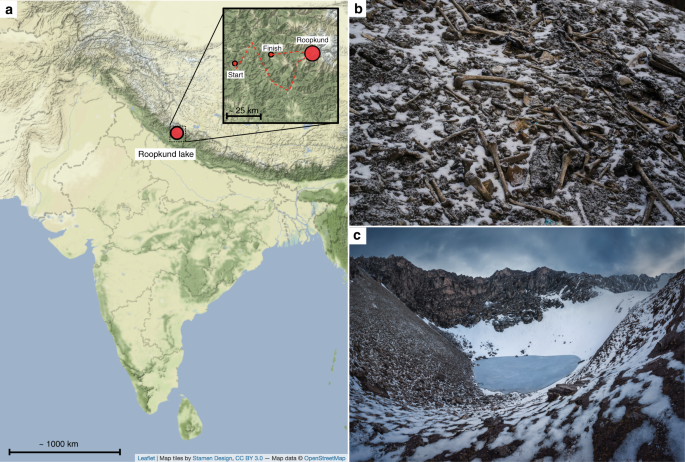
Context of Roopkund Lake. a Map showing the location of Roopkund Lake. The approximate route of the Nanda Devi Raj Jat pilgrimage relative to Roopkund Lake is shown in the inset. b Image of disarticulated skeletal elements scattered around the Roopkund Lake site. Photo by Himadri Sinha Roy. c Image of Roopkund Lake and surrounding mountains. Photo by Atish Waghwase
To shed light on the origin of the skeletons of Roopkund, we analyzed their remains using a series of bioarcheological analyses, including ancient DNA, stable isotope dietary reconstruction, radiocarbon dating, and osteological analysis. We find that the Roopkund skeletons belong to three genetically distinct groups that were deposited during multiple events, separated in time by approximately 1000 years. These findings refute previous suggestions that the skeletons of Roopkund Lake were deposited in a single catastrophic event.
Bioarcheological analysis of the Roopkund skeletons
We obtained genome-wide data from 38 individuals by extracting DNA from powder drilled from long bones, producing next-generation sequencing libraries, and enriching them for approximately 1.2 million single nucleotide polymorphisms (SNPs) from across the genome 6 , 7 , 8 , 9 , obtaining an average coverage of 0.51 × at targeted positions (Table 1 , Supplementary Data 1 ). We also obtained PCR-based mitochondrial haplogroup determinations for 71 individuals (35 of these were ones for whom we also obtained genome-wide data that confirmed the PCR-based determinations) (Table 2 , Supplementary Note 1 ). We generated stable isotope measurements (δ 13 C and δ 15 N) from 45 individuals, including 37 for whom we obtained genome-wide genetic data, and we obtained direct radiocarbon dates for 37 individuals for whom we also had both genetic and isotope data (Table 1 ).
In this study, we also present an osteological assessment of health and stature performed on a different set of bones from Roopkund; this report was drafted well before genetic results from Roopkund were available but was never formally published (an edited version of the original report is presented here as Supplementary Note 2 ). The analysis suggests that the Roopkund individuals were broadly healthy, but also identifies three individuals with unhealed compression fractures; the report hypothesizes that these injuries could have transpired during a violent hailstorm of the type that sometimes occurs in the vicinity of Roopkund Lake, while also recognizing that other scenarios are plausible. The report also identifies the presence of both very robust and tall individuals (outside the range of almost all South Asians), and more gracile individuals, and hypothesizes based on this the presence of at least two distinct groups of individuals, consistent with our genetic findings (Supplementary Note 2 ).
Our analysis of the genome-wide data from 38 Roopkund individuals shows that they include both genetic males ( n = 23) and females ( n = 15)—consistent with the physical anthropology evidence for the presence of both males and females (Supplementary Note 2 ). The relatively similar proportions of males and females is difficult to reconcile with the suggestion that these individuals might have been part of a military expedition. We detected no relative pairs (3rd degree or closer) among the sequenced individuals 10 , providing evidence against the idea that the Roopkund skeletons might represent the remains of groups of families. We also found no evidence that the individuals were infected with bacterial pathogens, providing no support for the suggestion that these individuals died in an epidemic, although we caution that failure to find evidence for pathogen DNA in long bone powder may simply reflect the fact that it was present at too low a concentration to detect (Supplementary Note 3 ) 11 .
Roopkund skeletons form three genetically distinct groups
We explored the genetic diversity of the 38 Roopkund individuals using a previously established Principal Component Analysis (PCA) that is effective at visualizing genetic variation of diverse present-day people from South Asia (a term we use to refer to the territories of the present day countries of India, Pakistan, Nepal, Bhutan, Bangladesh, and Bhutan) relative to West Eurasian-related groups (a term we use to refer to the cluster of ancestry types common in Europe, the Near East, and Iran) and East Asian-related groups (a term we apply to the cluster of ancestry types common in East Asia including China, Japan, Southeast Asia, and western Indonesia) 12 . We find that the Roopkund individuals cluster into three distinct groups, which we will henceforth refer to as Roopkund_A, Roopkund_B, and Roopkund_C (Fig. 2a ). Individuals in Roopkund_A ( n = 23) fall along a genetic gradient that includes most present-day South Asians. However, they do not fall in a tight cluster along this gradient, suggesting that they do not comprise a single endogamous group, and instead derive from a diversity of groups. Individuals belonging to the Roopkund_B cluster ( n = 14) do not fall along this gradient, and instead fall near present-day West Eurasians, suggesting that Roopkund_B individuals possess West Eurasian-related ancestry. A single individual, Roopkund_C, falls far from all other Roopkund individuals in the PCA, between the Onge (Andaman Islands) and Han Chinese, suggesting East Asian-related ancestry.
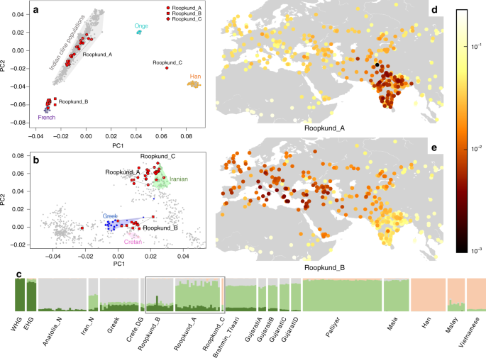
Genetic Structure of the Skeletons of Roopkund Lake. a Principal component analysis (PCA) of 1,453 present day individuals from selected groups throughout mainland South Asia (highlighted in gray). French individuals (representing the location where West Eurasian populations are known to cluster) are shown in purple, Chinese individuals are shown (representing the location where East Asian populations are known to cluster) in orange, and Andamanese individuals are shown in teal; the 38 Roopkund individuals are projected. b PCA of 988 present day West Eurasians with the Roopkund individuals projected. The PCA plot is truncated to remove Sardinians and southern Levantine groups; Present-day Greeks are shown in blue, Cretans in pink, Iranians in green, and all other West Eurasian populations in gray. A gray polygon encloses all the individuals in each Roopkund group with > 100,000 SNPs. c ADMIXTURE analysis of 2344 present-day and 1877 ancient individuals with K = 4 ancestral components. Only a subset of individuals with ancestries relevant to the interpretation of the Roopkund individuals are shown. Consistent with the PCA, Roopkund_A has ancestry most closely matching Indian groups; Roopkund_B has ancestry most closely matching Greek and Cretan groups; and Roopkund_C has ancestry most closely matching Southeast Asian groups. Genetic differentiation (F ST ) between Roopkund_A ( d ) and diverse present-day populations, and Roopkund_B ( e ) and diverse present-day populations. We only plotted present-day populations for which we have latitudes and longitudes; deeper red coloration indicates less differentiation to the Roopkund genetic cluster being analyzed. The plotted data are provided in a Source Data file
To further understand the West Eurasian-related affinity in the Roopkund_B cluster, we projected all the Roopkund individuals onto a second PCA designed to distinguish between sub-components of West Eurasian-related ancestry 13 , 14 (Fig. 2b ). Individuals assigned to the Roopkund_A and Roopkund_C groups cluster towards the top right of the PCA plot, close to present-day groups with Iranian ancestry, consistent with where populations with South Asian or East Asian ancestry cluster when projected onto such a plot 13 . Individuals belonging to the Roopkund_B group cluster toward the center of the plot, close to present-day people from mainland Greece and Crete 15 . We observe consistent patterns using the automated clustering software ADMIXTURE 16 (Fig. 2c ) and in pairwise F ST statistics (Fig. 2d, e , Supplementary Data 2 ). The visual evidence from the PCA suggests that two individuals from the Roopkund_B group might represent genetic outliers (Fig. 2b ). However, symmetry f 4 -statistics show that the two apparent outliers (one of which has relatively low coverage) are statistically indistinguishable in ancestry from individuals of the main Roopkund_B cluster relative to diverse comparison populations (Supplementary Data 3 ), and so we lump all the Roopkund_B individuals together in what follows.
Skeletons at Roopkund Lake were deposited in multiple events
The discovery of multiple, genetically distinct groups among the skeletons of Roopkund Lake raises the question of whether these individuals died simultaneously or during separate events. We used Accelerator Mass Spectrometry (AMS) radiocarbon dating to determine the age of the remains. We successfully generated radiocarbon dates from all but one of the individuals for which we have genetic data, using the same stocks of bone powder that we used for genetic analysis to ensure that the dates correspond directly to the genetic groupings. We find that the Roopkund_A and Roopkund_B groups are separated in time by ~1000 years, with the calibrated dates for individuals assigned to the Roopkund_A group ranging from the 7th–10th centuries CE, and the calibrated dates for individuals assigned to the Roopkund_B group ranging from the 17th–20th centuries CE (Table 1 ; Fig. 3a ; Supplementary Data 4 ). The single individual assigned to Roopkund_C also dates to this later period. These results demonstrate that the skeletons of Roopkund Lake perished in at least two separate events. For Roopkund_A, we detect non-overlapping 95% confidence intervals (for example individual I6943 dates to 675–769 CE, while individual I6941 dates to 894–985 CE), suggesting that even these individuals may not have died simultaneously (Fig. 3a ). In contrast, the calibrated dates obtained for 13 Roopkund_B individuals and the single Roopkund_C individual all have mutually overlapping 95% confidence intervals.
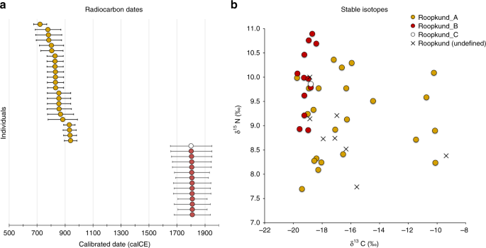
Radiocarbon and Isotopic Evidence of Distinct Origins of Roopkund Genetic Groups. a We generated 37 accelerator mass spectrometry radiocarbon dates and calibrated them using OxCal v4.3.2. The dating reveals that the individuals were deposited in at least two events ~1000 years apart. In fact, the Roopkund_A individuals (shown in yellow) may have been deposited over an extended period themselves, as the 95% confidence intervals for some of the radiocarbon dates (for example I6943 and I6941) do not overlap. Radiocarbon dates indicate that Roopkund_B (shown in red) and Roopkund_C (shown in white) individuals may have been deposited during a single event. Error bars indicate 95.4% confidence intervals. Calibration curves are shown in Supplementary Fig. 1 . b We show normalized δ 13 C and δ 15 N values for samples with isotopic data: 37 for which genetic data were generated (circles with colors indicating their cluster), and eight for which no genetic data were generated (labeled Roopkund_U). In cases where multiple measurements were obtained, we plot the average of all measurements. The plotted data are provided as a Source Data file
Differences in diet correlate with genetic groupings
We carried out carbon and nitrogen isotope analysis of femur bone collagen for 45 individuals. Femur bone collagen is determined by diet in the last 10–20 years of life 17 , and therefore is not necessarily correlated with the genetic ancestry of a population, which reflects processes occurring over generations. Nevertheless, we find evidence of dietary heterogeneity across the genetic ancestry groupings, providing additional support for the presence of multiple distinct groups at Roopkund Lake. We first observed that the Roopkund individuals are characterized by a range of δ 13 C values indicating diets reliant on both C 3 and C 4 plant sources, as well as δ 15 N values indicating varying degrees of consumption of protein derived from terrestrial animals (Fig. 3b and Supplementary Note 4 ). The δ 13 C values are non-randomly associated with the genetic groupings for the 37 individuals for whom we had both measurements. We find that all the Roopkund_B individuals (with typically eastern Mediterranean ancestry), as well as the Roopkund_C individual, have δ 13 C values between −19.7‰ and −18.2‰ reflecting consumption of terrestrial C 3 plants, such as wheat, barley, and rice (and/or animals foddered on such plants). In contrast, the Roopkund_A individuals (with typically South Asian ancestry) have much more varied δ 13 C values (−18.9‰ to −10.1‰), with some implying C 3 plant reliance and others reflecting either a mixed C 3 and C 4 derived diet, or alternatively consumption of C 3 plants along with animals foddered with millet, a C 4 plant (a practice that has been documented ethnographically in South Asia 17 ). The difference in the δ 13 C distribution between the Roopkund_A and Roopkund_B groupings is highly significant (p = 0.00022 from a two-sided Mann-Whitney test).
Genetic affinities of the Roopkund subgroups
We used qpWave 18 , 19 to test whether Roopkund_B is consistent with forming a genetic clade with any present-day population (that is, whether it is possible to model the two populations as descending entirely from the same ancestral population with no mixture with other groups since their split). We selected 26 present-day populations for comparison, with particular emphasis on West Eurasian-related groups (we analyzed the West Eurasian-related groups Basque, Crete, Cypriot, Egyptian, English, Estonian, Finnish, French, Georgian, German, Greek, Hungarian, Italian_North, Italian_South, Norwegian, Spanish, Syrian, Ukranian, and the non-West-Eurasian-related groups Brahmin_Tiwari, Chukchi, Han, Karitiana, Mala, Mbuti, Onge, and Papuan). We find that Roopkund_B is consistent with forming a genetic clade only with individuals from present-day Crete. These results by no means imply that the Roopkund_B individuals originated in the island of Crete itself, although they suggest that their recent ancestors or they themselves came from a nearby region (Supplementary Note 5 ; Supplementary Data 5 ).
We performed a similar analysis on individuals belonging to the Roopkund_A group and find that they cannot be modeled as deriving from a homogeneous group (Supplementary Note 6 ). Instead, Roopkund_A individuals vary significantly in their relationship to a diverse set of present-day South Asians, consistent with the heterogeneity evident in PCA (Fig. 2a ). We were unable to model the Roopkund_C individual as a genetic clade with any present-day populations, but we were able to model its ancestry as ~82% Malay-related and ~18% Vietnamese-related using qpAdm 7 , showing that this individual is consistent with being of Southeast Asian origin. We tested if any of the Roopkund groups show specific genetic affinity to present-day groups from the Himalayan region, including four neighboring villages in the northern Ladakh region for which we report new genome-wide sequence data, but we find no such evidence (Supplementary Note 7 ). Within the Roopkund_A group which has ancestry that falls within the variation of present-day South Asians, we observe a weakly significant difference in the proportion of West Eurasian-related ancestry in males and females ( p = 0.015 by a permutation test across individuals; Supplementary Note 8 ), with systematically lower proportions of West Eurasian-related ancestry in males than females. This suggests that the males and females were drawn from significantly different mixtures of groups within South Asia.
The genetically, temporally, and isotopically heterogeneous composition of the groups at Roopkund Lake was unanticipated from the context in which the skeletons were found. Radiocarbon dating reveals at least two key phases of deposition of human remains separated by around one thousand years and with significant heterogeneity in the dates for the earlier individuals indicating that they could not all have died in a single catastrophic event.
Combining multiple lines of evidence, we suggest a possible explanation for the origin of at least some of the Roopkund_A individuals. Roopkund Lake is not situated on any major trade route, but it is on a present-day pilgrimage route—the Nanda Devi Raj Jat pilgrimage which today occurs every 12 years (Fig. 1a ). As part of the event, pilgrims gather for worship and celebration along the route. Reliable descriptions of the pilgrimage ritual do not appear until the late-19 th century, but inscriptions in nearby temples dating to between the 8 th and 10 th centuries suggest potential earlier origins 20 . We view the hypothesis of a mass death during a pilgrimage event as a plausible explanation for at least some of the individuals in the Roopkund_A cluster.
The Roopkund_B cluster is more puzzling. It is tempting to hypothesize that the Roopkund_B individuals descend from Indo-Greek populations established after the time of Alexander the Great, who may have contributed ancestry to some present-day groups like the Kalash 21 . However, this is unlikely, as such a group would be expected to have admixture with groups with more typical South Asian ancestry (as the Kalash do), or would be expected to be inbred and to have relatively low genetic diversity. However, the Roopkund_B individuals have evidence for neither pattern (Supplementary Note 9 ). Combining different lines of evidence, the data suggest instead that what we have sampled is a group of unrelated men and women who were born in the eastern Mediterranean during the period of Ottoman political control. As suggested by their consumption of a predominantly terrestrial, rather than marine-based diet, they may have lived in an inland location, eventually traveling to and dying in the Himalayas. Whether they were participating in a pilgrimage, or were drawn to Roopkund Lake for other reasons, is a mystery. It would be surprising for a Hindu pilgrimage to be practiced by a large group of travelers from the eastern Mediterranean where Hindu practices have not been common; Hindu practice in this time might be more plausible for a southeast Asian individual with an ancestry type like that seen in the Roopkund_C individual. Given that the Roopkund_B and Roopkund_C individuals died only in the last few centuries, an important direction for future investigation will be to carry out archival research to determine if there were reports of large foreign traveling parties dying in the region over the last few hundred years.
Taken together, these results have produced meaningful insights about an enigmatic ancient site. More generally, this study highlights the power of biomolecular analyses to obtain rich information about the human story behind archaeological deposits that are so highly disturbed that traditional archaeological methods are not as informative.
The genetic analysis of Himalayan populations (described in Supplementary Note 7 ) was approved by the Institutional Ethical Committee of the Centre for Cellular and Molecular Biology in Hyderabad, India.
Ancient DNA laboratory Work
A total of 76 skeletal samples (72 long bones and four teeth) were sampled at the Anthropological Survey of India, Kolkata. Skeletal sampling was performed for all samples in dedicated ancient DNA facilities at the Centre for Cellular and Molecular Biology (CCMB) in Hyderabad, India. A subset of samples that underwent preliminary ancient DNA screening at CCMB, including three samples that did not yield sufficient data to assign mitochondrial DNA haplogroups during preliminary screening (see Supplementary Note 1 ), were further processed at Harvard Medical School, Boston, USA, consistent with recommendations in the ancient DNA literature for repeating analyses in two independent laboratories to increase confidence in results 22 .
At CCMB, samples were prepared for processing by wiping with a bleach solution, followed by deionized water. The samples were then subjected to UV irradiation for 30 min on each side to minimize surface DNA contamination. Bone powder was then produced using a sterile dentistry drill.
We successfully generated genome-wide DNA for 38 individuals (Supplementary Data 1 ). For each sample, approximately 75 mg of bone powder originally prepared at CCMB was further processed in dedicated ancient DNA clean rooms at Harvard Medical School using standard protocols, including DNA extraction optimized for ancient DNA recovery 23 , modified by replacing the Zymo extender/MinElute column assemblage with a preassembled spin column device 24 , followed by library preparation with partial UDG treatment 25 . The quality of authentic ancient DNA preservation in each sample was assessed by carrying out a preliminary screening of all libraries via targeted DNA enrichment, designed to capture mitochondrial DNA in addition to 50 nuclear targets 26 . We sequenced the enriched libraries on an Illumina NextSeq500 instrument for 2 × 76 cycles with an additional 2 × 7 cycles for identification of indices. Based on this preliminary assessment, libraries that were deemed promising underwent a further enrichment using a reagent that targeted ~1.2 million SNPs 6 , 7 , 8 , 9 , and then were sequenced using an Illumina NextSeq500 instrument.
Bioinformatic processing
We used SeqPrep to trim adapters and molecular barcodes, and then merged paired-end reads that overlapped by a minimum of 15 base pairs (with up to one mismatch allowed) and aligned to the mitochondrial rsrs genome 27 (for the mitochondrial screening analysis) or hg19 (for whole-genome analysis) using samse in bwa (v0.6.1) 28 . We identified duplicate sequences based on having the same start position, end position, orientation, and library-specific barcode, and only retained the copy with the highest quality sequence. We restricted to sequences with a minimum mapping quality (MAPQ ≥ 10) and minimum base quality (≥20) after excluding two bases from each end of the sequence. We obtained pseudo-haploid SNP calls by using a single randomly chosen sequence at SNPs covered by at least one sequence.
We subjected the resulting data to three tests of ancient DNA authenticity: (1) we analyzed the mitochondrial genome data to determine the rate of matching to the consensus sequence using contamMix, and excluded from analysis samples that exhibited a match rate less than 97% 8 . (2) We removed samples that exhibited a rate of C-to-T substitutions less than 3%: the minimum recommended threshold for authentic ancient DNA that has been subjected to partial UDG treatment 25 . (3) We used ANGSD 29 to determine the degree of heterogeneity on the X-chromosome in males (who should only have one X chromosome) and excluded from analysis individuals with contamination rates greater than 1.5%.
We determined the mitochondrial haplogroup of each individual in two ways. For individuals with whole mitochondrial genome data, we determined the mitochondrial haplogroups using haplogrep2 30 . We also determined mitochondrial haplogroups from mitochondrial DNA genotyping using multiplex PCR (see Supplementary Note 1 ).
We determined the genetic sex of the individuals by computing the ratio of the number of sequences that align to the X chromosome versus the Y chromosome. We searched for 1st, 2nd, and 3rd degree relative pairs in the dataset by analyzing patterns of allele sharing between pairs of individuals (we found none) 10 .
To identify Y-chromosome haplogroups in genetically male individuals, we used a modified version of the procedure reported in Poznik, et al. 31 , which performs a breadth-first search of the Y-chromosome tree. We made Y chromosome haplogroup calls using the ISOGG tree from 04.01.2016 [ http://isogg.org ], and recorded the derived and ancestral allele calls for each informative position on the tree. We counted the number of mismatches in the observed derived alleles on each branch of the tree and used this information to assign a score to each haplogroup, accounting for damage by down-weighting derived mutations that are the result of transitions to 1/3 of that of transversions. We assigned the closest matching Y-chromosome reference haplogroup to each male based on this score (Supplementary Data 6 ). We caution that males with fewer than 100,000 SNPs have too little data to confidently assign a haplogroup.
Population genetic analyses
We report data for 38 samples that passed contamination and quality control tests, with an average coverage of 0.51 × [range: 0.026–1.547] and 350088 SNPS covered at least once [range 30592–728448]. We processed the data in conjunction with published DNA obtained from ancient 6 , 9 , 13 , 14 , 15 , 32 , 33 , 34 , 35 , 36 , 37 , 38 , 39 , 40 , 41 , 42 , 43 , 44 , 45 , 46 , 47 , 48 , 49 , 50 , 51 , 52 , 53 , 54 , 55 , 56 , 57 , 58 , 59 , 60 , 61 and present-day groups from throughout the world 62 , 63 , 64 , 65 , 66 , 67 , 68 , including ~175 modern groups from the Indian subcontinent 12 . The resulting merged dataset included 1521 ancient and 7985 present-day individuals at 591,304 SNPs.
We used smartpca 69 to perform principal component analysis (PCA) using default parameters, with the settings lsqproject:YES and numoutlier:0. We projected the Roopkund individuals onto two PCA plots designed either to reveal a cline of West Eurasian-related ancestry in South Asian populations 18 , or to reveal the genetic substructure in present-day West Eurasians 13 . The first PCA (Fig. 2a ) included 1453 present-day populations 12 in addition to the Roopkund individuals, while the second PCA (Fig. 2b ) included 986 present-day populations 13 , in addition to the Roopkund individuals and two individuals from present-day Crete (population label Crete.DG). The PCA plots show that the samples cluster into three distinct groups, which we label Roopkund_A, Roopkund_B and Roopkund_C, and treat separately for subsequent analyses.
We used smartpca 69 to compute F ST between the two major Roopkund groups (Roopkund_A and Roopkund_B) and all other groups composed of at least 2 individuals in the dataset, using default parameters, with the settings inbreed:YES and fstonly:YES.
We performed clustering using ADMIXTURE 16 . We carried out this analysis on all samples used for the PCA analyses, although we display only selected populations for the sake of clarity. Prior to analysis, SNPs in linkage disequilibrium with one another were pruned in PLINK using the parameters–indep-pairwise 200 25 0.4. We performed an ADMIXTURE analysis on the remaining 344,363 SNPs in the pruned dataset for values of k between 2 and 10, and carried out 20 replicates at each value of k . We retained the highest likelihood replicate at each k and displayed results for k (k = 4), which we chose because we observed that it is most visually helpful for discriminating the ancestry of the groups of interest.
We used qpWave 18 , 19 , with default parameters and allsnps:YES, to determine if any of the Roopkund populations was consistent with being a clade with any present-day populations. We included a base set of nine populations in each test, chosen to represent diverse ancestry from throughout the world. We include an additional 5–15 populations of either South Asian, West Eurasian, or Southeast/East Asian ancestry in tests involving Roopkund_A, Roopkund_B and Roopkund_C respectively, chosen to provide additional resolution for each group based on their position in the previous PCA. Based on the observed genetic heterogeneity in the Roopkund_A population, we modeled each individual separately (Supplementary Note 6 ). For each test, the Left population set included the Roopkund population or individual of interest in addition to one of the selected present-day analysis populations, while the remaining populations were included in the Right population set. In the case of individuals belonging to the Roopkund_A and Roopkund_C groups, we also used qpAdm 7 , with default parameters and allsnps: YES, to determine whether these populations could be considered to be the product of a two-way admixture between any of the selected present-day populations (Supplementary Note 6 ). In this case, the Left population set included the Roopkund individual of interest in addition to all possible combinations of two of the selected present-day analysis populations, while the remaining populations were included in the Right population set.
AMS radiocarbon dating
We subjected bone powder from 37 samples to radiocarbon dating. We dated the remaining bone powder (360–750 mg) from the same samples that were processed for ancient DNA. We were unable to generate a radiocarbon date for individual I3401, as there was not enough remaining bone powder for analysis.
At the Pennsylvania State University AMS radiocarbon dating facility, bone collagen for 14 C and stable isotope analyses was extracted and purified using a modified Longin method with ultrafiltration 70 . Samples (200–400 mg) were demineralized for 24–36 h in 0.5 N HCl at 5 °C followed by a brief (<1 h) alkali bath in 0.1 N NaOH at room temperature to remove humates. The residue was rinsed to neutrality in multiple changes of Nanopure H 2 O, and then gelatinized for 12 h at 60 °C in 0.01 N HCl. The resulting gelatin was lyophilized and weighed to determine percent yield as a first evaluation of the degree of bone collagen preservation. Rehydrated gelatin solution was pipetted into pre-cleaned Centriprep 71 ultrafilters (retaining >30 kDa molecular weight gelatin) and centrifuged 3 times for 20 min, diluted with Nanopure H 2 O and centrifuged 3 more times for 20 min to desalt the solution.
In some instances, collagen samples were too poorly preserved and were pre-treated at Penn State using a modified XAD process 72 (Supplementary Data 4 shows that there were no systematic differences in the dates obtained based on the XAD and modified Longin pretreatment extraction methods.) Samples were demineralized in 0.5 N HCl for 2–3 days at 5 °C. The demineralized collagen pseudomorph was gelatinized at 60 °C in 1–2 mL 0.01 N HCl for 8–10 h. The gelatin was then lyophilized and percent gelatinization and yield determined by weight. The sample gelatin was then hydrolyzed in 2 mL 6 N HCl for 24 h at 110 °C. Supelco ENVI-Chrom® SPE (Solid Phase Extraction; Sigma-Aldrich) columns were prepped with 2 washes of methanol (2 mL) and rinsed with 10 mL DI H 2 O. Supelco ENVIChrom® SPE (Solid Phase Extraction; Sigma-Aldrich) columns with 0.45 µm Millex Durapore filters attached were equilibrated with 50 mL 6 N HCl and the washings discarded. 2 mL collagen hydrolyzate as HCl was pipetted onto the SPE column and driven with an additional 10 mL 6 N HCl dropwise with the syringe into a 20 mm culture tube. The hydrolyzate was finally dried into a viscous syrup by passing UHP N 2 gas over the sample heated at 50 °C for ~12 h.
For all bone samples that were subject to radiocarbon dating, carbon and nitrogen concentrations and stable isotope ratios of the ultrafiltered gelatin or XAD amino acid hydrolyzate were measured at the Yale Analytical and Stable Isotope Center with a Costech elemental analyzer (ECS 4010) and Thermo DeltaPlus analyzer. Sample quality was evaluated by percentage crude gelatin yield, %C, %N, and C/N ratios before AMS 14 C dating. C/N ratios for all samples fell between 2.9 and 3.6, indicating good collagen preservation 73 . Samples (~2.1 mg) were then combusted for 3 h at 900 °C in vacuum-sealed quartz tubes with CuO and Ag wires. Sample CO 2 was reduced to graphite at 550 °C using H 2 and a Fe catalyst, with reaction water drawn off with Mg(ClO 4 ) 2 74 .
Graphite samples were pressed into targets in Al boats and loaded on a target wheel with OX-1 (oxalic acid) standards, known-age bone secondaries, and a 14 C-free Pleistocene whale blank. 14 C measurements were performed at UCIAMS on a modified National Electronics Corporation compact spectrometer with a 0.5 MV accelerator (NEC 1.5SDH-1). The 14 C ages were corrected for mass-dependent fractionation with δ 13 C values 75 and compared with samples of Pleistocene whale bone (backgrounds, 48,000 14 C BP), late Holocene bison bone (~1850 14 C BP), late 1800s CE cow bone and OX-2 oxalic acid standards for calibration. All calibrated 14 C ages were computed using OxCal version 4.3 76 using the IntCal13 northern hemisphere curve 77 .
Stable isotope measurements
The isotopic measurement procedure at Yale University for the 37 samples for which we performed direct radiocarbon dating are described in the previous section.
We also obtained isotopic measurements for long bone samples from 19 individuals (including data from 11 of the same individuals that were also analyzed at Yale) at the Max Planck Institute for the Science of Human History. Bone samples of 1 g were subsequently cleaned using an air abrasive system with 5 μm aluminum oxide powder and then crushed into chunks. Collagen was extracted following standard procedures 78 . Approximately 1 g of pre-cleaned bone was demineralized in 10 mL aliquots of 0.5 M HCl at 4 °C, with changes of acid until CO 2 stopped evolving. The residue was then rinsed three times in deionized water before being gelatinized in pH 3 HCl at 80 °C for 48 h. The resulting solution was filtered, with the supernatant then freeze-dried over a period of 24 h.
Purified collagen samples (1 mg) were analyzed at the Department of Archaeology, Max Planck Institute for the Science of Human History, in duplicate by EA-IRMS on a ThermoFisher Elemental Analyzer coupled to a ThermoFisher Delta V Advantage Mass Spectrometer via a ConFloIV system. Accuracy was determined by measurements of international standard reference materials within each analytical run. These were USGS 40,40 δ 13 C raw = −26.4 ± 0.1, δ 13 C true = −26.4 ± 0.0, δ 15 N raw = −4.4 ± 0.1, δ 15 N true = −4.5 ± 0.2; IAEA N2, δ 15 N raw = 20.2 ± 0.1, δ 15 N true = 20.3 ± 0.2; IAEA C6 δ 13 C raw = -10.9 ± 0.1, δ 13 C true = −10.8 ± 0.0. An in-house fish gelatin sample was also used as a standard in each run. Reported δ 13 C values were measured against Vienna Pee Dee Belemnite (VPDB), while δ 15 N values are measured against ambient air.
Reporting summary
Further information on research design is available in the Nature Research Reporting Summary linked to this article.
Data availability
The aligned DNA sequences from the 38 individuals are available from the European Nucleotide Archive under accession number PRJEB29537 . Genotype files are available at https://reich.hms.harvard.edu/datasets . All other relevant data is available upon request.
Bengtsson, L. Ice covered lakes in Encyclopedia of Lakes and Reservoirs . 357–360 (Springer, Dordrecht, 2012).
Chapter Google Scholar
Pham, B. T., Pradhan, B., Bui, D. T., Prakash, I. & Dholakia, M. A comparative study of different machine learning methods for landslide susceptibility assessment: a case study of Uttarakhand area (India). Environ. Model Softw. 84 , 240–250 (2016).
Article Google Scholar
Gupta, S. Tourism in Garhwal Himalaya: Strategy for sustainable development in Domestic tourism in India. 199–218 (Indus Publishing Company, New Delhi, 1998).
Budhwar, K. Where gods dwell: central Himalayan folktales and legends . 19–27 (Penguin Books India, 2010).
“Skeleton Lake”. Riddles of the Dead. Television. (National Geographic, Hoggard Films, 2004).
Fu, Q. et al. An early modern human from Romania with a recent Neanderthal ancestor. Nature 524 , 216–219 (2015).
Article ADS CAS Google Scholar
Haak, W. et al. Massive migration from the steppe was a source for Indo-European languages in Europe. Nature 522 , 207–211 (2015).
Fu, Q. et al. DNA analysis of an early modern human from Tianyuan Cave, China. Proc. Natl Acad. Sci. USA 110 , 2223–2227 (2013).
Mathieson, I. et al. Genome-wide patterns of selection in 230 ancient Eurasians. Nature 528 , 499–512 (2015).
Kuhn, J. M. M., Jakobsson, M. & Günther, T. Estimating genetic kin relationships in prehistoric populations. PLoS ONE 13 , e0195491 (2018).
Herbig, A. et al. MALT: Fast alignment and analysis of metagenomic DNA sequence data applied to the Tyrolean Iceman. Preprint at https://www.biorxiv.org/content/10.1101/050559v050551 (2016).
Nakatsuka, N. et al. The promise of discovering population-specific disease-associated genes in South Asia. Nat. Genet. 49 , 1403–1407 (2017).
Article CAS Google Scholar
Lazaridis, I. et al. Genomic insights into the origin of farming in the ancient Near East. Nature 536 , 419–424 (2016).
Broushaki, F. et al. Early Neolithic genomes from the eastern Fertile Crescent. Science 353 , 499–503 (2016).
Lazaridis, I. et al. Genetic origins of the Minoans and Mycenaeans. Nature 548 , 214–218 (2017).
Alexander, D. H., Novembre, J. & Lange, K. Fast model-based estimation of ancestry in unrelated individuals. Genome Res. 19 , 1655–1664 (2009).
Hedges, R. E., Clement, J. G., Thomas, C. D. L. & O’Connell, T. C. Collagen turnover in the adult femoral mid‐shaft: Modeled from anthropogenic radiocarbon tracer measurements. Am. J. Phys. Anthropol. 133 , 808–816 (2007).
Moorjani, P. et al. Genetic evidence for recent population mixture in India. Am. J. Hum. Genet. 93 , 422–438 (2013).
Reich, D. et al. Reconstructing native American population history. Nature 488 , 370–374 (2012).
Sax, W. From Procession to Heritage: The Royal Procession of the Goddess Shri Nanda in Prozessionen, Wallfahrten, Aufmärsche : Bewegung zwischen Religion und Politik in Europa und Asien seit dem Mittelalter 4 , 277–287 (Böhlau Verlag, Köln, 2008).
Hellenthal, G. et al. A genetic atlas of human admixture history. Science 343 , 747–751 (2014).
Cooper, A. & Poinar, H. N. Ancient DNA: do it right or not at all. Science 289 , 1139–1139 (2000).
Dabney, J. et al. Complete mitochondrial genome sequence of a Middle Pleistocene cave bear reconstructed from ultrashort DNA fragments. Proc. Natl Acad. Sci. USA 110 , 15758–15763 (2013).
Korlević, P. et al. Reducing microbial and human contamination in DNA extractions from ancient bones and teeth. Biotechniques 59 , 87–93 (2015).
Rohland, N., Harney, E., Mallick, S., Nordenfelt, S. & Reich, D. Partial uracil–DNA–glycosylase treatment for screening of ancient. Dna. Philos. Trans. R. Soc. B 370 , 20130624 (2015).
Maricic, T., Whitten, M. & Pääbo, S. Multiplexed DNA sequence capture of mitochondrial genomes using PCR products. PLoS ONE 5 , e14004 (2010).
Article ADS Google Scholar
Behar, D. M. et al. A “Copernican” reassessment of the human mitochondrial DNA tree from its root. Am. J. Hum. Genet. 90 , 675–684 (2012).
Li, H. & Durbin, R. Fast and accurate short read alignment with Burrows–Wheeler transform. Bioinformatics 25 , 1754–1760 (2009).
Korneliussen, T. S., Albrechtsen, A. & Nielsen, R. ANGSD: analysis of next generation sequencing data. BMC Bioinforma. 15 , 356 (2014).
Weissensteiner, H. et al. HaploGrep 2: mitochondrial haplogroup classification in the era of high-throughput sequencing. Nucleic Acids Res. 44 , W58–W63 (2016).
Poznik, G. D. et al. Punctuated bursts in human male demography inferred from 1,244 worldwide Y-chromosome sequences. Nat. Genet. 48 , 593 (2016).
Allentoft, M. E. et al. Population genomics of bronze age Eurasia. Nature 522 , 167–172 (2015).
de Barros Damgaard, P. et al. 137 ancient human genomes from across the Eurasian steppes. Nature 557 , 369 (2018).
de Barros Damgaard, P. et al. The first horse herders and the impact of early Bronze Age steppe expansions into Asia. Science 360 , eaar7711 (2018).
Fu, Q. et al. Genome sequence of a 45,000-year-old modern human from western Siberia. Nature 514 , 445–449 (2014).
Fu, Q. et al. The genetic history of ice age Europe. Nature 534 , 200 (2016).
Haber, M. et al. Continuity and admixture in the last five millennia of Levantine history from ancient Canaanite and present-day Lebanese genome sequences. Am. J. Hum. Genet. 101 , 274–282 (2017).
Jeong, C. et al. Long-term genetic stability and a high-altitude East Asian origin for the peoples of the high valleys of the Himalayan arc. Proc. Natl Acad. Sci USA 113 , 7485–7490 (2016).
Jones, E. R. et al. Upper Palaeolithic genomes reveal deep roots of modern Eurasians. Nat. Commun. 6 , 8912 (2015).
Jones, E. R. et al. The Neolithic transition in the Baltic was not driven by admixture with early European farmers. Curr. Biol. 27 , 576–582 (2017).
Lazaridis, I. et al. Ancient human genomes suggest three ancestral populations for present-day Europeans. Nature 513 , 409–413 (2014).
Lipson, M. et al. Parallel palaeogenomic transects reveal complex genetic history of early European farmers. Nature 551 , 368 (2017).
Llorente, M. G. et al. Ancient Ethiopian genome reveals extensive Eurasian admixture in Eastern Africa. Science 350 , 820–822 (2015).
Malaspinas, A.-S. et al. Two ancient human genomes reveal Polynesian ancestry among the indigenous Botocudos of Brazil. Curr. Biol. 24 , R1035–R1037 (2014).
Martiniano, R. et al. Genomic signals of migration and continuity in Britain before the Anglo-Saxons. Nat. Commun. 7 , 10326 (2016).
Mathieson, I. et al. The genomic history of southeastern Europe. Nature 555 , 197 (2018).
Meyer, M. et al. A high-coverage genome sequence from an archaic Denisovan individual. Science 338 , 222–226 (2012).
Mittnik, A. et al. The genetic prehistory of the Baltic Sea region. Nat. Commun. 9 , 442 (2018).
Olalde, I. et al. Derived immune and ancestral pigmentation alleles in a 7,000-year-old Mesolithic European. Nature 507 , 225 (2014).
Olalde, I. et al. The Beaker phenomenon and the genomic transformation of northwest Europe. Nature 555 , 190 (2018).
Prüfer, K. et al. The complete genome sequence of a Neanderthal from the Altai Mountains. Nature 505 , 43 (2014).
Raghavan, M. et al. Upper Palaeolithic Siberian genome reveals dual ancestry of Native Americans. Nature 505 , 87 (2014).
Raghavan, M. et al. Genomic evidence for the Pleistocene and recent population history of Native Americans. Science 349 , aab3884 (2015).
Rasmussen, S. et al. Early divergent strains of Yersinia pestis in Eurasia 5,000 years ago. Cell 163 , 571–582 (2015).
Schiffels, S. et al. Iron age and Anglo-Saxon genomes from East England reveal British migration history. Nat. Commun. 7 , 10408 (2016).
Schuenemann, V. J. et al. Ancient Egyptian mummy genomes suggest an increase of Sub-Saharan African ancestry in post-Roman periods. Nat. Commun. 8 , 15694 (2017).
Skoglund, P. et al. Genetic evidence for two founding populations of the Americas. Nature 525 , 104 (2015).
Skoglund, P. et al. Genomic insights into the peopling of the Southwest Pacific. Nature 538 , 510 (2016).
Skoglund, P. et al. Reconstructing prehistoric African population structure. Cell 171 , 59–71. e21 (2017).
Veeramah, K. R. et al. Population genomic analysis of elongated skulls reveals extensive female-biased immigration in Early Medieval Bavaria. Proc. Natl Acad. Sci. USA 115 , 3494–3499 (2018).
Harney, É. et al. Ancient DNA from Chalcolithic Israel reveals the role of population mixture in cultural transformation. Nat. Commun. 9 , 3336 (2018).
1000 Genomes Project Consortium. A global reference for human genetic variation. Nature 526 , 68 (2015).
Mallick, S. et al. The Simons genome diversity project: 300 genomes from 142 diverse populations. Nature 538 , 201 (2016).
Mondal, M. et al. Genomic analysis of Andamanese provides insights into ancient human migration into Asia and adaptation. Nat. Genet. 48 , 1066–1070 (2016).
Patterson, N. et al. Ancient admixture in human history. Genetics 192 , 1065–1093 (2012).
Pickrell, J. K. et al. The genetic prehistory of southern Africa. Nat. Commun. 3 , 1143 (2012).
Qin, P. & Stoneking, M. Denisovan ancestry in East Eurasian and native American populations. Mol. Biol. Evol. 32 , 2665–2674 (2015).
Vyas, D. N., Al‐Meeri, A. & Mulligan, C. J. Testing support for the northern and southern dispersal routes out of Africa: an analysis of Levantine and southern Arabian populations. Am. J. Phys. Anthropol. 164 , 736–749 (2017).
Patterson, N., Price, A. L. & Reich, D. Population structure and eigenanalysis. PLoS Genet. 2 , e190 (2006).
Kennett, D. J. et al. Archaeogenomic evidence reveals prehistoric matrilineal dynasty. Nat. Commun. 8 , 14115 (2017).
McClure, S. B., Puchol, O. G. & Culleton, B. J. AMS dating of human bone from Cova de la Pastora: new evidence of ritual continuity in the prehistory of eastern Spain. Radiocarbon 52 , 25–32 (2010).
Lohse, J. C., Culleton, B. J., Black, S. L. & Kennett, D. J. A precise chronology of middle to late Holocene Bison exploitation in the Far Southern Great Plains. J. Tex. Archeol. Hist. 1 , 94–126 (2014).
Google Scholar
Van Klinken, G. J. Bone collagen quality indicators for palaeodietary and radiocarbon measurements. J. Archaeol. Sci. 26 , 687–695 (1999).
Santos, G. M., Southon, J. R., Druffel-Rodriguez, K. C., Griffin, S. & Mazon, M. Magnesium perchlorate as an alternative water trap in AMS graphite sample preparation: a report on sample preparation at KCCAMS at the University of California, Irvine. Radiocarbon 46 , 165–173 (2004).
Stuiver, M. & Polach, H. A. Discussion reporting of 14 C data. Radiocarbon 19 , 355–363 (1977).
Ramsey, C. B. & Lee, S. Recent and planned developments of the program OxCal. Radiocarbon 55 , 720–730 (2013).
Reimer, P. J. et al. IntCal13 and Marine13 radiocarbon age calibration curves 0–50,000 years cal BP. Radiocarbon 55 , 1869–1887 (2013).
Richards, M. P. & Hedges, R. E. Stable isotope evidence for similarities in the types of marine foods used by Late Mesolithic humans at sites along the Atlantic coast of Europe. J. Archaeol. Sci. 26 , 717–722 (1999).
Download references
Acknowledgements
We acknowledge the people living and dead whose samples we analyzed in this study. We thank Professor Subhash Walimbe of the Deccan College Post-Graduate and Research Institute in Pune India (retired) who prepared the physical anthropology report on the Roopkund skeletons that was used as the basis of a National Geographic documentary; we reprint it in edited form in Supplementary Note 2 , updated in light of the genetic findings. We are grateful to Dr. Lalji Singh (deceased) for his longstanding support for this project, to the Lucknow University anthropology department and the Anthroplogical Survey of India for permission to analyze their skeletal collection, and to Iosif Lazaridis, Michael McCormick, Arie Shaus, John Wakeley for critical comments. E.H. was supported by a graduate student fellowship from the Max Planck-Harvard Research Center for the Archaeoscience of the Ancient Mediterranean (MHAAM). A.N., P.R., and N.B. are funded by the Max Planck Society. D.J.K. and B.J.C. were supported by NSF BCS-1460367. K.T. was supported by grant NCP (MLP0117) from the Council of Scientific and Industrial Research (CSIR), Government of India. D.R. was supported by the U.S. National Science Foundation HOMINID grant BCS-1032255, the U.S. National Institutes of Health grant GM100233, by an Allen Discovery Center grant, by grant 61220 from the John Templeton Foundation, and is an investigator of the Howard Hughes Medical Institute.
Author information
These authors jointly directed this work: Ayushi Nayak, Douglas J. Kennett, Kumarasamy Thangaraj, David Reich, Niraj Rai.
Authors and Affiliations
Department of Organismic and Evolutionary Biology, Harvard University, Cambridge, MA, 02138, USA
Éadaoin Harney
The Max Planck-Harvard Research Center for the Archaeoscience of the Ancient Mediterranean, Cambridge, MA, 02138, USA
Éadaoin Harney & David Reich
Department of Genetics, Harvard Medical School, Boston, MA, 02115, USA
Éadaoin Harney, Swapan Mallick, Nadin Rohland, Jakob Sedig, Nicole Adamski, Rebecca Bernardos, Nasreen Broomandkhoshbacht, Matthew Ferry, Megan Michel, Jonas Oppenheimer, Kristin Stewardson, Zhao Zhang & David Reich
Department of Archaeology, Max Planck Institute for the Science of Human History, D-07745, Jena, Germany
Ayushi Nayak, Patrick Roberts & Nicole Boivin
Broad Institute of Harvard and MIT, Cambridge, MA, 02142 USA, USA
Nick Patterson, Swapan Mallick & David Reich
Department of Human Evolutionary Biology, Harvard University, Cambridge, MA, 02138, USA
Nick Patterson
Deccan College, Pune, 411006, India
Pramod Joglekar & Veena Mushrif-Tripathy
Howard Hughes Medical Institute, Harvard Medical School, Boston, MA, 02115, USA
Swapan Mallick, Nicole Adamski, Nasreen Broomandkhoshbacht, Matthew Ferry, Megan Michel, Jonas Oppenheimer, Kristin Stewardson & David Reich
Institutes of Energy and the Environment, The Pennsylvania State University, University Park, PA, 16802, USA
Brendan J. Culleton
Department of Anthropology, The Pennsylvania State University, University Park, PA, 16802, USA
Brendan J. Culleton & Thomas K. Harper
The Max Planck-Harvard Research Center for the Archaeoscience of the Ancient Mediterranean, D-07745, Jena, Germany
Megan Michel
Anthropological Survey of India, North West Regional Centre, Dehradun, 248195, India
Harashawaradhana & Maanwendra Singh Bartwal
CSIR Centre for Cellular and Molecular Biology, Hyderabad, Telangana, 500007, India
Sachin Kumar, Kumarasamy Thangaraj & Niraj Rai
Birbal Sahni Institute of Palaeosciences, Lucknow, Uttar Pradesh, 226007, India
Sachin Kumar & Niraj Rai
Gautam Budh Health Care Foundation, Noida, Uttar Pradesh, 201301, India
Subhash Chandra Diyundi
Department of Anthropology, University of California, Santa Barbara, CA, 93106, USA
Douglas J. Kennett
You can also search for this author in PubMed Google Scholar
Contributions
N.B., K.T., D.R., and N.Ra. conceived the study. N.Ro., D.R., and K.T. supervised the ancient DNA work and analysis of DNA from modern population samples. P.J., H., M.S.B., and V.M.-T. collected the archaeological samples. S.C.D. and S.K. collected blood samples from present-day Indian populations. J.S. assisted with interpretations of archaeological background and isotopic data. N.Ro., N.A., R.B., M.F., M.M., J.O. and K.S. performed or supervised wet laboratory work. S.M. and Z.Z. performed bioinformatics analyses. A.N. performed the sampling, pretreatment, and interpretation for the stable isotope analysis under the supervision of P.R., N.Ra., and N.B. D.J.K., B.J.C. and T.K.H. supervised or performed the AMS radiocarbon dating analysis. E.H. performed statistical analyses, with guidance from N.P. and D.R. The paper was written by E.H., D.R., and N.Ra. with input from all coauthors.
Corresponding author
Correspondence to David Reich .
Ethics declarations
Competing interests.
The authors declare no competing interests.
Additional information
Peer review information: Nature Communications thanks Carles Lalueza-Fox and other anonymous reviewer(s) for their contribution to the peer review of this work. Peer reviewer reports are available.
Publisher’s note: Springer Nature remains neutral with regard to jurisdictional claims in published maps and institutional affiliations.

Supplementary information
Supplementary information, peer review file, reporting summary, description of additional supplementary files, supplementary data 1, supplementary data 2, supplementary data 3, supplementary data 4, supplementary data 5, supplementary data 6, supplementary data 7, supplementary data 8, supplementary data 9, supplementary data 10, supplementary data 11, supplementary data 12, supplementary data 13, supplementary data 14, supplementary data 15, supplementary data 16, supplementary data 17, supplementary data 18, supplementary data 19, supplementary data 20, source data, rights and permissions.
Open Access This article is licensed under a Creative Commons Attribution 4.0 International License, which permits use, sharing, adaptation, distribution and reproduction in any medium or format, as long as you give appropriate credit to the original author(s) and the source, provide a link to the Creative Commons license, and indicate if changes were made. The images or other third party material in this article are included in the article’s Creative Commons license, unless indicated otherwise in a credit line to the material. If material is not included in the article’s Creative Commons license and your intended use is not permitted by statutory regulation or exceeds the permitted use, you will need to obtain permission directly from the copyright holder. To view a copy of this license, visit http://creativecommons.org/licenses/by/4.0/ .
Reprints and permissions
About this article
Cite this article.
Harney, É., Nayak, A., Patterson, N. et al. Ancient DNA from the skeletons of Roopkund Lake reveals Mediterranean migrants in India. Nat Commun 10 , 3670 (2019). https://doi.org/10.1038/s41467-019-11357-9
Download citation
Received : 18 April 2019
Accepted : 26 June 2019
Published : 20 August 2019
DOI : https://doi.org/10.1038/s41467-019-11357-9
Share this article
Anyone you share the following link with will be able to read this content:
Sorry, a shareable link is not currently available for this article.
Provided by the Springer Nature SharedIt content-sharing initiative
This article is cited by
The allen ancient dna resource (aadr) a curated compendium of ancient human genomes.
- Swapan Mallick
- David Reich
Scientific Data (2024)
correctKin: an optimized method to infer relatedness up to the 4th degree from low-coverage ancient human genomes
- Emil Nyerki
- Tibor Kalmár
- Zoltán Maróti
Genome Biology (2023)
Validation of whole genome sequencing from dried blood spots
- Pooja Agrawal
- Shanmukh Katragadda
- David E. Bloom
BMC Medical Genomics (2021)
Origin of ethnic groups, linguistic families, and civilizations in China viewed from the Y chromosome
Molecular Genetics and Genomics (2021)
Beyond broad strokes: sociocultural insights from the study of ancient genomes
- Fernando Racimo
- Martin Sikora
- Carles Lalueza-Fox
Nature Reviews Genetics (2020)
By submitting a comment you agree to abide by our Terms and Community Guidelines . If you find something abusive or that does not comply with our terms or guidelines please flag it as inappropriate.
Quick links
- Explore articles by subject
- Guide to authors
- Editorial policies
Sign up for the Nature Briefing newsletter — what matters in science, free to your inbox daily.
- Open access
- Published: 19 February 2020
The exoskeleton expansion: improving walking and running economy
- Gregory S. Sawicki 1 , 2 , 3 ,
- Owen N. Beck 1 , 2 ,
- Inseung Kang 1 &
- Aaron J. Young 1 , 3
Journal of NeuroEngineering and Rehabilitation volume 17 , Article number: 25 ( 2020 ) Cite this article
36k Accesses
228 Citations
301 Altmetric
Metrics details
Since the early 2000s, researchers have been trying to develop lower-limb exoskeletons that augment human mobility by reducing the metabolic cost of walking and running versus without a device. In 2013, researchers finally broke this ‘metabolic cost barrier’. We analyzed the literature through December 2019, and identified 23 studies that demonstrate exoskeleton designs that improved human walking and running economy beyond capable without a device. Here, we reviewed these studies and highlighted key innovations and techniques that enabled these devices to surpass the metabolic cost barrier and steadily improve user walking and running economy from 2013 to nearly 2020. These studies include, physiologically-informed targeting of lower-limb joints; use of off-board actuators to rapidly prototype exoskeleton controllers; mechatronic designs of both active and passive systems; and a renewed focus on human-exoskeleton interface design. Lastly, we highlight emerging trends that we anticipate will further augment wearable-device performance and pose the next grand challenges facing exoskeleton technology for augmenting human mobility.
Exoskeletons to augment human walking and running economy: previous predictions and recent milestones
The day that people move about their communities with the assistance of wearable exoskeletons is fast approaching. A decade ago, Ferris predicted that this day would happen by 2024 [ 1 ] and Herr foresaw a future where people using exoskeletons to move on natural terrain would be more common than them driving automobiles on concrete roads [ 2 ]. Impressively, Ferris and Herr put forth these visions prior to the field achieving the sought-after goal of developing an exoskeleton that breaks the ‘metabolic cost barrier’. That is, a wearable assistive device that alters user limb-joint dynamics, often with the intention of reducing user metabolic cost during natural level-ground walking and running compared to not using a device. When the goal is to reduce effort, metabolic cost is the gold-standard for assessing lower-limb exoskeleton performance since it is an easily attainable, objective measure of effort, and relates closely to overall performance within a given gait mode [ 3 , 4 ]. For example, reducing ‘exoskeleton’ mass improves user running economy, and in turn running performance [ 4 ]. Further, enhanced walking performance is often related to improved walking economy [ 3 ] and quality of life [ 5 , 6 ]. To augment human walking and running performance, researchers seriously began attempting to break the metabolic cost barrier using exoskeletons in the first decade of this century, shortly after the launch of DARPA’s Exoskeletons for Human Performance Augmentation program [ 7 , 8 , 9 , 10 ].
It was not until 2013 that an exoskeleton broke the metabolic cost barrier [ 11 ]. In that year, Malcolm and colleagues [ 11 ] were the first to break the barrier when they developed a tethered active ankle exoskeleton that reduced their participants’ metabolic cost during walking (improved walking economy) by 6% (Fig. 1 ). In the following 2 years, both autonomous active [ 12 ] and passive [ 13 ] ankle exoskeletons emerged that also improved human walking economy (Fig. 1 ). Shortly after those milestones, Lee and colleagues [ 14 ] broke running’s metabolic cost barrier using a tethered active hip exoskeleton that improved participants’ running economy by 5% (Fig. 1 ). Since then, researchers have also developed autonomous active [ 15 , 16 ] and passive [ 17 , 18 ] exoskeletons that improve human running economy (Fig. 1 ).
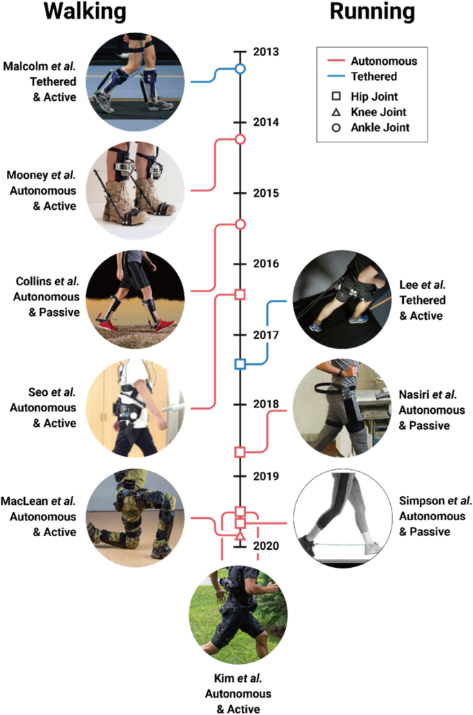
Milestones illustrating the advancement of exoskeleton technology. Tethered (blue) and autonomous (red) exoskeletons assisting at the ankle (circle), knee (triangle), and hip (square) joint to improve healthy, natural walking (left) and running (right) economy versus using no device are shown
In seven short years, our world went from having zero exoskeletons that could reduce a person’s metabolic cost during walking or running to boasting many such devices (Fig. 2 ). Continued progress to convert laboratory-constrained exoskeletons to autonomous systems hints at the possibility that exoskeletons may soon expand their reach beyond college campuses and clinics, and improve walking and running economy across more real-world venues. If research and development continues its trajectory, lower-limb exoskeletons will soon augment human walking and running during everyday life – hopefully, fulfilling Ferris’s and Herr’s predictions.
“What a time to be alive” – Aubrey Drake Graham.
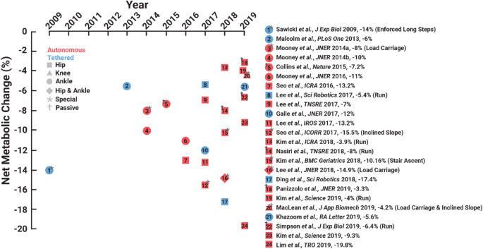
The year that each exoskeleton study was published versus the change in net metabolic cost versus walking or running without using the respective device. Red indicates autonomous and blue indicates a tethered exoskeletons. Different symbols indicate the leg joint(s) that each device directly targets. Asterisk indicates special case and cross indicates a passive exoskeleton
Exoskeleton user performance: insights and trends
To highlight the recent growth of exoskeleton technology, we compiled peer-reviewed publications that reported that an exoskeleton improved user walking or running economy versus without using a device through December 2019. We indexed Web of Science for articles in the English language that included the following topic: (exoskeleton or exosuit or exotendon or assist robot) and (metabolic or energetic or economy) and (walking or running or walk or run). Of the 235 indexed articles, we only included publications that reported that an exoskeleton statistically improved their cohort’s walking and/or running economy versus an experimental no exoskeleton condition. We excluded studies that did not experimentally compare exoskeleton assisted walking or running to a no device condition, choosing to focus on devices that have been shown to break the metabolic cost barrier in the strictest sense. In total, 23 publications satisfied our criteria, and six of these articles improved walking economy during “special” conditions: load carriage [ 19 , 20 , 21 ], inclined slope [ 21 , 22 ], stair ascent [ 23 ], and with enforced long steps [ 24 ] (Fig. 2 and Table 1 ). We categorized exoskeletons into a special category, when researchers increased their participant’s metabolic cost above natural level-ground locomotion (e.g. by adding mass to the user’s body), and subsequently used an exoskeleton to reduce the penalized metabolic cost.
Seventeen publications presented improved human walking and/or running economy using an exoskeleton versus without using a device during preferred level-ground conditions: twelve exoskeletons improved walking economy [ 11 , 12 , 13 , 25 , 26 , 27 , 28 , 29 , 30 , 31 , 32 , 33 ], four improved running economy [ 14 , 15 , 17 , 18 ], and one improved both walking and running economy [ 16 ] versus using no device (Fig. 2 ). These studies demonstrate that exoskeletons improved net metabolic cost during walking by 3.3 to 19.8% versus using no device. For context, improving walking economy by 19.8% is equivalent to the change in metabolic cost due to a person shedding a ~ 25 kg rucksack while walking [ 34 ]. Moreover, four exoskeletons improved net metabolic cost during running by 3.9 to 8.0% versus the no device condition (Table 1 ). Theoretically, improving running economy by 8% would enable the world’s fastest marathoner to break the current marathon world record by over 6 min [ 35 ] – How about a 1:50 marathon challenge?
We labeled six studies as “special” due to an added metabolic penalty placed on the user such as load carriage [ 19 , 20 , 21 ], enforced unnaturally long steps [ 24 ], inclined ground slope [ 21 , 22 ], and/or stair ascent [ 23 ] (Fig. 1 ). Each of these exoskeletons mitigated the negative penalty by reducing metabolic cost. Yet, in some cases [ 21 , 24 ], the authors also performed a comparison at level ground walking without an added “special” penalty. In these cases, the exoskeleton did not significantly mitigate (and may have increased) metabolic cost. For other “special” cases [ 19 , 22 , 23 ], exoskeletons have achieved a metabolic cost benefit in other relevant studies using the same device [ 12 , 26 ]. However, in such cases, there were differences in the experimental setup such as the utilized controller, recruited cohort, and testing conditions.
Despite the popular notion that devices with greater power density (e.g. , tethered exoskeletons with powerful off-board motors and lightweight interfaces) would reduce user metabolic cost beyond that capable by autonomous devices, to date tethered systems have not improved user walking/running economy beyond that of autonomous systems (t-test: p = 0.90) (Fig. 2 ). Namely, tethered exoskeletons have improved user net metabolic cost during walking by 5.4 to 17.4% and autonomous exoskeletons have improved net metabolic cost during walking by 3.3 to 19.8%. These data are from a variety of devices (Table 1 ), walking speeds, and control systems, and thus more rigorous comparisons between autonomous and tethered systems may reveal a more stark performance benefit of tethered systems due to their inherently smaller added mass penalty.
Even though distal leg muscles are thought to be more economical/efficient than proximal leg muscles [ 36 , 37 ], ankle exoskeletons broke the metabolic cost barrier before hip exoskeletons. Perhaps that is because researchers initially targeted the ankles because they yield the greatest positive mechanical power output of any joint [ 37 ]. Notably, only one knee exoskeleton has improved walking economy [ 21 ] (Fig. 2 ). Finally, hip exoskeletons (17.4% metabolic reduction for a tethered device and 19.8% for an autonomous device) have numerically improved metabolic cost by more than ankle exoskeletons (12% metabolic reduction for a tethered case and 11% for an autonomous device), perhaps due to the physiological differences between ankle and hip morphology [ 37 , 38 ] and/or due to the location of the device’s added mass [ 39 ].
A closer examination of the subset of exoskeletons that have yielded the greatest metabolic benefit provides insight into the factors that may maximize users’ benefits with future devices. One emerging factor is the exoskeleton controller. There are numerous methods to command [ 40 ] and control exoskeleton torque profiles. For example, myoelectric controllers depend on the user’s muscle activity [ 41 , 42 ] and impedance controllers depend on the user’s joint kinematics [ 43 ]. Time-based controllers do not take the state of the user as direct input, and only depend on the resolution offered by the chosen torque versus time parameterization [ 27 , 30 , 44 ]. Recent exoskeleton studies indicate that both magnitude [ 45 , 46 ] and perhaps more importantly, timing of assistance [ 11 , 47 , 48 ], affect user metabolism. Additionally, time-based controllers have the flexibility to generate a generalized set of assistive torque patterns that can be optimized on the fly and considerably improve walking and running economy over zero-torque conditions [ 30 , 44 ]. Interestingly, the optimal exoskeleton torque patterns that emerge do not correspond to physiological torques in either their timing or magnitude [ 14 , 44 ]. But, at least at the ankle, getting the timing right seems paramount, as data from optimized exoskeleton torque patterns show lower variability in the timing versus magnitude of the peak torque across many users [ 44 ]. Finally, regarding the magnitude of exoskeleton torque and the net mechanical energy transfer from the device to the user, more is not always better with respect to improving user locomotion economy [ 13 , 27 , 44 , 46 ].
Leading approaches and technologies for advancing exoskeletons
Exoskeleton testbeds enable systematic, high throughput studies on human physiological response.
Tethered exoskeleton testbeds have accelerated device development. In the first decade of the twenty-first century, most exoskeletons were portable, but also cumbersome and limited natural human movement. In addition, these devices were typically designed for one-off, proof of concept demonstrations; not systematic, high-throughput research [ 49 , 50 , 51 , 52 ]. As researchers began focusing on studies that aimed to understand the user’s physiological response to exoskeleton assistance, a key innovation emerged - the laboratory-based exoskeleton testbed. Rather than placing actuators on the exoskeleton’s end-effector, researchers began placing them off-board and attached them through tethers (e.g. , air hoses and Bowden cables) to streamlined exoskeleton end-effectors [ 45 , 53 , 54 ]. This approach enabled researchers to conduct high throughput, systematic studies during treadmill walking and running to determine optimal exoskeleton assistance parameters (e.g. , timing and magnitude of mechanical power delivery [ 27 , 55 ]) for improving walking and running economy. Furthermore, the high-performance motors on recent tethered exoskeleton testbeds have relatively high torque control bandwidth that can be leveraged to render the dynamics of existing or novel design concepts [ 43 , 56 ]. Testing multiple concepts prior to the final device development could enable researchers to quickly diagnose the independent effects of design parameters on current products and test novel ideas [ 57 ]. Thus, we reason that exoskeleton testbeds have progressed exoskeleton technology by enabling researchers to optimize a high number of device parameters [ 58 ], test new ideas, and then iterate designs without having to build one-off prototypes.
Embedding ‘smart mechanics’ into passive exoskeletons provides an alternative to fully powered designs
Laboratory-based exoskeletons are moving into the real-world through the use of small, transportable energy supplies [ 59 ] and/or by harvesting mechanical energy to power the device [ 60 ]. Despite these improvements, another way to circumnavigate the burden of lugging around bulky energy sources is by developing passive exoskeletons [ 13 , 17 , 18 , 31 ]. Passive exoskeletons have been able to assist the user by storing and subsequently returning mechanical energy to the user without injecting net positive mechanical work. Passive exoskeletons are typically cheaper and lighter than active devices (e.g. , Collins et al.’s ankle exoskeleton is 400 g [ 13 ]) and, like active devices, are hypothesized to primarily improve walking and running economy by reducing active muscle volume [ 61 ]. However, due to their simplified designs, passive exoskeletons are in some ways less adaptable than powered devices. Passive devices can only offer fixed mechanical properties that are at best only switchable between locomotion bouts. Thus, while passive systems may be adequate for providing assistance during stereotyped locomotion tasks such as running on a track or hiking downhill at fixed speed, they may not be able to handle variable conditions. On the other hand, active devices offer the opportunity to apply any generic torque-time profile, but require bulky motors and/or gears that need a significant source of power to do so. Thus, combining features from active and passive exoskeletons to create a new class of pseudo-passive (or semi-active) devices may yield a promising future direction for exoskeleton technology [ 59 ]. For example, rather than continuously modulating the assistance torque profile, a pseudo-passive device might inject small amounts of power to change the mechanical properties of an underlying passive structure during periods when it is unloaded [ 62 ]. The pseudo-passive approach likely benefits from the streamlined structural design (e.g. , small motors) and adaptability that requires only small amounts of energy input (e.g. , small batteries).
Providing comfort at the human-exoskeleton interface
Regardless of active or passive exoskeleton design, researchers struggle to effectively and comfortably interface exoskeletons to the human body [ 63 ]. That is primarily due to the human body having multiple degrees of freedom, deforming tissues, and sensitive points of pressure. Accordingly, many researchers utilize custom orthotic fabrication techniques [ 46 , 64 , 65 ], and/or malleable textiles (commonly referred to as exo-suits) [ 16 , 66 , 67 , 68 ] to tackle this challenge. Textile-based exoskeletons may be superior to traditional rigid exoskeletons due to their lower mass, improved comfort, fewer kinematic restrictions, and better translation to practical-use [ 16 , 67 , 68 ]. Reaffirming soft technology, the tethered exoskeleton that best improves walking economy versus not using a device is currently an exoskeleton with a soft, malleable user-device interface [ 67 ] (Fig. 2 ).
Exoskeleton controllers using artificial intelligence and on-line optimization to adapt to both user and environment may facilitate the transition to ‘real-world’ functionality
Researchers are also developing smart controllers that constantly update exoskeleton characteristics to optimize user walking and running economy. This is exemplified by Zhang and colleagues [ 44 ], who developed a controller that rapidly estimates metabolic profiles and adjusts ankle exoskeleton torque profiles to optimize human walking and running economy. We foresee smart controllers enabling exoskeletons to move beyond conventional fixed assistance parameters, and steering user physiology in-a-closed-loop with the device to maintain optimal exoskeleton assistance across conditions [ 30 , 69 ]. Since measuring metabolic cost throughout everyday life is unrealistic, future exoskeletons may incorporate embedded wearable sensors (e.g. , electromyography surface electrodes, pulse oximetry units, and/or low-profile ultrasonography probes) that inform the controller of the user’s current physiological state [ 70 , 71 ] and thereby enable continuous optimizing of device assistance [ 20 , 72 , 73 ] to minimize the user’s estimated metabolic cost.
At a high level of control, researchers are using techniques to detect user intent, environmental parameters, and optimize exoskeleton assistance across multiple tasks [ 15 , 16 , 68 , 74 , 75 ]. An early version of this techniques paradigm was implementing proportional myoelectric control into exoskeletons [ 76 , 77 , 78 ]. This strategy directly modulates exoskeleton torque based on the timing and magnitude of a targeted muscle’s activity, which can adapt the device to the users changing biomechanics. However, this strategy has yielded mixed results [ 42 , 79 , 80 ] and is challenging to effectively use due to quick adaptations that occur to accommodate various tasks as well as slower changes that occur due to learning the device [ 41 ]. Scientists have made exciting advances using machine learning and artificial intelligence techniques to fuse information from both sensors on the user and device to better merge the user and exoskeleton [ 81 , 82 ], but these techniques have not yet been commercially translated to exoskeleton technology to the authors’ knowledge. These strategies have the potential to enable exoskeletons to discern user locomotion states (such as running, walking, descending ramps, and ascending stairs) and alter device parameters to meet the respective task demands.
Closing remarks and vision for the future of exoskeleton technology
In the near-term, we predict that the exoskeleton expansion will break researchers out of laboratory confinement. Doing so will enable studies that directly address how exoskeleton-assistance affects real-world walking and running performance without relying on extrapolated laboratory-based findings. By escaping the laboratory, we expect that exoskeleton technology will expand beyond improving human walking and running economy over the next decade and begin optimizing other aspects of locomotor performance that influence day-to-day mobility in natural environments. To list a few grand-challenges, exoskeletons may begin to augment user stability, agility, and robustness of gait. For example, exoskeletons may make users,
· More stable by modulating the sensorimotor response of their neuromuscular system to perturbations [ 83 , 84 , 85 ].
· More agile and faster by increasing the relative force capacity of their muscles [ 86 ].
· More robust by dissipating mechanical energy to prevent injury during high impact activities like rapid cutting maneuvers or falling from extreme heights [ 87 ].
To make these leaps, engineers will need to continue to improve exoskeleton technology, physiologists will need to refine the evaluation of human performance, clinicians will need to consider how exoskeletons can further rehabilitation interventions, psychologists will need to better understand how user’s interact with and embody exoskeletons, designers will need to account for exoskeletons in space planning, and healthcare professionals may need to update their exercise recommendations to account for the use of exoskeletons. Combined, these efforts will help establish a ‘map’ that can be continuously updated to help navigate the interaction between human, machine, and environment. Such guidelines will set the stage for exoskeletons that operate in symbiosis with the user to blur lines between human and machine. Closing the loop between exoskeleton hardware, software, and the user’s biological systems (e.g. , both musculoskeletal and neural tissues) will enable a new class of devices capable of steering human neuromechanical structure and function over both short and long timescales during walking and running. On the shortest of time scales, exoskeletons that have access to body state information have the potential to modify sensory feedback from mechanoreceptors and augment dynamic balance. On the longest of timescales, exoskeletons that have access to biomarkers indicating tissue degradation [ 88 ] could modify external loads to shape the material properties of connective tissues and maintain homeostasis.
Until then, we focus our attention on the ability of exoskeletons to improve human walking and running economy. So far, 17 studies have reported that exoskeletons improve natural human walking and running economy (Fig. 2 ). As these devices evolve and become more available for public use, they will not only continue to improve walking and running economy of young adults, but they will also augment elite athlete performance, allow older adults to keep up with their kinfolk, enable people with disability to outpace their peers, and take explorers deeper into the wilderness.
Availability of data and materials
Not applicable.
Ferris DP. The exoskeletons are here. J Neuroeng Rehabil. 2009;6:17.
Article PubMed PubMed Central Google Scholar
Herr H. Exoskeletons and orthoses: classification, design challenges and future directions. J Neuroeng Rehabil. 2009;6:21.
Beneke R, Meyer K. Walking performance and economy in chronic heart failure patients pre and post exercise training. Eur J Appl Physiol Occup Physiol. 1997;75(3):246–51.
Article CAS PubMed Google Scholar
Hoogkamer W, et al. Altered running economy directly translates to altered distance-running performance. Med Sci Sports Exerc. 2016;48(11):2175–80.
Article PubMed Google Scholar
Newman AB, et al. Association of long-distance corridor walk performance with mortality, cardiovascular disease, mobility limitation, and disability. JAMA. 2006;295(17):2018–26.
Gerardi D, et al. Variables related to increased mortality following out-patient pulmonary rehabilitation. Eur Respir J. 1996;9(3):431–5.
Norris JA, et al. Effect of augmented plantarflexion power on preferred walking speed and economy in young and older adults. Gait Posture. 2007;25(4):620–7.
Walsh CJ, Endo K, Herr H. A quasi-passive leg exoskeleton for load-carrying augmentation. Int J Humanoid Robot. 2007;4(3):487–506.
Article Google Scholar
Sawicki GS, Ferris DP. Mechanics and energetics of level walking with powered ankle exoskeletons. J Exp Biol. 2008;211:1402–13.
Gregorczyk KN, et al. Effects of a lower-body exoskeleton device on metabolic cost and gait biomechanics during load carriage. Ergonomics. 2010;53:1263–75.
Malcolm P, et al. A simple exoskeleton that assists plantarflexion can reduce the metabolic cost of human walking. PLoS One. 2013;8:e56137.
Article CAS PubMed PubMed Central Google Scholar
Mooney LM, Rouse EJ, Herr HM. Autonomous exoskeleton reduces metabolic cost of human walking. J Neuroeng Rehabil. 2014;11(1):151.
Collins SH, Wiggin MB, Sawicki GS. Reducing the energy cost of human walking using an unpowered exoskeleton. Nature. 2015;522:212–5 advance online publication.
Lee G, et al. Reducing the metabolic cost of running with a tethered soft exosuit. Sci Robot. 2017;2(6):eaan6708.
Kim J, et al. Autonomous and portable soft exosuit for hip extension assistance with online walking and running detection algorithm. In: 2018 IEEE International Conference on Robotics and Automation (ICRA); 2018.
Google Scholar
Kim J, et al. Reducing the metabolic rate of walking and running with a versatile, portable exosuit. Science. 2019;365(6454):668.
Nasiri R, Ahmadi A, Ahmadabadi MN. Reducing the energy cost of human running using an unpowered exoskeleton. IEEE Trans Neural Syst Rehabil Eng. 2018;26(10):2026–32.
Simpson CS, et al. Connecting the legs with a spring improves human running economy. J Exp Biol. 2019;222(17):jeb202895.
Mooney LM, Rouse EJ, Herr HM. Autonomous exoskeleton reduces metabolic cost of human walking during load carriage. J Neuroeng Rehabil. 2014;11(1):80.
Lee S, et al. Autonomous multi-joint soft exosuit with augmentation-power-based control parameter tuning reduces energy cost of loaded walking. J Neuroeng Rehabil. 2018;15(1):66.
MacLean MK, Ferris DP. Energetics of walking with a robotic knee exoskeleton. J Appl Biomech. 2019;35(5):320.
Seo K, Lee J, Park YJ. Autonomous hip exoskeleton saves metabolic cost of walking uphill. In: 2017 IEEE International Conference on Rehabilitation Robotics (ICORR); 2017.
Kim D-S, et al. A wearable hip-assist robot reduces the cardiopulmonary metabolic energy expenditure during stair ascent in elderly adults: a pilot cross-sectional study. BMC Geriatr. 2018;18(1):230.
Sawicki GS, Ferris DP. Powered ankle exoskeletons reveal the metabolic cost of plantar flexor mechanical work during walking with longer steps at constant step frequency. J Exp Biol. 2009;212:21–31.
Mooney LM, Herr HM. Biomechanical walking mechanisms underlying the metabolic reduction caused by an autonomous exoskeleton. J Neuroeng Rehabil. 2016;13:4.
Seo K, et al. Fully autonomous hip exoskeleton saves metabolic cost of walking. In: 2016 IEEE International Conference on Robotics and Automation (ICRA); 2016.
Galle S, et al. Reducing the metabolic cost of walking with an ankle exoskeleton: interaction between actuation timing and power. J Neuroeng Rehabil. 2017;14(1):35.
Lee Y, et al. A flexible exoskeleton for hip assistance. In: 2017 IEEE/RSJ International Conference on Intelligent Robots and Systems (IROS); 2017.
Lee H, et al. A wearable hip assist robot can improve gait function and cardiopulmonary metabolic efficiency in elderly adults. IEEE Trans Neural Syst Rehabil Eng. 2017;25(9):1549–57.
Ding Y, et al. Human-in-the-loop optimization of hip assistance with a soft exosuit during walking. Science Robotics. 2018;3(15):eaar5438.
Panizzolo FA, et al. Reducing the energy cost of walking in older adults using a passive hip flexion device. J Neuroeng Rehabil. 2019;16(1):117.
Lim B, et al. Delayed outputf feedback control for gait assistance with a robotic hip exoskeleton. IEEE Trans Robot. 2019;35(4):1055–62.
Khazoom C, et al. Design and control of a multifunctional ankle exoskeleton powered by magnetorheological actuators to assist walking, jumping, and landing. IEEE Robot Automation Lett. 2019;4(3):3083–90.
Farley CT, McMahon TA. Energetics of walking and running: insights from simulated reduced-gravity experiments. J Appl Physiol. 1992;73(6):2709–12.
Kipp S, Kram R, Hoogkamer W. Extrapolating metabolic savings in running: implications for performance predictions. Front Physiol. 2019;10:79.
Umberger BR, Rubenson J. Understanding muscle energetics in locomotion: new modeling and experimental approaches. Exerc Sport Sci Rev. 2011;39(2):59–67.
Sawicki GS, Lewis CL, Ferris DP. It pays to have a spring in your step. Exerc Sport Sci Rev. 2009;37(3):130.
Chen W, et al. On the biological mechanics and energetics of the hip joint muscle–tendon system assisted by passive hip exoskeleton. Bioinspir Biomim. 2018;14(1):016012.
Browning RC, et al. The effects of adding mass to the legs on the energetics and biomechanics of walking. Med Sci Sports Exerc. 2007;39(3):515–25.
Yan T, et al. Review of assistive strategies in powered lower-limb orthoses and exoskeletons. Robot Auton Syst. 2015;64:120–36.
Koller JR, et al. Learning to walk with an adaptive gain proportional myoelectric controller for a robotic ankle exoskeleton. J Neuroeng Rehabil. 2015;12(1):1.
Young AJ, Gannon H, Ferris DP. A biomechanical comparison of proportional electromyography control to biological torque control using a powered hip exoskeleton. Front Bioeng Biotechnol. 2017;5:37.
Zhang J, Cheah CC, Collins SH. Torque Control in Legged Locomotion. In: Sharbafi MA, Seyfarth A, editors. Bioinspired Legged Locomotion. Amsterdam: Elsevier; 2017. p. 347–400.
Zhang J, et al. Human-in-the-loop optimization of exoskeleton assistance during walking. Science. 2017;356(6344):1280.
Quinlivan BT, et al. Assistance magnitude versus metabolic cost reductions for a tethered multiarticular soft exosuit. Sci Robot. 2017;2(2):1–10.
Kang I, Hsu H, Young A. The effect of hip assistance levels on human energetic cost using robotic hip exoskeletons. IEEE Robot Automation Lett. 2019;4(2):430–7.
Jackson RW, Collins SH. An experimental comparison of the relative benefits of work and torque assistance in ankle exoskeletons. J Appl Physiol. 2015;119(5):541–57.
Ding Y, et al. Effect of timing of hip extension assistance during loaded walking with a soft exosuit. J Neuroeng Rehabil. 2016;13(1):87.
Guizzo E, Goldstein H. The rise of the body bots [robotic exoskeletons]. IEEE Spectr. 2005;42(10):50–6.
Zoss AB, Kazerooni H, Chu A. Biomechanical design of the Berkeley lower extremity exoskeleton (BLEEX). IEEE/ASME Trans Mechatronics. 2006;11(2):128–38.
Walsh CJ, Pasch K, Herr H. An autonomous, underactuated exoskeleton for load-carrying augmentation. In: 2006 IEEE/RSJ International Conference on Intelligent Robots and Systems (IROS); 2006.
Raytheon XOS 2 exoskeleton, second-generation robotics suit. 2010; Available from: http://www.army-technology.com/projects/raytheon-xos-2-exoskeleton-us/ .
Caputo JM, Collins SH. An experimental robotic testbed for accelerated development of ankle prostheses. In: 2013 IEEE International Conference on Robotics and Automation; 2013.
Ding Y, et al. Multi-joint actuation platform for lower extremity soft exosuits. In: 2014 IEEE International Conference on Robotics and Automation (ICRA); 2014.
Young A, et al. Influence of power delivery timing on the energetics and biomechanics of humans wearing a hip exoskeleton. Front Bioeng Biotechnol. 2017;5:4.
PubMed PubMed Central Google Scholar
Witte KA, Collins SH. Design of Lower-Limb Exoskeletons and Emulator Systems. In: Rosen J, Ferguson PW, editors. Wearable Robotics. Amsterdam: Elsevier; 2020. p. 251–74.
Chapter Google Scholar
Caputo JM, Collins SH, Adamczyk PG. Emulating prosthetic feet during the prescription process to improve outcomes and justifications. In: 2014 IEEE International Workshop on Advanced Robotics and its Social Impacts; 2014.
Kim M, et al. Human-in-the-loop bayesian optimization of wearable device parameters. PLoS One. 2017;12(9):e0184054.
Article PubMed PubMed Central CAS Google Scholar
Diller S, Majidi C, Collins SH. A lightweight, low-power electroadhesive clutch and spring for exoskeleton actuation. In: 2016 IEEE International Conference on Robotics and Automation (ICRA); 2016.
Donelan JM, et al. Biomechanical energy harvesting: generating electricity during walking with minimal user effort. Science. 2008;319(5864):807–10.
Beck ON, et al. Exoskeletons improve locomotion economy by reducing active muscle volume. Exerc Sport Sci Rev. 2019;47(4):237–45.
Braun DJ, et al. Variable stiffness spring actuators for low-energy-cost human augmentation. IEEE Trans Robot. 2019;35(6):1435–49.
Yandell MB, et al. Physical interface dynamics alter how robotic exosuits augment human movement: implications for optimizing wearable assistive devices. J Neuroeng Rehabil. 2017;14(1):40.
Giovacchini F, et al. A light-weight active orthosis for hip movement assistance. Robot Auton Syst. 2015;73:123–34.
Lv G, Zhu H, Gregg RD. On the design and control of highly backdrivable lower-limb exoskeletons: a discussion of past and ongoing work. IEEE Control Syst Mag. 2018;38(6):88–113.
Asbeck AT, et al. Biologically-inspired soft exosuit. In: IEEE International Conference on Rehabilitation Robotics; 2013.
Panizzolo FA, et al. A biologically-inspired multi-joint soft exosuit that can reduce the energy cost of loaded walking. J Neuroeng Rehabil. 2016;13(1):43.
Lee S, et al. Autonomous Multi-Joint Soft Exosuit for Assistance with Walking Overground. In: 2018 IEEE International Conference on Robotics and Automation (ICRA); 2018.
Felt W, et al. “Body-in-the-loop”: optimizing device parameters using measures of instantaneous energetic cost. PLoS One. 2015;10:e0135342.
Ingraham KA, Ferris DP, Remy CD. Evaluating physiological signal salience for estimating metabolic energy cost from wearable sensors. J Appl Physiol. 2019;126(3):717–29.
Slade P, et al. Rapid energy expenditure estimation for ankle assisted and inclined loaded walking. J Neuroeng Rehabil. 2019;16(1):67.
Huang H, et al. A cyber expert system for auto-tuning powered prosthesis impedance control parameters. Ann Biomed Eng. 2016;44(5):1613–24.
Kumar S, et al. Extremum seeking control for model-free auto-tuning of powered prosthetic legs. In: IEEE Transactions on Control Systems Technology; 2019. p. 1–16.
Huang H, et al. Continuous locomotion-mode identification for prosthetic legs based on neuromuscular-mechanical fusion. IEEE Trans Biomed Eng. 2011;58(10):2867–75.
Young AJ, Hargrove LJ. A classification method for user-independent intent recognition for transfemoral amputees using powered lower limb prostheses. IEEE Trans Neural Syst Rehabil Eng. 2016;24(2):217–25.
Ferris DP, et al. An improved powered ankle-foot orthosis using proportional myoelectric control. Gait Posture. 2006;23(4):425–8.
Sawicki GS, Ferris DP. A pneumatically powered knee-ankle-foot orthosis (KAFO) with myoelectric activation and inhibition. J Neuroeng Rehabil. 2009;6:23.
Ferris DP, Lewis CL. Robotic lower limb exoskeletons using proportional myoelectric control. In: 2009 Annual international conference of the Ieee engineering in medicine and biology society, vol. 1-20; 2009. p. 2119–24.
Koller JR, Remy CD, Ferris DP. Biomechanics and energetics of walking in powered ankle exoskeletons using myoelectric control versus mechanically intrinsic control. J Neuroeng Rehabil. 2018;15(1):42.
Grazi L, et al. Gastrocnemius myoelectric control of a robotic hip exoskeleton can reduce the user's lower-limb muscle activities at push off. Front Neurosci. 2018;12:71.
Young A, Kuiken T, Hargrove L. Analysis of using EMG and mechanical sensors to enhance intent recognition in powered lower limb prostheses. J Neural Eng. 2014;11(5):056021.
Kang I, et al. Electromyography (EMG) signal contributions in speed and slope estimation using robotic exoskeletons. In: 2019 IEEE 16th International Conference on Rehabilitation Robotics (ICORR); 2019.
Steele KM, et al. Muscle recruitment and coordination with an ankle exoskeleton. J Biomech. 2017;59:50–8.
Kao P-C, Lewis CL, Ferris DP. Short-term locomotor adaptation to a robotic ankle exoskeleton does not alter soleus Hoffmann reflex amplitude. J Neuroeng Rehabil. 2010;7:33.
Kao P-C, Lewis CL, Ferris DP. Joint kinetic response during unexpectedly reduced plantar flexor torque provided by a robotic ankle exoskeleton during walking. J Biomech. 2010;43(7):1401–7.
Weyand PG, et al. Faster top running speeds are achieved with greater ground forces not more rapid leg movements. J Appl Physiol. 2000;89(5):1991–9.
Sutrisno A, Braun DJ. Enhancing mobility with quasi-passive variable stiffness exoskeletons. IEEE Trans Neural Syst Rehabil Eng. 2019;27(3):487–96.
Lane AR, et al. Body mass index and type 2 collagen turnover in individuals after anterior cruciate ligament reconstruction. J Athl Train. 2019;54(3):270–5.
Download references
Acknowledgements
This work was funded in part by NSF National Robotics Initiative (award # 1830215) to A.J.Y., U.S. Army Natick Soldier Research, Development and Engineering Center (W911QY18C0140) to G.S.S, and an NIH National Institute on Aging fellowship award (F32AG063460) to O.N.B. The content is solely the responsibility of the authors and does not necessarily represent the official views of the funding agencies listed.
Author information
Authors and affiliations.
The George W. Woodruff School of Mechanical Engineering, Georgia Institute of Technology, Atlanta, GA, USA
Gregory S. Sawicki, Owen N. Beck, Inseung Kang & Aaron J. Young
School of Biological Sciences, Georgia Institute of Technology, Atlanta, GA, USA
Gregory S. Sawicki & Owen N. Beck
Institute for Robotics and Intelligent Machines, Georgia Institute of Technology, Atlanta, GA, USA
Gregory S. Sawicki & Aaron J. Young
You can also search for this author in PubMed Google Scholar
Contributions
All authors contributed to writing the manuscript. G. Sawicki and A. Young jointly conceived of the review paper idea, extracted the trends, and determined the leading approaches and technologies. O. Beck and I. Kang performed a literature search to benchmark progress in exoskeletons vs. time to improve the economy of human locomotion. They logged all the studies and categorized them by joint and gait mode. All authors drafted, edited, and approved the final manuscript.
Corresponding authors
Correspondence to Gregory S. Sawicki or Aaron J. Young .
Ethics declarations
Ethics approval and consent to participate, consent for publication, competing interests.
The authors declare that they have no competing interests.
Additional information
Publisher’s note.
Springer Nature remains neutral with regard to jurisdictional claims in published maps and institutional affiliations.
Rights and permissions
Open Access This article is distributed under the terms of the Creative Commons Attribution 4.0 International License ( http://creativecommons.org/licenses/by/4.0/ ), which permits unrestricted use, distribution, and reproduction in any medium, provided you give appropriate credit to the original author(s) and the source, provide a link to the Creative Commons license, and indicate if changes were made. The Creative Commons Public Domain Dedication waiver ( http://creativecommons.org/publicdomain/zero/1.0/ ) applies to the data made available in this article, unless otherwise stated.
Reprints and permissions
About this article
Cite this article.
Sawicki, G.S., Beck, O.N., Kang, I. et al. The exoskeleton expansion: improving walking and running economy. J NeuroEngineering Rehabil 17 , 25 (2020). https://doi.org/10.1186/s12984-020-00663-9
Download citation
Received : 05 August 2019
Accepted : 13 February 2020
Published : 19 February 2020
DOI : https://doi.org/10.1186/s12984-020-00663-9
Share this article
Anyone you share the following link with will be able to read this content:
Sorry, a shareable link is not currently available for this article.
Provided by the Springer Nature SharedIt content-sharing initiative
- Wearable robotics
- Assistive devices
- Metabolic cost
- Augmentation
Journal of NeuroEngineering and Rehabilitation
ISSN: 1743-0003
- Submission enquiries: [email protected]
- Social Science
- Abnormal Psychology
Research Paper Skeleton
Related documents
Add this document to collection(s)
You can add this document to your study collection(s)
Add this document to saved
You can add this document to your saved list
Suggest us how to improve StudyLib
(For complaints, use another form )
Input it if you want to receive answer
We use cookies to enhance our website for you. Proceed if you agree to this policy or learn more about it.
- Essay Database >
- Essays Samples >
- Essay Types >
- Research Paper Example
Skeleton Research Papers Samples For Students
16 samples of this type
Regardless of how high you rate your writing skills, it's always an appropriate idea to check out an expertly written Research Paper example, especially when you're dealing with a sophisticated Skeleton topic. This is precisely the case when WowEssays.com collection of sample Research Papers on Skeleton will come in handy. Whether you need to think up a fresh and meaningful Skeleton Research Paper topic or examine the paper's structure or formatting peculiarities, our samples will provide you with the necessary data.
Another activity area of our write my paper company is providing practical writing assistance to students working on Skeleton Research Papers. Research help, editing, proofreading, formatting, plagiarism check, or even crafting entirely original model Skeleton papers upon your request – we can do that all! Place an order and buy a research paper now.
Good Research Paper On Nursing: Skeletal Diseases
The musculoskeletal system is responsible for all body movements. The skeletal component of the muscular system provides a structural framework for the body, which protects the internal organs and provides the shape for the body. It acts as the storage for calcium and phosphorus and plays a part in mineral homeostasis(Davidson, 2002)
Functions of the skeletal system(Haywood, 2008)
Forensic anthropology research paper samples, good research paper about cartilage and bone growth stimulation by growth hormone.
Don't waste your time searching for a sample.
Get your research paper done by professional writers!
Just from $10/page
Lipids Research Paper Examples
Introduction, hopi legend and culture research paper.
“How the Great Chiefs Made the Moon and the Sun” is a Hopi legend. As its name suggests, it is a myth surrounding the creation of the moon and the sun. Many Hopi legends involve the sun as it is a central symbol in Hopi culture. The Hopi Indians, meaning good, peaceful, or wise, come from a group of South-western people called Pueblo.
A-Level Research Paper On Animation Technologies In The Incredibles For Free Use
19th century buildings research paper example.
Slavick (4) explains that Chicago School refers to building designs in the early 19th Century. Turak (60) claim that Chicago is indeed the birthplace of skyscrapers. In 1880’s Chicago manages to develop architects referred as the First Chicago School (Turak 60). At this time, Chicago is the second largest metropolis with a high population growth in America. People consider building large commercial buildings since low-level buildings are inefficient. Chicago experiences a booming economy and a lot of innovation that enables the building of skyscrapers to allow for more space for business people.
Free Research Paper On Temperature & Great Bear Reef
Impact of climatic change on the great barrier reef, the struggle for wealth race in toni morrisons song of solomon research paper sample, homo erectus research paper, homo erectus, gerard john schaefer research papers examples, evolution of the genus homo research papers examples, australopithecus afarensis research papers example, corallium rubrum (linnaeus, 1758) research paper example.
Kingdom: Animalia Phylum: Cnidaria Subclass: Alcyonacea Order: Gorgonacea Family: Coralliidae Genus: Corallium Species: Corallium rubrum Synonyms: Red coral, Precious coral, Reed coral, noble coral, Sardinia coral, midway coral, angel skin coral (Arkive, para. 1) Changes in Taxonomy: Madrepora rubra Linnaeus, 1758; Isis nobills Pallas, 1766; Gorgonia nobills Linnaeus, 1789; Corallium rubrum Lamarck, 1816. (FAO, para. 1)
Introduction:
Example of phylogenetic family tree research paper, anthropology and anecdote witchcraft and spiritual healing as ethnography and literature research paper example.
Password recovery email has been sent to [email protected]
Use your new password to log in
You are not register!
By clicking Register, you agree to our Terms of Service and that you have read our Privacy Policy .
Now you can download documents directly to your device!
Check your email! An email with your password has already been sent to you! Now you can download documents directly to your device.
or Use the QR code to Save this Paper to Your Phone
The sample is NOT original!
Short on a deadline?
Don't waste time. Get help with 11% off using code - GETWOWED
No, thanks! I'm fine with missing my deadline
Cavers and scientists unearth near-complete short-nosed kangaroo fossil from East Gippsland cave
Science Cavers and scientists unearth near-complete short-nosed kangaroo fossil from East Gippsland cave
Joshua Van Dyk will happily abseil into a pitch black cave, or squeeze through crevices no one has ever gone into before.
"It feels comforting," he laughs, "I don't mind tight spots."
Although that might sound terrifying, he's had plenty of experience.
He's from a multi-generational caving family in Buchan, Victoria who have been working with palaeontologists at Museums Victoria since the 1990s to investigate caves in their local area.
And he's also part of a team of scientists, rangers and citizen cavers who recently uncovered a 50,000-year-old short-faced kangaroo skeleton in an undisturbed cave in East Gippsland.
The fossil of Simosthenurus occidentalis is one of the most complete found in Australia, and was extracted after more than 58 hours of caving in a two-year period.
"The rarity of finding a single, near complete example of an individual like this [...] it's the dream for any palaeontologist," said Tim Ziegler, Museums Victoria vertebrate palaeontology collections manager.
"The last and first time a comparably complete example of this species of kangaroo was found was in the mid-1970s. It's been nearly 50 years."
The discovery highlights the role of non-scientists in finding and extracting fossils like this — particularly recreational cavers who dig out, abseil down, and crawl through caves in their spare time.
What was found?
Scientists uncovered 150 bones of the extinct kangaroo species in Nightshade Cave near Buchan.
The skeleton has 71 per cent of its bones, which makes it the most complete fossil skeleton ever discovered in a Victorian cave.
"Most of the [bones] that are missing are very small elements, like knuckles from the hands, and small bones in the feet," Mr Ziegler said.
The short-faced kangaroo specimen found by the researchers is a juvenile, but it may have weighed as much as 80 kilograms.
An adult of the ancient species would have been about the size of a large grey or red kangaroo today, but would weigh twice as much.
Researchers have suggested that this heavier structure might mean that S. occidentalis and other species of short-faced kangaroos may not have hopped, and instead walked in a similar way to humans.
"The entire skeleton — from its shoulders and its arms, through its backbone to his hips and its legs and its tail — tells you something very different from what a living kangaroo tells you," Mr Ziegler said.
To find out the age of the specimen, the researchers sent a small sample to the Australian Nuclear Science and Technology Organisation (ANSTO) to radiocarbon date the remains.
"We tried first to extract any collagen from the bones but we found it was quite old," said Vladimir Levchenko, a research scientist at ANSTO who dated the specimen.
"But there are other ways to date things. In particular, with this kangaroo, it was associated with a layer of charcoal. In Australia, fires are quite common, so charcoal is very present in the Australian environment."
Charcoal just above the kangaroo was found and dated, providing a minimum age for the specimen of 49,400 years old.
How was the kangaroo extracted?
The fossil was first noticed in July 2011, when a group of five recreational cavers, including Mr Van Dyk, opened a cave near Buchan in East Gippsland.
According to him, the area was almost completely closed off, with "just a tiny depression and a tiny hole about the size of a 50 cent piece".
After about 20 hours of digging, the team exposed an entrance that allowed the group to abseil into the cave about 10 metres down, before belly crawling through a tight U-turn crevice into the cave.
At the time, only a small amount of the skeleton was exposed, and the remainder was under a large boulder, but the team let Museums Victoria know there was something there.
"The impression at the time was that it would be difficult, maybe impossible, to attempt the retrieval of the rest of it," Mr Ziegler said.
Being in a secluded cave, it was thought the bones would be safe from the elements, but during a follow up visit over 10 years later, the team discovered the fossil — which was not just a few bones, but an entire skeleton — had begun to decay.
"We hadn't acquired the direct dating, but we knew from national records that it had to be at least 45,000 years old," Mr Ziegler said.
"What a tragedy it would be if it had just decayed away after all of that time."
Over the next two years, Mr Ziegler worked with rangers, cavers and scientists to get into the tight spot, and painstakingly extract the skeleton piece by piece.
"Carefully cleaning and exposing bones in place, documenting, collecting samples from sediment, and individually collecting, packing and removing each element from the cave," he said.
" [You're] working at the end of your reach into this tiny little crevice. It's a very uncomfortable place to be, particularly in your all-in-one caving suit and helmets and lights.
"It was a really physically challenging process."
What do cavers do?
It's not normally scientists or museum collection managers who shimmy through caves to find bones.
Instead, it's citizen cavers, like Mr Van Dyk, who explore and chart cave systems during their spare time, mostly on weekends and days off.
"Especially with new caves, no one's ever been there before. You're the first one," he said.
"You don't know what's around the next corner, or down the next hole."
Gavin Prideaux, a palaeontology researcher at Flinders University who was not involved in the research, said cavers were "absolutely critical" to finding new fossils.
"Often times, at least in the first instance, you're relying on the people with eyes on — or under — the ground reporting finds to you, even the bad things," Professor Prideaux said.
"They're not usually in contexts where you can just casually wander in."
He suggests cavers take photos of the bones instead of touching them, as the location and surrounding area near the bones can also hold valuable information.
Most of the time the bones will likely be from a recent animal, and aren't helpful for researchers, but occasionally you strike fossil gold.
"Sometimes in cave deposits — the Naracoorte Caves are a good example — you get thousands upon thousands of bones, but it's all different individuals jumbled up," Professor Prideaux said.
While cavers are some of the few groups who have the speciality knowledge to be able to get into these difficult environments, they're not doing it for the bones.
The Victorian Speleological Association (VSA) says their aim is to "explore and chart the extensive cave systems in Victoria".
Wayne Revell, the president of the VSA, said although most scientists who need their help end up catching the caving bug themselves, they do sometimes help non-caving scientists dig out, rig up, and enter caves safely.
"A lot of cavers are volunteers and are just doing it for the fun of caving. Whether there's a paper released or not, whether it mentions the cavers or not, I don't think it's a big deal, although it might be for some," Mr Revell said.
"Tim [Ziegler] has been really good with this. He's put my name on papers and I thought, 'wow, I hardly did anything. I was just there for the enjoyment'."
As an example of a scientist who caught the caving bug, Mr Ziegler is now the vice president of the VSA.
How scientists will use the fossil
While museum-goers are likely accustomed to seeing full skeletons of dinosaurs and ancient Australian mammals, they're not actually what they seem.
"Most fossil skeletons that are put on public display in museum galleries are replicas, and replicas are often composites," Mr Ziegler said.
This means that usually one ancient animal was not the source of all the bones in a full skeleton. Instead, multiple fossils are brought together to design one creature.
This is fine for public display, but it's not as useful for scientists, who may need to investigate how certain bones change over time or location.
Megan Jones is a PhD student looking at the vertebral column (known as the backbone) in both extinct and living species of kangaroo to better understand how they walked or hopped.
"I need to find specimens which have fairly complete vertebral columns, which the Simosthenurus occidentalis skeleton is a very good example of," Ms Jones said.
"Vertebrae are smaller bones and there's a lot of them, so the chances are they're going to get damaged."
Ms Jones also said small bones like vertebrae are sometimes not noticed among the other, more recognisable bones.
Dozens of the small bones that form the vertebral column need to be found, compared to just one easily recognisable skull.
"It's really a challenge to find good specimens," she added.
The S. occidentalis fossil will be on public display in the Research Institute Gallery at Melbourne Museum from June 24.
Science in your inbox
- X (formerly Twitter)
Related Stories
Scientists discover extinct marsupial double the size of the red kangaroo.
How fast can wombats run? We dug into the truth of claims they can beat Usain Bolt
The mountains of Papua New Guinea were once home to kangaroos with 'unique teeth'
- Science and Technology
- Share full article
Advertisement
Supported by
New Study Bolsters Idea of Athletic Differences Between Men and Trans Women
Research financed by the International Olympic Committee introduced new data to the unsettled and fractious debate about bans on transgender athletes.

By Jeré Longman
A new study financed by the International Olympic Committee found that transgender female athletes showed greater handgrip strength — an indicator of overall muscle strength — but lower jumping ability, lung function and relative cardiovascular fitness compared with women whose gender was assigned female at birth.
That data, which also compared trans women with men, contradicted a broad claim often made by proponents of rules that bar transgender women from competing in women’s sports. It also led the study’s authors to caution against a rush to expand such policies, which already bar transgender athletes from a handful of Olympic sports.
The study’s most important finding, according to one of its authors, Yannis Pitsiladis, a member of the I.O.C.’s medical and scientific commission, was that, given physiological differences, “Trans women are not biological men.”
Alternately praised and criticized, the study added an intriguing data set to an unsettled and often politicized debate that may only grow louder with the Paris Olympics and a U.S. presidential election approaching.
The authors cautioned against the presumption of immutable and disproportionate advantages for transgender female athletes who compete in women’s sports, and they advised against “precautionary bans and sport eligibility exclusions” that were not based on sport-specific research.
Outright bans, though, continue to proliferate. Twenty-five U.S. states now have laws or regulations barring transgender athletes from competing in girls and women’s sports, according to the Movement Advancement Project , a nonprofit that focuses on gay, lesbian, bisexual and transgender parity. And the National Association of Intercollegiate Athletics , the governing body for smaller colleges, this month barred transgender athletes from competing in women’s sports unless their sex was assigned female at birth and they had not undergone hormone therapy.
Two of the most visible sports at this summer’s Paris Games — swimming and track and field — along with cycling have effectively barred transgender female athletes who went through puberty as males. Rugby has instituted a total ban on trans female athletes, citing safety concerns, and those permitted to participate in other sports often face stricter requirements in suppressing their levels of testosterone.
The International Olympic Committee has left eligibility rules for transgender female athletes up to the global federations that govern individual sports. And while the Olympic committee provided financing for the study — as it does on a variety of topics through a research fund — Olympic officials had no input or influence on the results, Dr. Pitsiladis said.
In general, the argument for the bans has been that profound advantages gained from testosterone-fueled male puberty — broader shoulders, bigger hands, longer torsos, and greater muscle mass, strength, bone density and heart and lung capacity — give transgender female athletes an inequitable and largely irreversible competitive edge.
The new laboratory-based, peer-reviewed and I.O.C.-funded study at the University of Brighton, published this month in the British Journal of Sports Medicine , tested 19 cisgender men (those whose gender identity matches the sex they were assigned at birth) and 12 trans men, along with 23 trans women and 21 cisgender women.
All of the participants played competitive sports or underwent physical training at least three times a week. And all of the trans female athletes had undergone at least a year of treatment suppressing their testosterone levels and taking estrogen supplementation, the researchers said. None of the participants were athletes competing at the national or international level.
The study found that transgender female participants showed greater handgrip strength than cisgender female participants but lower lung function and relative VO2 max, the amount of oxygen used when exercising. Transgender female athletes also scored below cisgender women and men on a jumping test that measured lower-body power.
The study acknowledged some limitations, including its small sample size and the fact that the athletes were not followed over the long term as they transitioned. And, as previous research has indicated, it found that transgender female athletes did retain at least one advantage over cisgender female athletes — a measurement of handgrip strength .
But it is a combination of factors, not a single parameter, that determines athletic performance, said Dr. Pitsiladis, a professor of sport and exercise science.
Athletes who grow taller and heavier after going through puberty as males must “carry this big skeleton with a smaller engine” after transitioning, he said. He cited volleyball as an example, saying that, for transgender female athletes, “the jumping and blocking will not be to the same height as they were doing before. And they may find that, overall, their performance is less good.”
But Michael J. Joyner, a doctor at the Mayo Clinic who studies the physiology of male and female athletes, said that, based on his research and the research of others, science supports the bans in elite sports, where events can be decided by the smallest of margins.
“We know testosterone is performance enhancing,” Dr. Joyner said. “And we know testosterone has residual effects.” Additionally, he added, declines in performance by trans women after taking drugs to suppress their testosterone levels do not fully reduce the typical differences in athletic performance between men and women.
Supporters of transgender athletes, and some scientists who disagree with bans, have accused governing bodies and lawmakers of enacting solutions for a problem that doesn’t exist. There are few elite trans female athletes, they have noted. And there has been limited scientific study of presumed unalterable advantages in strength, power and aerobic capacity gained by experiencing puberty as a male.
For those who have competed in the Olympics, results have varied widely. At the 2021 Tokyo Games, Quinn , a soccer player who is trans nonbinary and was assigned female at birth, helped Canada’s team win a gold medal. But Laurel Hubbard , a transgender weight lifter from New Zealand, failed to complete a lift in her event.
“The idea that trans women are going to take over women’s sports is ludicrous,” said Joanna Harper, a leading researcher of trans athletes and a postdoctoral scholar at Oregon Health & Science University.
Dr. Harper, who is transgender, said it was important for sports to consider physiological differences between transgender women and cisgender women and that she supported certain restrictions, such as requiring the suppression of testosterone levels. But she called blanket bans “unnecessary and unjustified” and said she welcomed the I.O.C.-funded study.
“This fear that trans women aren’t really women, that they’re men who are invading women’s sports, and that trans women will carry all of their male athleticism, their athletic capabilities, into women’s sports — neither of those things are true,” Dr. Harper said.
Sebastian Coe, the president of World Athletics, which governs global track and field, acknowledged that the science remains unresolved. But the organization decided to bar transgender female athletes from international track and field, he said, because “I’m not going to take a risk on this.”
“We think this is in the best interest of preserving the female category,” Mr. Coe said.
In at least two prominent cases, the fight over transgender bans has moved to the courts. The former University of Pennsylvania swimmer Lia Thomas is challenging a ban imposed by World Aquatics, swimming’s global governing body, after she won the 500-yard freestyle race at the 2022 N.C.A.A. championships. That victory made Thomas, who had been among the best men’s swimmers in the Ivy League, the first known trans athlete to win a women’s championship event in college sports’ top division.
Thomas did not dominate all of her races, though, finishing tied for fifth in a second race and eighth in a third. Her winning time in the 500 was more than nine seconds slower than the N.C.A.A. record. Her case, filed at the Swiss-based Court of Arbitration for Sport, is not expected to be resolved before the Paris Olympics begin in July.
Meanwhile, more than a dozen current and former U.S. college athletes, including at least one who competed against Thomas, sued the N.C.A.A. last month . They claimed that, by letting Thomas participate in the national championships, the organization had violated their rights under Title IX, the law that prohibits sex discrimination at institutions that receive federal funding. (Title IX has also been relied upon to argue in favor of transgender female athletes.)
Outsports , a website that reports on L.G.B.T.Q. issues, hailed the I.O.C.-funded study as a “landmark” that concluded that “blanket sports bans are a mistake.” But some scientists and athletes called the study deeply flawed in an article in The Telegraph , which labeled the suggestion that transgender women are at a disadvantage in sports a “new low” for the I.O.C.
So heated is the debate that Dr. Pitsiladis said he and his research team have received threats. That, he warned, could lead other scientists to shy away from pursuing research on the topic.
“Why would any scientist do this if you’re going to get totally slammed and character-assassinated?” he said. “This is no longer a science matter. Unfortunately, it’s become a political matter.”
Jeré Longman covers international sports, focusing on competitive, social, cultural and political issues around the world. More about Jeré Longman

IMAGES
VIDEO
COMMENTS
The outline is the skeleton of your research paper. Simply start by writing down your thesis and the main ideas you wish to present. This will likely change as your research progresses; therefore, do not worry about being too specific in the early stages of writing your outline. Organize your papers in one place. Try Paperpile.
A skeleton is the assemblage of a given paper's first and last sentences of each paragraph. Why Should I Use a Skeleton? A skeleton can be used to address a bunch of different elements of a paper: precision of topic and concluding sentences, transitions, arrangement, repetition -- you name it. Mostly, it forces us to think of these sentences ...
Bone function and bone-derived factors. The conventional function of the skeleton is as a static structural organ supporting body movement, protecting the internal organs, and as a reservoir of minerals 2. The skeleton is an important organ for the support of the body and for the attachment of muscles and tendons, as well as body movement.
Abstract. The human body is a complex organism, the gross mechanical properties of which are enabled by an interconnected musculoskeletal network controlled by the nervous system. The nature of musculoskeletal interconnection facilitates stability, voluntary movement, and robustness to injury. However, a fundamental understanding of this ...
Step 2: Write the key sentences. For each paper section, I gave you a key sentence skeleton. Writing these sentences before starting to fully dig into writing each individual section will help you to refine the paper outline and define the main message of your paper. Step 3: Write the full research paper.
Skeleton articles from across Nature Portfolio. The skeleton is a rigid framework of bones that forms the supporting structure of the musculoskeletal system, and also functions in movement ...
Bone articles from across Nature Portfolio. Bone is a mineralized connective tissue from which bones, the main component of the vertebrate skeleton, are formed. Bone tissue is composed of cells ...
Decades of research in skeletal muscle physiology have provided multiscale insights into the structural and functional complexity of this important anatomical tissue, designed to accomplish the task of generating contraction, force and movement. ... The interested reader is directed to outstanding papers, of research and reviews, for in‐depth ...
The skeleton is of fundamental importance in research in comparative vertebrate morphology, paleontology, biomechanics, developmental biology, and systematics. Motivated by research questions that require computational access to and comparative reasoning across the diverse skeletal phenotypes of vertebrates, we developed a module of anatomical concepts for the skeletal system, the Vertebrate ...
At the gross level, bone can be broadly categorized into five. types: long bones (femur, tibia, ulna and radius), short bones. (carpal bones of the hand), flat bones (skull, sternum and. scapula ...
Musculoskeletal system articles from across Nature Portfolio. The musculoskeletal system is an organ system comprising the bones, muscles and connective tissues (including cartilage, tendons and ...
The skeleton of a thesis A PhD by research produces, and is examined by, a dissertation. It is commonly called a "thesis": i.e. it is supposed to present an argument, and one that advances knowledge. ... If you are really stuck about what your story essentially is, try this: I often do it when writing a paper, though I don't remember doing it ...
Hence human remains refer to not just a complete skeleton, but also a part of a bone or tooth, hair and mummified remains. In more recent forensic, police or medico-legal cases, human skeletal remains can be found in a number of contexts, such as fire scenes, natural disasters, clandestine graves, or on the surface in open areas (e.g. a woodland).
The Paper's Skeleton; Search this Guide Search. My First College Paper (MLA) - South Bend-Elkhart: The Paper's Skeleton ... Summary (Good for longer, research papers) Question (Gives readers something to think about) Call to Action (Good for persuasive essays) Quote (A good ending quote will make your paper memorable)
human skeleton, the internal skeleton that serves as a framework for the body. This framework consists of many individual bones and cartilages.There also are bands of fibrous connective tissue—the ligaments and the tendons—in intimate relationship with the parts of the skeleton. This article is concerned primarily with the gross structure and the function of the skeleton of the normal ...
The musculoskeletal system provides structure, support, stability, and movement to the body. It is made. up of the muscles, bones of the skeleton, cartilage, tendons, ligaments, joints, and other ...
OUTLINE - This is a skeleton of your research paper; it . Organizes the paper by determining order and highlighting facts. Is used as a guide to keep the writer on the right path. Tells what the reader will be reading. An outline will continually change as you research your paper. However, in its final form, it is a . table of contents, and
We thank Professor Subhash Walimbe of the Deccan College Post-Graduate and Research Institute in Pune India (retired) who prepared the physical anthropology report on the Roopkund skeletons that ...
Since the early 2000s, researchers have been trying to develop lower-limb exoskeletons that augment human mobility by reducing the metabolic cost of walking and running versus without a device. In 2013, researchers finally broke this 'metabolic cost barrier'. We analyzed the literature through December 2019, and identified 23 studies that demonstrate exoskeleton designs that improved human ...
Research Paper Skeleton. advertisement ... The introduction of an academic research paper is not a place to discuss personal reasons for being interested in the topic, but rather to make a case for why your topic is important and currently relevant. 3. Conclusion Write the substantive, coherent, perfectly grammatical conclusion that would serve ...
1. Introduction. In recent years, wearable exoskeleton devices have widespread development in medical rehabilitation, assisted movement and military fields, which are widely used in scientific research, industrial production and daily life [1,2,3].In the field of medical rehabilitation, assisted exoskeletons are mainly used in basic rehabilitation training, disabled walking, medical care, and ...
Another activity area of our write my paper company is providing practical writing assistance to students working on Skeleton Research Papers. Research help, editing, proofreading, formatting, plagiarism check, or even crafting entirely original model Skeleton papers upon your request - we can do that all! Place an order and buy a research ...
The skeleton has 71 per cent of its bones, which makes it the most complete fossil skeleton ever discovered in a Victorian cave. "Most of the [bones] that are missing are very small elements, like ...
Robotic exoskeletons may prove an attractive rehabilitation tool not only to restore locomotion but also to improve the level of physical activity years after injury [ 6, 7 ]. Robotic exoskeletons may decrease seated time, increase standing and walking time as well as social engagements with family and friends [ 6, 7 ].
Research financed by the International Olympic Committee introduced new data to the unsettled and fractious debate about bans on transgender athletes. By Jeré Longman A new study financed by the ...