The Library wishes you a nice holiday break. Buildings will be closed from 12/23/22 to 12/31/22. For a full list of closing and opening times, please visit the library hours page.
- Undergraduate Students
- Graduate & Medical Students
- Medical & Clinical Faculty
- Visiting Scholars
- Special Collections Researchers
- Library Staff

Brown University Theses and Dissertations
Brown University Library archives dissertations in accordance with the Brown Graduate School policy .
For dissertations published prior to 2008, please consult the following Dissertation LibGuide
Active filters
Refine your results.
- 5 Biology and Medicine
- 5 Biomedical Engineering
- 4 Chemistry
- 1 Computer Science
- 3 Engineering
- Show More...
- 2 Bachelors Thesis
- 14 Doctoral Dissertation
- 1 Aluminum alloys
- 1 Aluminum oxide
- 1 Aluminum--Anodic oxidation
- 1 Antibacterial agents
- 1 Fluid, Thermal, and Chemical Processes
- 1 Mechanics of Solids
- 1 Artificial intelligence
- 1 Bio-sensors
- 1 Biodegradation
- 1 Bioengineering
- 1 Biomedical engineering
- 1 Biomedical engineering--Research
- 1 Biomimetics
- 1 Biosensing
- 2 Biosensors
- 1 Biotechnology
- 1 Blood-vessels
- 1 Bone Implants
- 1 Cancer cells
- 1 Cell functions
- 1 Chemical engineering
- 1 Coded Computation
- 1 Computational science and engineering
- 1 Contrast media (Diagnostic imaging)
- 1 Deep learning (Machine learning)
- 1 Defective Graphene
- 1 Detection
- 1 Diagnosis
- 1 Diagnostics
- 1 Drug Delivery
- 1 Drug delivery systems
- 1 Drug-delivery
- 1 Electrokinetics
- 1 Emerging Nanomaterials
- 1 Engineering
- 1 Engineering design
- 1 Environment
- 1 Environmental degradation
- 1 Fabry-Perot interferometers
- 1 Fault-Tolerance
- 1 Functionalization
- 1 Gadolinium
- 1 Health risk assessment
- 1 Integrated circuits--Fault tolerance
- 1 Ion Batteries
- 1 Magnetic Nanoparticles
- 1 Magnetic resonance imaging
- 1 Material Science
- 1 Materials science
- 1 Medical instruments and apparatus
- 1 Microelectromechanical systems
- 1 Microelectronics
- 1 Microfluidic devices
- 1 Microfluidics
- 1 Molecular diagnosis
- 1 Molybdenum disulfide
- 1 Nano-Selenium
- 2 Nanomaterials
- 1 Nanomechanics
- 1 Nanomedicine
- 1 Nanoparticle Characterization
- 1 Nanoparticles
- 1 Nanopores
- 1 Nanoscience
- 3 Nanostructured materials
- 16 Nanotechnology
- 1 Nanowire Decoders
- 1 Nanowires
- 1 Orthopedic implants
- 1 Power resources
- 1 Probabilistic Analysis
- 1 Radiographic contrast media
- 1 Remediation
- 1 Self-assembly (Chemistry)
- 1 Single molecular test tube
- 1 Solid Mechanics
- 1 Stochastic Assembly
- 1 Surface chemistry
- 1 Surface properties
- 1 Targeting
- 1 Theranostics
- 1 Thin films--Optical properties
- 1 Toxicology
- 1 Vascular stents
- 1 anodic alumina
- 1 anodization
- 1 anodized aluminum
- 1 antibacterial
- 1 antibiotic resistance
- 1 biomaterial
- 1 bone tissue engineering
- 1 cancer screening
- 1 coloration
- 1 data science
- 1 diagnostic test
- 1 diagnostics
- 1 fabry perot
- 1 filamentation
- 1 metabolic simulation
- 1 nanocrystalline hydroxyapatite
- 2 nanofabrication
- 1 nanomaterials
- 1 nanorough surface
- 1 optical cavity
- 1 optical rectification
- 1 optical resonator
- 1 physical coloration
- 1 resistive switching
- 1 rosette nanotubes
- 1 self-assembly
- 1 structural coloration
- 1 superparamagnetic iron oxide nanoparticles (SPION)
- 1 tissue engineering
Items (1-16) out of 16 results
- title (A-Z)
- title (Z-A)
- date (old to new)
- date (new to old)
- new to the BDR
- 10 per page
- 20 per page
- 50 per page
- 100 per page
Anodic alumina as a scalable platform for structural coloration and optical rectification
Biological Application of Magnetic Nanoparticles in Targeted Therapeutics and Diagnostics
Biologically Inspired Rosette Nanotube Nanocomposites for Bone Tissue Engineering, Orthopedic and Vascular Applications
Biologically Relevant Degradation of 2D Nanomaterials: Kinetics, Hazard Classification and Biomedical Applications
Enhanced Efficacy of Nanotechnology-Driven Approaches against Antibiotic-Resistant Biofilms in the Presence of Metabolites
Evaluating the Human Health and Environmental Impacts of Exposure to Two-Dimensional Manganese Dioxide Nanosheets
Nano-fabrication and Characterization of Novel Titanium Surfaces for Vascular Stent Application
Nano-Selenium: Novel Formulations for Biological and Environmental Applications
Nanopatterned PLGA for Anti-cancer Implant Applications
Novel Devices, Physical Mechanisms, and Analytical Techniques for Use in Next Generation Cellular Diagnostics
Novel Polymers as Phase Transfer Agents for Gadolinium Oxide Nanoplates: Improving Magnetic Resonance Imaging Contrast
Reliable Computing at the Nanoscale
Select Nanofabricated Titanium Materials for Enhancing Bone and Skin Growth of Intraosseous Transcutaneous Amputation Prostheses
Structural and optical characterization of diamond nanowires
The Use of Entropic Cages For Trapping DNA and Controlling its Configurations in Nanopore Studies
Topics in Nanomechanics, Energy Storage Systems, and Emerging Nanomaterials
- Reference Manager
- Simple TEXT file
People also looked at
Mini review article, nanotechnology in plant science: to make a long story short.

- 1 Faculty of Engineering and the Environment, University of Southampton, Southampton, United Kingdom
- 2 Department of Pharmacy, University of Salerno, Fisciano, Italy
This mini-review aims at gaining knowledge on basic aspects of plant nanotechnology. While in recent years the enormous progress of nanotechnology in biomedical sciences has revolutionized therapeutic and diagnostic approaches, the comprehension of nanoparticle-plant interactions, including uptake, mobilization and accumulation, is still in its infancy. Deeper studies are needed to establish the impact of nanomaterials (NMs) on plant growth and agro-ecosystems and to develop smart nanotechnology applications in crop improvement. Herein we provide a short overview of NMs employed in plant science and concisely describe key NM-plant interactions in terms of uptake, mobilization mechanisms, and biological effects. The major current applications in plants are reviewed also discussing the potential use of polymeric soft NMs which may open new and safer opportunities for smart delivery of biomolecules and for new strategies in plant genetic engineering, with the final aim to enhance plant defense and/or stimulate plant growth and development and, ultimately, crop production. Finally, we envisage that multidisciplinary collaborative approaches will be central to fill the knowledge gap in plant nanotechnology and push toward the use of NMs in agriculture and, more in general, in plant science research.
Introduction
Nanomaterials have unique physicochemical properties and provide versatile scaffolds for functionalization with biomolecules. Moreover, certain NMs such as gold and magnetic nanoparticles as well as polymeric or hybrid NMs have shown to respond to external stimuli achieving a spatiotemporal controlled release of macromolecules. For these reasons, over the last two decades, engineered nanomaterials have been successfully tested and applied in medicine and pharmacology, especially for diagnostic or therapeutic purposes ( Bruchez et al., 1998 ; Tang et al., 2006 ; Perrault et al., 2009 ). More recently, the field of nanotechnology is gaining an increased interest in plant science, especially for the application of nanomaterials (NMs) as vehicles of agrochemicals or biomolecules in plants, and the great potential to enhance crop productivity ( Khan et al., 2017 ).
It is reasonable to argue that the potentiality and the benefits of the application of NMs in plant sciences and agriculture are still not fully exploited, due to some bottlenecks, which can be briefly summarized as follows: (i) the need to design and synthesis safe NMs which do not interfere negatively with plant growth and development ( Sabo-Attwood et al., 2012 ); (ii) the lack of knowledge on the exact mechanisms of NMs uptake and mobilization in plants ( Ranjan et al., 2017 ) and, (iii) the lack of multidisciplinary approaches, necessary for the design and the implementation of nanotechnology applications in plants.
Nanomaterials in Plant Science
According to ASTM standards, Nanomaterials (NMs) can be defined as natural or manufactured materials, typically ranging between 1 and100 nm ( Astm E2456 - 06 , 2012 ). NMs have a small size and a high surface-to-volume ratio, which confer to them remarkable chemical and physical properties in comparison to their bulk counterparts ( Roduner, 2006 ). NMs have unique and versatile physicochemical properties, which makes their use suitable in different fields, such as life science, electronics and chemical engineering ( Jeevanandam et al., 2018 ). Recently, nanotechnology is gaining interest also in plant science, due to the need to develop miniaturized efficient systems to improve seed germination, growth and plant protection to abiotic and biotic stresses ( Wang et al., 2016 ).
Metallic nanoparticles (NPs), such as gold (Au), and silver (Ag) NPs, have been widely introduced in plant science for different applications ( Figure 1A ). Their chemical synthesis is quite costly and requires the use of hazardous chemicals ( Viswanath and Kim, 2015 ; Rastogi et al., 2019 ). However, greener approaches based on the use of plant extract as well as ionizing radiation chemistry in aqueous solutions have been developed ( Abedini et al., 2013 ). Also oxidized NMs, such as MgO, CaO, ZnO, and TiO 2 NMs, have been widely proposed, thanks to their superior electrical, catalytic and light absorption properties ( Jahan et al., 2018 ). Over the recent years, the interest in polymeric nanomaterials is predominantly increasing due to their biocompatibility, low-cost synthesis and capability to response to external stimuli ( Baskar et al., 2018 ). Core/Shell NPs are also available and can be manufactured with a variety of combination of materials such as inorganic/inorganic, inorganic/organic, organic/inorganic, and organic/organic materials. The choice of the shell of the NPs strongly depends on the end application and use ( Ghosh Chaudhuri and Paria, 2012 ). For example, polymeric shells have been proposed to improve the biocompatibility of the NPs ( Nath et al., 2008 ). NPs with a nanostructured shell have been also synthesized, such as mesoporous silica nanoparticles (NPs) made from a mesoporous structure with a highly functionalizable surface area ( Torney et al., 2007 ).
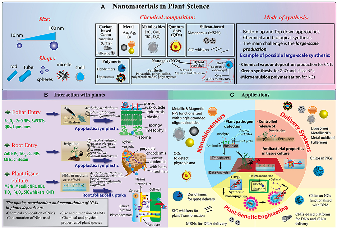
Figure 1. (A) Illustration of NMs grouped into several categories: carbon-based NMs such as fullerenes and carbon nanotubes, including single-walled carbon nanotubes (SWCNTs) or multi-walled carbon nanotubes (MWCNTs); metallic NPs, including metals such as gold (Au), silver (Ag), aluminum (Al); metal oxides (ZnO, CuO, TiO 2 , Fe 2 O3, SiO 2 , etc.); quantum dots (QDs); dendrimers, which are three dimensional polymer network immensely branched with low polydispersity and liposomes and nanogels. With the development of new techniques for chemical synthesis, it is possible to synthesize NMs not only with a symmetrical (spherical) shape but also having a variety of different nanoforms, such as nanoclays (polypropylene nanoclay systems) and nanoemulsions (lipophilic nanoemulsions), tubes, rods, disks, bars, and sheets. (B) Schematization of different NP delivery methods and translocation in plants. Nanoparticle can be administered both at foliar and root system. Once penetrated the external layers, they move through the symplastic or apoplastic routes and reach different organs and tissues. (C) Currently, the main focus of the publications in plant science deals with the use of NPs as biosensors or biomolecules nanocarriers for crop production and protection under controlled conditions. New advances in DNA/miRNA/siRNA delivery have found limited application in plant so far, while new nanotechnology tools addressing technical concerns in genome editing strategies are strongly demanded.
Nanogels (NGs) are a new category of NM with a growing interest in the nanotechnology community. They have excellent physicochemical properties, colloidal stability, high encapsulation capacity of biomolecules (bioconjugation), and stimuli-responsiveness (pH, temperature, etc.). NGs are defined as nano-sized ionic and non-ionic hydrogels made of synthetic or natural polymeric chains, chemically or physically cross-linked ( Molina et al., 2015 ; Neamtu et al., 2017 ). NGs possess a high water content (70–90% of the entire structure), a high degree of porosity and high load capacity. The most common NGs are chitosan, alginate, poly(vinyl alcohol), poly(ethylene oxide), poly(ethyleneimine), poly (vinylpyrrolidone), poly(N-isopropylacrylamide). NGs with hybrid structures, made of polymeric or non-polymeric materials can be obtained ( Molina et al., 2015 ). Hybrid NGs have been classified in: (i) nanomaterial– nanogel, which are synthesized by incorporation of nanosized materials such as magnetic or carbonaceous nanoparticles, and (ii) polymer–nanogel composites, which include interpenetrated networks (IPNs), copolymer, and core-shell particles ( Molina et al., 2015 ). The main advantage of IPNs and copolymer NGs relies on their stimuli-responsiveness, whereas core-shell NGs are more promising for encapsulating biomolecules and drug delivery.
Nanoparticle Uptake, Translocation, and Biological Impact in Plants
Applications of nanotechnology strategies in plants need a preventive accurate evaluation of nanoparticle-plant interactions, including the comprehension of the mechanisms of their uptake, translocation and accumulation, together with the assessment of potential adverse effects on plant growth and development. Plant uptake of NPs is hardly predictable, depending on multiple factors related to the nanoparticle itself (size, chemical composition, net charge and surface functionalization), but also on the application routes, the interactions with environmental components (soil texture, water availability, microbiota), the constraint due to the presence of a cell wall, the physiology and the multifaceted anatomy of individual plant species. Most of the previous studies in plants deal with the uptake of small metal and metal oxide NPs, due to the wide use in industry and to the easy detection and tracking by microscopy techniques ( González-Melendi et al., 2008 ). However, compared to the great wealth of information available in metazoans, only a handful of integrated comparative analyses have been conceived to establish the contribution of the physicochemical features (e.g., size, charge, coatings, etc.) of NPs in plant-nanoparticle interaction ( Zhu et al., 2012 ; Song et al., 2013 ; Moon et al., 2016 ; Vidyalakshmi et al., 2017 ; García-Gómez et al., 2018 ).
Delivery Methods and Primary Interactions at the Plant Surface
Basically, engineered nanomaterials can be applied either to the roots or to the vegetative part of plants, preferentially to the leaves ( Figure 1B ). At the shooting surface, NPs can be taken up passively through natural plant openings with nano- or microscale exclusion size, such as stomata, hydathodes, stigma and bark texture ( Eichert et al., 2008 ; Kurepa et al., 2010 ). However, additional plant anatomical and physiological aspects need to be considered to better understand the dynamics of NP-plant interactions. For instance, shoot surfaces are generally covered by a cuticle made of biopolymers (e.g., cutin, cutan) and associated waxes, which function as a lipophilic barrier to protect above-ground plant primary organs, leaving access only through natural openings ( Figure 1B ). Dynamics of NPs at the cuticle level are poorly investigated, but at present, this barrier appears to be an almost impenetrable layer to nanoparticles, although nano-TiO2 has been shown to be able to produce holes in the cuticle ( Larue et al., 2014 ; Schwab et al., 2016 ). Trichomes on plant organs can affect dynamics at the plant surface by entrapping NP on the plant surface and thus increasing the permanence time of exogenous materials on tissues. Damages and wounds may also function as viable routes for NP internalization in plants in both aerial and hypogeal parts ( Al-Salim et al., 2011 ). Delivery methods also seem to influence NP uptake efficiency in plants. As recently reported, the aerosol application promotes higher internalization rates of different nanoparticles with respect to NP drop cast in watermelon ( Raliya et al., 2016 ). Also, leaf lamina infiltration strategies may force NM penetration in plant tissues as reported for single-walled carbon nanotubes ( Giraldo et al., 2014 ) and resulted to be functional for gene delivery ( Demirer et al., 2018 ). At the root level, rhizodermis lateral root junctions may provide easy access to NMs, especially near the root tip, while upper parts are impermeable due to the presence of suberin ( Chichiriccò and Poma, 2015 ). Generally, the dynamics of NP uptake appear to be more complex in the soil compared to the plant aerial part. Several factors, as the presence of mucilage and exudates, symbiotic organisms, and soil organic matter may influence NPs availability. For instance, root mucilage and exudates normally excreted into the rhizosphere play a dual role: they may promote NP adhesion to the root surface, which in turn may enhance NP internalization rate or, conversely, these gel-like substances may also trigger NP trapping and aggregation ( Avellan et al., 2017 ; Milewska-Hendel et al., 2017 ). Recent observations, by means of X-ray computed nanotomography and enhanced dark-field microscopy combined with hyperspectral imaging, have demonstrated that root border cells and associated mucilage tend to trap gold NPs irrespective of particle charge, while negatively charged NPs are not sequestered by the mucilage of Arabidopsis thaliana root cap and translocate directly into the root tissue ( Avellan et al., 2017 ).
The presence of symbiotic bacteria and fungi in the soil have been demonstrated to play controversial roles as well; in general, they enhance accumulation of different types of heavy metal NPs in true grasses, but reduce nano-Ag and nano-FeO uptake in legumes ( Whiteside et al., 2009 ; Feng et al., 2013 ; Guo and Chi, 2014 ).
Nanoparticle Mobilization in Plant
Once penetrated the plant outer protective layers and regardless of aerial or hypogeal exposure, NMs have two mobilization routes in the plant: apoplastic and symplastic paths ( Figure 1B ). Apoplastic transport occurs outside the plasma membrane through the cell wall and extracellular spaces, whereas symplastic movements involve the transport of water and solutes between the cytoplasm of adjacent cells connected by plasmodesmata and sieve plate pores.
Apoplastic transport has been demonstrated to promote radial movement of NMs, which may move NPs to the root central cylinder and the vascular tissues, and promoting their movement upwards the aerial part ( González-Melendi et al., 2008 ; Larue et al., 2014 ; Sun et al., 2014 ; Zhao et al., 2017 ). This manner of NP translocation is instrumental for applications requiring systemic NP delivery. However, the Casparian strip, a longitudinally oriented layer made of lignin-like structures, prevent the completion of this radial movement in the root endodermis ( Sun et al., 2014 ; Lv et al., 2015 ). To bypass this natural barrier, water and another solute switch from apoplastic to the simplastic path. Similar abilities to circumvent the block at Casparian strip have been documented for different kinds of NPs as reviewed in Schwab et al. (2016) . This may happen especially in those anatomical regions where the Casparian strip is not yet properly formed, such as root tips and root lateral junctions ( Lv et al., 2019 ).
The symplastic transport of NPs requires that at some point NPs penetrate inside the cells. The presence of a rigid plant cell wall creates a physical barrier to the cell entry and makes the intracellular delivery of NPs in plants much more difficult with respect to animal cells. Basically, the cell wall is a multi-layered framework of primarily cellulose/hemicellulose microfibrils and scaffold proteins, creating a porous milieu which acts as a narrow selective filter with a mean diameter <10 nm, with some exception up to 20 nm ( Carpita et al., 1979 ). Actually, this is a critical point and currently represents one of the main hurdles to the design and the implementation of bioengineering tools in plants ( Cunningham et al., 2018 ). However, different types of nanoparticles with a mean diameter between 3 and 50 nm and carbon nanotubes have been demonstrated to easily pass through the cell wall in many plant species ( Liu et al., 2009 ; Kurepa et al., 2010 ; Chang et al., 2013 ; Etxeberria et al., 2019 ).
Subsequent cell internalization may occur preferentially by endocytosis ( Valletta et al., 2014 ; Palocci et al., 2017 ), although alternative cell entry mechanisms, such as those based on pore formation, membrane translocation or carrier proteins already described in cells ( Nel et al., 2009 ; Lin et al., 2010 ; Wang et al., 2012 ) and in invertebrate models ( Marchesano et al., 2013 ) need to be further elucidated in plant cells. For instance, it has been demonstrated that Multi-Walled Carbon nanotubes (MWCNTs) may enter in Catharanthus roseus protoplasts by an endosome-escaping uptake mode ( Serag et al., 2011 ).
Once in the cytoplasm, cell to cell movements of NPs are facilitated by plasmodesmata, membrane-lined cytoplasmic bridges with a flexible diameter (20–50 nm), which ensure membrane and cytoplasmic continuity among cells throughout plant tissues. Transport of NPs with variable sizes through plasmodesmata has been described in Arabidopsis, rice, and poplar plant species ( Lin et al., 2009 ; Geisler-Lee et al., 2013 ; Zhai et al., 2014 ).
Through the symplastic and apoplastic pathways, small particles can reach the xylem and phloem vessels and translocate in the whole plant to different tissues and organs. Remarkably, organs like flowers, fruits and seeds normally have a strong capability to import fluids from the phloem (sink activity) and tend to accumulate NMs. Besides plant toxicity, NP accumulation in specialized organs raises another important issue related to their safe use in human and animal consumption ( Pérez-de-Luque, 2017 ).
Worth mentioning from an application perspective, studies in different crops, such as maize, spinach, cabbage, reported the ability of metal-NPs to penetrate seeds and translocate into the seedlings, without significant effects on seed viability, germination rate, and shoot development. These data suggest the possible use of functional NPs for seed priming and plant growth stimulation, also in limiting environmental conditions ( Zheng et al., 2005 ; Rǎcuciu and Creangǎ, 2009 ; Pokhrel and Dubey, 2013 ).
Nanoparticle Phytotoxicity
The comprehension of NM toxicity in crop plants is still at dawn, but it is crucial for the implementation of innovative agro-nanotech tools and products ( Servin and White, 2016 ). Current NP studies in plants have investigated unrealistic scenarios, such as short-term and high dose exposure, often in model media and plant species, gathering contradictory results ( Miralles et al., 2012 ). Basically, most of the studies have demonstrated that in cultivated species (e.g., tomato, wheat, onion, and zucchini) excess of metal-based NPs trigger an oxidative burst by interfering with the electron transport chain as well as by impairing the reactive oxygen species (ROS) detoxifying machinery, with genotoxic implications ( Dimkpa et al., 2013 ; Faisal et al., 2013 ; Pakrashi et al., 2014 ; Pagano et al., 2016 ). As a consequence, plant secondary metabolism, hormonal balance and growth are often negatively affected. Interestingly, recent transcriptome analyses revealed that exposures to different types of NPs (e.g., zinc oxide, fullerene soot, or titanium dioxide) exposure represses a significant number of genes involved in phosphate-starvation, pathogen and stress responses, with possible negative effects on plant root development and defense mechanisms in A. thaliana . A recent systems biology approach, including omics data from tobacco, rice, rocket salad, wheat, and kidney beans, confirmed that metal NMs provoke a generalized stress response, with the prevalence of oxidative stress components ( Ruotolo et al., 2018 ). These data suggest that further studies based on high-throughput analysis of genetic and metabolic responses, triggered by NP exposure, are necessary to shed light on many aspects of NP phytotoxicity in crops, even in absence of overt toxicity at the phenotypic level ( Majumdar et al., 2015 ). In light of these evidence, it appears fair to exploit for future applications in plants engineered NMs for which a safe profile has been already established in animal systems, such as soft polymeric NPs.
Current Applications in Plant Science
As mentioned above, while nanotechnology innovation is running fast in many fields of life science, smart applications in plant and agricultural science still lag behind ( Wang et al., 2016 ). In this section, we review the most significant current approaches (schematized in Figure 1C ), in particular, those inherent to biosensing, delivery of agrochemicals and genetic engineering. Representative applications for different types of NPs are also listed in Table 1 together with a brief description of their positive effects and drawbacks in plant species.
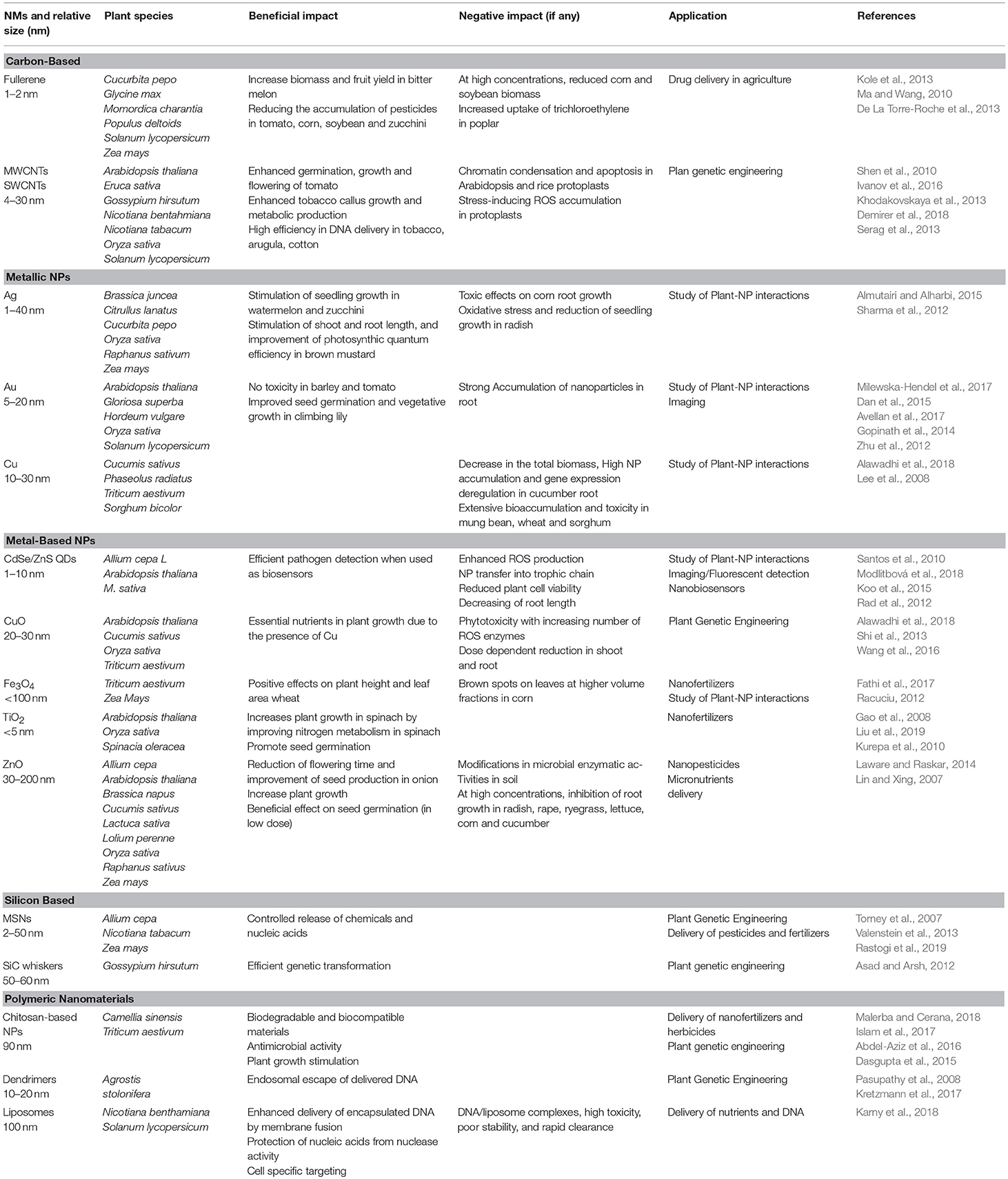
Table 1 . Major applications of different nanomaterials in plant and respective positive/negative impact.
NMs have been applied to develop biosensors or they have been used as “sensing materials” in the fields of crop biotechnology, agriculture, and food industry ( Duhan et al., 2017 ; Chaudhry et al., 2018 ). Different categories of nanosensor types have been tested in plants, including plasmonic nanosensors, fluorescence resonance energy transfer (FRET)-based nanosensors, carbon-based electrochemical nanosensors, nanowire nanosensors and antibody nanosensors. Although the use of nanosensors in plants is at an initial stage ( Rai et al., 2012 ), interesting reports have proposed the use of NMs as tools for detection and quantification of plant metabolic flux, residual of pesticides in food and bacteria, viral and fungal pathogens. Recently, it has been reported the fabrication of a fluorometric optical onion membrane-based sensor for detection of sucrose based on the synthesis of invertase-nanogold clusters embedded in plant membranes ( Bagal-Kestwal et al., 2015 ). In addition, single-walled carbon nanotubes (SWNTs) have been exploited for near-infrared fluorescence monitoring of nitric oxide in A. thaliana ( Giraldo et al., 2014 ). FRET probes conjugated to polystyrene NPs have been also designed to quantify and recognize the phytoalexins ( Dumbrepatil et al., 2010 ).
As above mentioned, NMs-based biosensors are very promising as they allow rapid detection and precise quantification of fungi, bacteria and viruses in plants ( Duhan et al., 2017 ). For example, fluorescent silica NPs combined with antibody was designed for diagnosing Xanthomonas axonopodis pv. vesicatoria , which causes bacterial spot disease in Solanaceae plants ( Yao et al., 2009 ). Recently, Au NPs have been proposed from Lau et al. as DNA biochemical labels to detect Pseudomonas syringae in A. thaliana by differential pulse voltammetry (DPV) on disposable screen-printed carbon electrodes ( Lau et al., 2017 ). Similarly, fluorescently labeled-DNA oligonucleotide conjugated to Au NPs were employed in the diagnosis of the phytoplasma associated with the flavescence dorée disease of grapevine ( Firrao et al., 2005 ). Finally, smart nanosensors are also available for mycotoxin detection; for instance, the 4mycosensor is a competitive antibody-based assay successfully introduced in the market to test the presence of ZEA, T-2/HT-2, DON, and FB1/FB2 mycotoxin residues in corn, wheat, oat and barley ( Lattanzio and Nivarlet, 2017 ).
Controlled Release of Agrochemicals and Nutrients
NMs can be applied to the soil as nanostructured fertilizers (nanofertilizers, as for Fe, Mn, Zn, Cu, Mo NPs) or can be used as enhanced delivery systems to improve the uptake and the performance of conventional fertilizers (nutrients and phosphates) ( Liu and Lal, 2015 ). Even though nanofertilizers and NM-enhanced fertilizers are very promising for agriculture, the use of nanotechnology in fertilizer supply is very scanty ( DeRosa et al., 2010 ).
Hydroxyapatite nanoparticles, used as phosphorous nanofertilizers, enhance the soybean growth rate and seed yield by 33 and 20%, compared to a regular P fertilizer ( Liu and Lal, 2015 ). In addition, nanofertilizers can be released at slower rates which may contribute to maintain the soil fertility by reducing the transport of these nutrients into a runoff or ground water and decreasing the risks of environmental pollution and toxic effects due to their over-application ( Liu and Lal, 2015 ).
Metallic nanoparticles based on Iron oxide, ZnO, TiO 2 , and copper have been directly applied as nanofertilizers in soil by irrigation or via foliar applications in different plants, such as mung bean plant, cucumber and rape ( Gao et al., 2006 ; Tarafdar et al., 2014 ; Saharan et al., 2016 ; Verma et al., 2018 ). Similarly, MWNTs used as soil supplements increased twice the number of flowers and fruits in tomato plants likely through the activation of genes/proteins essential for plant growth and development ( Khodakovskaya et al., 2013 ). Despite these intriguing evidence, the use of nanofertilizers is still debatable. Accumulation in treated soils may pose a threat to soil microbial communities such as small invertebrates, bacteria and fungi ( Frenk et al., 2013 ; Waalewijn-Kool et al., 2013 ; Shen et al., 2015 ; Simonin et al., 2016 ; Goncalves et al., 2017 ). This impact on the agro-ecosystem reasonably discourages the use of metallic nanoparticles in agriculture.
Only recently, a natural polymer, such as chitosan NPs, have been used for controlled release of nitrogen, phosphorus and potassium in wheat by foliar uptake ( Abdel-Aziz et al., 2016 ). The use of organic NPs is more acceptable in terms of environmental pollution. However, their effective advantages for nutrient supply over traditional fertilization methods need more robust evidence ( Liu and Lal, 2015 ).
On the other hand, pesticides delivered by nanomaterials generally have increased stability and solubility and enable slow release and effective targeted delivery in pest management ( Duhan et al., 2017 ). Organic and polymeric NPs in the form of nanospheres or nanocapsules have been used as nanocarriers for herbicide distribution ( Tanaka et al., 2012 ). In particular, polymeric NPs, such as Poly(epsilon-caprolactone), present good properties of biocompatibility and have been repeatedly used for the encapsulation of atrazine herbicide ( Tanaka et al., 2012 ). In another study, chitosan nanoparticles loaded with three triazine herbicides have shown reduced environmental impact and low genotoxic effects in Allium cepa ( Grillo et al., 2015 ).
Nanomaterials for Plant Genetic Engineering
As stated above, the cell wall represents a barrier to the delivery of exogenous biomolecules in plant cells. To overcome this barrier and achieve plant genetic transformation, different strategies based on Agrobacterium transformation or biolistic methods are worldwide used for DNA delivery in plant cells. Limitations to these approaches rely on narrow host range and plant extensive damages, which often inhibit plant development.
Most of the pioneering studies for nanomaterial-based plant genetic engineering have been conducted in plant cell cultures. For example, Silicon Carbide-Mediated Transformation has been reported as a successful approach to deliver DNA in different calli (tobacco, maize, rice, soybean and cotton) ( Armstrong and Green, 1985 ; Wang et al., 1995 ; Serik et al., 1996 ; Asad and Arsh, 2012 ; Lau et al., 2017 ).
Although lagged behind the advancements achieved in animal systems, results reported recently in plants are proving that NMs may overcome the barrier of the cell wall in adult plants and reduce the drawbacks associated with current transgene delivery systems.
One seminal study proved that dsRNA of different plant viruses can be loaded on non-toxic, degradable, layered double hydroxide (LDH) clay nanosheets or BioClay. The dsRNAs and/or their RNA breakdown products provide protection against the Cauliflower Mosaic Virus (CMV) in sprayed tobacco leaves, but they also confer systemic protection to newly emerged, unsprayed leaves on viral challenge 20 days after a single spray treatment in tobacco ( Mitter et al., 2017 ). More in general, this is a proof of concept for species-independent and passive delivery of genetic material, without transgene integration, into plant cells for different biotechnology applications in plants.
A successful stable genetic transformation has been achieved in cotton plants via magnetic nanoparticles (MNPs). β-glucuronidase (GUS) reporter gene- MNP complex were infiltrated into cotton pollen grains by magnetic force, without compromising pollen viability. Through pollination with magnetofected pollen, cotton transgenic plants were successfully generated and exogenous DNA was successfully integrated into the genome, effectively expressed, and stably inherited in the offspring obtained by selfing ( Zhao et al., 2017 ).
In another recent paper, carbon nanotubes scaffolds applied to external plant tissue by infusion were used to deliver linear and plasmid DNA, as well as siRNA, in Nicotiana benthamiana, Eruca sativa, Triticum aestivum , and Gossypium hirsutum leaves and in E. sativa protoplasts, resulting in a strong transient Green Fluorescent Protein (GFP) expression. Moreover, the same authors reported that small interfering RNA (siRNA) was delivered to N . benthamiana plants constitutively expressing GFP, causing a 95% silencing of this gene ( Demirer et al., 2018 ).
The first and promising approach of genome editing mediated by mesoporous silica nanoparticles (MSNs) has been recently proposed. MSNs have used as carriers to deliver Cre recombinase in Zea mays immature embryos, carrying loxP sites integrated into chromosomal DNA. After the biolistic introduction of engineered MSNs in plant tissues, the loxP was correctly recombined establishing a successful genome editing ( Valenstein et al., 2013 ).
Conclusions and Future Perspectives
Herein, we have discussed various facets of using NMs in plant sciences. In the last years, it has been demonstrated that nanotechnology has made huge progress in the synthesis of NMs and their application in medicine for diagnosis and therapy. On the other side, the application of NMs for plants is still poor. Recent outcomes and current applications suggest that more studies are necessary for this direction to optimize the synthesis and biofunctionalization of NMs for plant applications, but also to elucidate deeper the mechanisms of plant uptake and improving the sustainability for agro-ecosystems and human health. Interestingly, applications need to be extended to address uncovered important aspects of plant physiology. For instance, nanobiosensors for detecting secondary metabolites or phytoregulators in real time may provide advances in monitoring plant development and interactions with the environment, especially in limiting growth conditions.
Despite the huge progress in plant genetics, the delivery of exogenous DNA and/or enzymes for genome editing remain a big challenge. New strategies based on nanoparticle-mediated clustered regularly interspersed palindromic repeats—CRISPR associated proteins (CRISPR-Cas9) technology, as those tested in other biological systems ( Lee et al., 2017 ; Glass et al., 2018 ), would provide ground-breaking innovation in plant genetics.
On the base of consolidated evidence reported in cell and animal models, soft materials, like nanogels, and polymeric nanostructures should be further exploited as favorable candidates to develop new strategies for controlled release of biomolecules and plant genome editing. Owing to their safe profile, high loading capacity and excellent cargo protection from degradation polymeric and hydrogel-based NPs have shown undeniable advantages in drug delivery. Moreover, this kind of NMs has been elegantly employed to achieve a controlled (spatial and temporal) release of cargos triggered by external stimuli (e.g., UV, NIR, acoustic waves etc.) ( Ma et al., 2013 ; Ambrosone et al., 2016 ; Linsley and Wu, 2017 ) in cell and animal models. These outstanding results suggest that the huge potential of soft nanomaterials remains almost unexplored in plants. Besides a few successful attempts for agrochemicals delivery above-mentioned and listed in Table 1 , more efforts are needed to design strategies and smart tools based on polymeric or hybrid materials for applications in plants. Of course, a careful analysis of manufacturing scalability and cost-effectiveness needs to be undertaken before the extensive use of polymeric nanomaterials in agriculture.
As a final remark, the delay in plant nanotechnology might be overcome by encouraging the activation of multidisciplinary approaches for the design and the synthesis of smart nanomaterials. To this aim, joint collaborative initiatives, merging complementary professional competencies such those of plant biologists, geneticists, chemists, biochemists, and engineers, may disclose new horizons in phytonanotechnology.
Author Contributions
IS and AA conceived the idea and organized this mini review. All authors wrote the manuscript and approved the contents for publication.
Conflict of Interest Statement
The authors declare that the research was conducted in the absence of any commercial or financial relationships that could be construed as a potential conflict of interest.
Abdel-Aziz, H. M. M., Hasaneen, M. N. A., and Omer, A. M. (2016). Nano chitosan-NPK fertilizer enhances the growth and productivity of wheat plants grown in sandy soil. Spanish J. Agric. Res. 14, 1–9. doi: 10.5424/sjar/2016141-8205
CrossRef Full Text | Google Scholar
Abedini, A., Daud, A. R., Hamid, M. A. A., Othman, N. K., and Saion, E. (2013). A review on radiation-induced nucleation and growth of colloidal metallic nanoparticles. Nanoscale Res. Lett. 8, 1–10. doi: 10.1186/1556-276X-8-474
PubMed Abstract | CrossRef Full Text | Google Scholar
Alawadhi, H., Ramamoorthy, K., Mosa, K. A., Elnaggar, A., Ibrahim, E., El-Naggar, M., et al. (2018). Copper nanoparticles induced genotoxicty, oxidative stress, and changes in Superoxide Dismutase (SOD) gene expression in cucumber ( Cucumis sativus ) plants. Front. Plant Sci. 9:872. doi: 10.3389/fpls.2018.00872
Almutairi, Z. M., and Alharbi, A. (2015). Effect of silver nanoparticles on seed germination of crop plants. Int. J. Biol. Biomol. Agric. Food Biotechnol. Eng. 9, 572–576. doi: 10.24297/jaa.v4i1.4295
Al-Salim, N., Barraclough, E., Burgess, E., Clothier, B., Deurer, M., Green, S., et al. (2011). Quantum dot transport in soil, plants, and insects. Sci. Total Environ. 409:3237–3248.doi: 10.1016/j.scitotenv.2011.05.017
Ambrosone, A., Marchesano, V., Carregal-Romero, S., Intartaglia, D., Parak, W. J., and Tortiglione, C. (2016). Control of Wnt/β-catenin signaling pathway in vivo via light responsive capsules. ACS Nano 10, 4828–4834. doi: 10.1021/acsnano.5b07817
Armstrong, C. L., and Green, C. E. (1985). Establishment and maintenance of friable, embryogenic maize callus and the involvement of L-proline. Planta 164:207–214. doi: 10.1007/BF00396083
Asad, S., and Arsh, M. (2012). “Silicon carbide whisker-mediated plant transformation,” in Properties and Applications of Silicon Carbide , ed R. Gerhardt (Rijeka: BoD-Books on Deman), 1–16. doi: 10.5772/15721
Astm E2456 - 06 (2012). (2006)Standard Terminol. Relat. to Nanotechnol. 06, 5–6. doi: 10.1520/E2456-06R12
Avellan, A., Schwab, F., Masion, A., Chaurand, P., Borschneck, D., Vidal, V., et al. (2017). Nanoparticle uptake in plants: gold nanomaterial localized in roots of Arabidopsis thaliana by X-ray computed nanotomography and hyperspectral imaging. Environ. Sci. Technol. 51, 8682–8691. doi: 10.1021/acs.est.7b01133
Bagal-Kestwal, D., Kestwal, R. M., and Chiang, B. H. (2015). Invertase-nanogold clusters decorated plant membranes for fluorescence-based sucrose sensor. J. Nanobiotechnology 13:30. doi: 10.1186/s12951-015-0089-1
Baskar, V., Meeran, S., Shabeer, S. T. K, and Subramani Sruthi, Ali, J. (2018). Historic review on modern herbal nanogel formulation and delivery methods. Int. J. Pharm. Pharm. Sci. 10, 1–10. doi: 10.22159/ijpps.2018v10i10.23071
Bruchez, M., Moronne, M., Gin, P., Weiss, S., and Alivisatos, A. P. (1998). Semiconductor nanocrystals as fluorescent biological labels. Science 281, 2013–2016. doi: 10.1126/science.281.5385.2013
Carpita, N., Sabularse, D., Montezinos, D., and Delmer, D. P. (1979). Determination of the pore size of cell walls of living plant cells. Science 205, 1144–1147. doi: 10.1126/science.205.4411.1144
Chang, F. P., Kuang, L. Y., Huang, C. A., Jane, W. N., Hung, Y., Hsing, Y. I. C., et al. (2013). A simple plant gene delivery system using mesoporous silica nanoparticles as carriers. J. Mater. Chem. B 1, 5279–5287. doi: 10.1039/c3tb20529k
Chaudhry, N., Dwivedi, S., Chaudhry, V., Singh, A., Saquib, Q., Azam, A., et al. (2018). Bio-inspired nanomaterials in agriculture and food: current status, foreseen applications and challenges. Microb. Pathog. 123, 196–200. doi: 10.1016/j.micpath.2018.07.013
Chichiriccò, G., and Poma, A. (2015). Penetration and toxicity of nanomaterials in higher plants. Nanomaterials 5, 851–873. doi: 10.3390/nano5020851
Cunningham, F. J., Goh, N. S., Demirer, G. S., Matos, J. L., and Landry, M. P. (2018). Nanoparticle-mediated delivery towards advancing plant genetic engineering. Trends Biotechnol. 36, 882–897. doi: 10.1016/j.tibtech.2018.03.009
Dan, Y., Zhang, W., Xue, R., Ma, X., Stephan, C., and Shi, H. (2015). Characterization of gold nanoparticle uptake by tomato plants using enzymatic extraction followed by single-particle inductively coupled plasma-mass spectrometry analysis. Environ. Sci. Technol. 49, 3007–3014. doi: 10.1021/es506179e
Dasgupta, A., Chakraborty, N., Panda, K., Chandra, S., Acharya, K., and Sarkar, J. (2015). Chitosan nanoparticles: a positive modulator of innate immune responses in plants. Sci. Rep. 5:15195. doi: 10.1038/srep15195
De La Torre-Roche, R., Hawthorne, J., Deng, Y., Xing, B., Cai, W., Newman, L. A., et al. (2013). Multiwalled carbon nanotubes and C60 fullerenes differentially impact the accumulation of weathered pesticides in four agricultural plants. Environ. Sci. Technol. 47, 12539–12547. doi: 10.1021/es4034809
Demirer, G. S., Zhang, H., Matos, J., Goh, N., Cunningham, F. J., Sung, Y., et al. (2018). High aspect ratio nanomaterials enable delivery of functional genetic material without DNA integration in mature plants. bioRxiv 10, 1–32. doi: 10.1101/179549
DeRosa, M. C., Monreal, C., Schnitzer, M., Walsh, R., and Sultan, Y. (2010). Nanotechnology in fertilizers. Nat. Nanotechnol. 5, 91–91. doi: 10.1038/nnano.2010.2
Dimkpa, C. O., McLean, J. E., Martineau, N., Britt, D. W., Haverkamp, R., and Anderson, A. J. (2013). Silver nanoparticles disrupt wheat ( Triticum aestivum L.) growth in a sand matrix. Environ. Sci. Technol. 47, 1082–1090. doi: 10.1021/es302973y
Duhan, J. S., Kumar, R., Kumar, N., Kaur, P., Nehra, K., and Duhan, S. (2017). Nanotechnology: the new perspective in precision agriculture. Biotechnol. Rep. 15, 11–23. doi: 10.1016/j.btre.2017.03.002
Dumbrepatil, A. B., Lee, S. G., Chung, S. J., Lee, M. G., Park, B. C., Kim, T. J., et al. (2010). Development of a nanoparticle-based FRET sensor for ultrasensitive detection of phytoestrogen compounds. Analyst 135, 2879–2886. doi: 10.1039/c0an00385a
Eichert, T., Kurtz, A., Steiner, U., and Goldbach, H. E. (2008). Size exclusion limits and lateral heterogeneity of the stomatal foliar uptake pathway for aqueous solutes and water-suspended nanoparticles. Physiol. Plant. 134, 151–160. doi: 10.1111/j.1399-3054.2008.01135.x
Etxeberria, E., Gonzalez, P., Bhattacharya, P., Sharma, P., and Ke, P. C. (2019). Determining the size exclusion for nanoparticles in citrus leaves. Hort Sci. 51, 732–737. doi: 10.21273/HORTSCI.51.6.732
Faisal, M., Saquib, Q., Alatar, A. A., Al-Khedhairy, A. A., Hegazy, A. K., and Musarrat, J. (2013). Phytotoxic hazards of NiO-nanoparticles in tomato: a study on mechanism of cell death. J. Hazard. Mater. 250–251, 318–332. doi: 10.1016/j.jhazmat.2013.01.063
Fathi, A., Zahedi, M., Torabian, S., and Khoshgoftar, A. (2017). Response of wheat genotypes to foliar spray of ZnO and Fe2O3 nanoparticles under salt stress. J. Plant Nutr. 40, 1376–1385. doi: 10.1080/01904167.2016.1262418
Feng, Y., Cui, X., He, S., Dong, G., Chen, M., Wang, J., et al. (2013). The role of metal nanoparticles in influencing arbuscular mycorrhizal fungi effects on plant growth. Environ. Sci. Technol. 47, 9496–9504. doi: 10.1021/es402109n
Firrao, G., Moretti, M., Ruiz Rosquete, M., Gobbi, E., and Locci, R. (2005). Nanobiotransducer for detecting flavescence dorée phytoplasma. J. Plant Pathol. 87, 101–107. doi: 10.4454/jpp.v87i2.903
Frenk, S., Ben-Moshe, T., Dror, I., Berkowitz, B., and Minz, D. (2013). Effect of metal oxide nanoparticles on microbial community structure and function in two different soil types. PLoS ONE 8:e84441. doi: 10.1371/journal.pone.0084441
Gao, F., Hong, F., Liu, C., Zheng, L., Su, M., Wu, X., et al. (2006). Mechanism of nano-anatase TiO2 on promoting photosynthetic carbon reaction of spinach: inducing complex of Rubisco-Rubisco activase. Biol. Trace Elem. Res. 111, 239–253. doi: 10.1385/BTER:111:1:239
Gao, F., Liu, C., Qu, C., Zheng, L., Yang, F., Su, M., et al. (2008). Was improvement of spinach growth by nano-TiO2 treatment related to the changes of Rubisco activase? BioMetals 21, 211–217. doi: 10.1007/s10534-007-9110-y
García-Gómez, C., Obrador, A., González, D., Babín, M., and Fernández, M. D. (2018). Comparative study of the phytotoxicity of ZnO nanoparticles and Zn accumulation in nine crops grown in a calcareous soil and an acidic soil. Sci. Total Environ. 644, 770–780. doi: 10.1016/j.scitotenv.2018.06.356
Geisler-Lee, J., Wang, Q., Yao, Y., Zhang, W., Geisler, M., Li, K., et al. (2013). Phytotoxicity, accumulation and transport of silver nanoparticles by Arabidopsis thaliana . Nanotoxicology 7, 323–337. doi: 10.3109/17435390.2012.658094
Ghosh Chaudhuri, R., and Paria, S. (2012). Core/shell nanoparticles: classes, properties, synthesis mechanisms, characterization, and applications. Chem. Rev. 112, 2373–2433. doi: 10.1021/cr100449n
Giraldo, J. P., Landry, M. P., Faltermeier, S. M., McNicholas, T. P., Iverson, N. M., Boghossian, A. A., et al. (2014). Plant nanobionics approach to augment photosynthesis and biochemical sensing. Nat. Mater. 13, 400–408. doi: 10.1038/nmat3890
Glass, Z., Lee, M., Li, Y., and Xu, Q. (2018). Engineering the delivery system for CRISPR-based genome editing. Trends Biotechnol. 36, 173–185. doi: 10.1016/j.tibtech.2017.11.006
Goncalves, M. F. M., Gomes, S. I. L., Scott-Fordsmand, J. J., and Amorim, M. J. B. (2017). Shorter lifetime of a soil invertebrate species when exposed to copper oxide nanoparticles in a full lifespan exposure test. Sci. Rep. 7:1355. doi: 10.1038/s41598-017-01507-8
González-Melendi, P., Fernández-Pacheco, R., Coronado, M. J., Corredor, E., Testillano, P. S., Risueño, M. C., et al. (2008). Nanoparticles as smart treatment-delivery systems in plants: assessment of different techniques of microscopy for their visualization in plant tissues. Ann. Bot. 101, 187–195. doi: 10.1093/aob/mcm283
Gopinath, K., Gowri, S., Karthika, V., and Arumugam, A. (2014). Green synthesis of gold nanoparticles from fruit extract of Terminalia arjuna , for the enhanced seed germination activity of Gloriosa superba . J. Nanostructure Chem. 4:115. doi: 10.1007/s40097-014-0115-0
Grillo, R., Clemente, Z., Oliveira, J. L., de Campos, E. V. R., Chalupe, V. C., Jonsson, C. M., et al. (2015). Chitosan nanoparticles loaded the herbicide paraquat: the influence of the aquatic humic substances on the colloidal stability and toxicity. J. Hazard. Mater. 286, 562–572. doi: 10.1016/j.jhazmat.2014.12.021
Guo, J., and Chi, J. (2014). Effect of Cd-tolerant plant growth-promoting rhizobium on plant growth and Cd uptake by Lolium multiflorum Lam. and Glycine max (L.) Merr. in Cd-contaminated soil. Plant Soil 375, 205–214. doi: 10.1007/s11104-013-1952-1
Islam, P., Water, J. J., Bohr, A., and Rantanen, J. (2017). Chitosan-based nano-embedded microparticles: impact of Nanogel composition on physicochemical properties. Pharmaceutics 9, 1–12. doi: 10.3390/pharmaceutics9010001
Ivanov, I., Khodakovskaya, M., Dervishi, E., Lahiani, M. H., and Chen, J. (2016). Comparative study of plant responses to carbon-based nanomaterials with different morphologies. Nanotechnology 27:265102. doi: 10.1088/0957-4484/27/26/265102
Jahan, S., Alias, Y. B., Bakar, A. F. B. A., and Yusoff, I. B. (2018). Toxicity evaluation of ZnO and TiO2 nanomaterials in hydroponic red bean ( Vigna angularis ) plant: physiology, biochemistry and kinetic transport. J. Environ. Sci. 72, 140–152. doi: 10.1016/j.jes.2017.12.022
Jeevanandam, J., Barhoum, A., Chan, Y. S., Dufresne, A., and Danquah, M. K. (2018). Review on nanoparticles and nanostructured materials: history, sources, toxicity and regulations. Beilstein J. Nanotechnol. 9, 1050–1074. doi: 10.3762/bjnano.9.98
Karny, A., Zinger, A., Kajal, A., Shainsky-Roitman, J., and Schroeder, A. (2018). Therapeutic nanoparticles penetrate leaves and deliver nutrients to agricultural crops. Sci. Rep. 8:7589. doi: 10.1038/s41598-018-25197-y
Khan, M. N., Mobin, M., Abbas, Z. K., AlMutairi, K. A., and Siddiqui, Z. H. (2017). Role of nanomaterials in plants under challenging environments. Plant Physiol. Biochem. 110, 194–209. doi: 10.1016/j.plaphy.2016.05.038
Khodakovskaya, M. V., Kim, B. S., Kim, J. N., Alimohammadi, M., Dervishi, E., Mustafa, T., et al. (2013). Carbon nanotubes as plant growth regulators: effects on tomato growth, reproductive system, and soil microbial community. Small 9, 115–123. doi: 10.1002/smll.201201225
Kole, C., Kole, P., Randunu, K. M., Choudhary, P., Podila, R., Ke, P. C., et al. (2013). Nanobiotechnology can boost crop production and quality: first evidence from increased plant biomass, fruit yield and phytomedicine content in bitter melon ( Momordica charantia ). BMC Biotechnol. 13:37. doi: 10.1186/1472-6750-13-37
Koo, Y., Wang, J., Zhang, Q., Zhu, H., Chehab, E. W., Colvin, V. L., et al. (2015). Fluorescence reports intact quantum dot uptake into roots and translocation to leaves of arabidopsis thaliana and subsequent ingestion by insect herbivores. Environ. Sci. Technol. 49, 626–632. doi: 10.1021/es5050562
Kretzmann, J. A., Ho, D., Evans, C. W., Plani-Lam, J. H. C., Garcia-Bloj, B., Mohamed, A. E., et al. (2017). Synthetically controlling dendrimer flexibility improves delivery of large plasmid DNA. Chem. Sci. 8, 2923–2930. doi: 10.1039/C7SC00097A
Kurepa, J., Paunesku, T., Vogt, S., Arora, H., Rabatic, B. M., Lu, J., et al. (2010). Uptake and distribution of ultrasmall anatase TiO2 alizarin red s nanoconjugates in arabidopsis thaliana. Nano Lett. 10, 2296–2302. doi: 10.1021/nl903518f
Larue, C., Castillo-Michel, H., Sobanska, S., Cécillon, L., Bureau, S., Barthès, V., et al. (2014). Foliar exposure of the crop Lactuca sativa to silver nanoparticles: evidence for internalization and changes in Ag speciation. J. Hazard. Mater. 264, 98–106. doi: 10.1016/j.jhazmat.2013.10.053
Lattanzio, V. M. T., and Nivarlet, N. (2017). Multiplex dipstick immunoassay for semiquantitative determination of fusarium mycotoxins in oat in Methods Mol Biol. 1536, 137–142. doi: 10.1007/978-1-4939-6682-0_10
Lau, H. Y., Wu, H., Wee, E. J. H., Trau, M., Wang, Y., and Botella, J. R. (2017). Specific and sensitive isothermal electrochemical biosensor for plant pathogen DNA detection with colloidal gold nanoparticles as probes. Sci. Rep. 7:38896. doi: 10.1038/srep38896
Laware, S. L., and Raskar, S. (2014). Influence of zinc oxide nanoparticles on growth, flowering and seed productivity in onion. Int. J. Curr. Microbiol. App. Sci. 3, 874–881.
Google Scholar
Lee, K., Conboy, M., Park, H. M., Jiang, F., Kim, H. J., Dewitt, M. A., et al. (2017). Nanoparticle delivery of Cas9 ribonucleoprotein and donor DNA in vivo induces homology-directed DNA repair. Nat. Biomed. Eng. 1, 889–901. doi: 10.1038/s41551-017-0137-2
Lee, W. M., An, Y.-J., Yoon, H., and Kweon, H.-S. (2008). Toxicity and bioavailability of copper nanoparticles to the terrestrial plants mung bean ( Phaseolus radiatus ) and wheat. Environ. Toxicol. Chem. 27, 1915–1921. doi: 10.1897/07-481.1
Lin, D., and Xing, B. (2007). Phytotoxicity of nanoparticles: Inhibition of seed germination and root growth. Environ. Pollut. 150, 243–250. doi: 10.1016/j.envpol.2007.01.016
Lin, J., Zhang, H., Chen, Z., and Zheng, Y. (2010). Penetration of lipid membranes by gold nanoparticles: insights into cellular uptake, cytotoxicity, and their relationship. ACS Nano 4, 5421–5429. doi: 10.1021/nn1010792
Lin, S., Reppert, J., Hu, Q., Hudson, J. S., Reid, M. L., Ratnikova, T. A., et al. (2009). Uptake, translocation, and transmission of carbon nanomaterials in rice plants. Small 5, 1128–1132. doi: 10.1002/smll.200801556
Linsley, C. S., and Wu, B. M. (2017). Recent advances in light-responsive on-demand drug-delivery systems. Ther. Deliv. 8, 89–107. doi: 10.4155/tde-2016-0060
Liu, J., Williams, P. C., Goodson, B. M., Geisler-Lee, J., Fakharifar, M., and Gemeinhardt, M. E. (2019). TiO2nanoparticles in irrigation water mitigate impacts of aged Agnanoparticles on soil microorganisms, Arabidopsis thalianaplants , and Eisenia fetida earthworms. Environ. Res. 172, 202–215. doi: 10.1016/j.envres.2019.02.010
Liu, Q., Chen, B., Wang, Q., Shi, X., Xiao, Z., Lin, J., et al. (2009). Carbon nanotubes as molecular transporters for walled plant cells. Nano Lett. 9, 1007–1010. doi: 10.1021/nl803083u
Liu, R., and Lal, R. (2015). Potentials of engineered nanoparticles as fertilizers for increasing agronomic productions. Sci. Total Environ. 514, 131–139. doi: 10.1016/j.scitotenv.2015.01.104
Lv, J., Christie, P., and Zhang, S. (2019). Uptake, translocation, and transformation of metal-based nanoparticles in plants: recent advances and methodological challenges. Environ. Sci. Nano 6, 41–59. doi: 10.1039/C8EN00645H
Lv, J., Zhang, S., Luo, L., Zhang, J., Yang, K., and Christied, P. (2015). Accumulation, speciation and uptake pathway of ZnO nanoparticles in maize. Environ. Sci. Nano 2, 68–77. doi: 10.1039/C.4E.N.00064A
Ma, J., Du, L. F., Chen, M., Wang, H. H., Xing, L. X., Jing, L. F., et al. (2013). Drug-loaded nano-microcapsules delivery system mediated by ultrasound-targeted microbubble destruction: a promising therapy method. Biomed. Rep. 1, 506–510. doi: 10.3892/br.2013.110
Ma, X., and Wang, C. (2010). Fullerene nanoparticles affect the fate and uptake of trichloroethylene in phytoremediation systems. Environ. Eng. Sci. 27, 989–992. doi: 10.1089/ees.2010.0141
Majumdar, S., Almeida, I. C., Arigi, E. A., Choi, H., VerBerkmoes, N. C., Trujillo-Reyes, J., et al. (2015). Environmental effects of nanoceria on seed production of common bean ( Phaseolus vulgaris ): a proteomic analysis. Environ. Sci. Technol. 49, 13283–13293. doi: 10.1021/acs.est.5b03452
Malerba, M., and Cerana, R. (2018). Recent advances of chitosan applications in plants. Polymers. 10, 1–10. doi: 10.3390/polym10020118
Marchesano, V., Hernandez, Y., Salvenmoser, W., Ambrosone, A., Tino, A., Hobmayer, B., et al. (2013). Imaging inward and outward trafficking of gold nanoparticles in whole animals. ACS Nano 7, 2431–2442. doi: 10.1021/nn305747e
Milewska-Hendel, A., Zubko, M., Karcz, J., Stróz, D., and Kurczynska, E. (2017). Fate of neutral-charged gold nanoparticles in the roots of the Hordeum vulgare L. cultivar Karat. Sci. Rep. 7:3014. doi: 10.1038/s41598-017-02965-w
Miralles, P., Church, T. L., and Harris, A. T. (2012). Toxicity, uptake, and translocation of engineered nanomaterials in vascular plants. Environ. Sci. Technol. 46, 9224–9239. doi: 10.1021/es202995d
Mitter, N., Worrall, E. A., Robinson, K. E., Li, P., Jain, R. G., Taochy, C., et al. (2017). Clay nanosheets for topical delivery of RNAi for sustained protection against plant viruses. Nat. Plants 3:16207. doi: 10.1038/nplants.2016.207
Modlitbová, P., Porízka, P., Novotný, K., Drbohlavová, J., Chamradová, I., Farka, Z., et al. (2018). Short-term assessment of cadmium toxicity and uptake from different types of Cd-based quantum dots in the model plant Allium cepa L. Ecotoxicol. Environ. Saf. 153, 23–31. doi: 10.1016/j.ecoenv.2018.01.044
Molina, M., Asadian-Birjand, M., Balach, J., Bergueiro, J., Miceli, E., and Calderón, M. (2015). Stimuli-responsive nanogel composites and their application in nanomedicine. Chem. Soc. Rev. 44, 6161–6186. doi: 10.1039/C5CS00199D
Moon, J. W., Phelps, T. J., Fitzgerald, C. L., Lind, R. F., Elkins, J. G., Jang, G. G., et al. (2016). Manufacturing demonstration of microbially mediated zinc sulfide nanoparticles in pilot-plant scale reactors. Appl. Microbiol. Biotechnol. 100, 7921–7931. doi: 10.1007/s00253-016-7556-y
Nath, S., Kaittanis, C., Tinkham, A., and Perez, J. M. (2008). Dextran-coated gold nanoparticles for the assessment of antimicrobial susceptibility. Anal. Chem. 80, 1033–1038. doi: 10.1021/ac701969u
Neamtu, I., Rusu, A. G., Diaconu, A., Nita, L. E., and Chiriac, A. P. (2017). Basic concepts and recent advances in nanogels as carriers for medical applications. Drug Deliv. 24, 539–557. doi: 10.1080/10717544.2016.1276232
Nel, A. E., Mädler, L., Velegol, D., Xia, T., Hoek, E. M. V., Somasundaran, P., et al. (2009). Understanding biophysicochemical interactions at the nano-bio interface. Nat. Mater. 8, 543–557. doi: 10.1038/nmat2442
Pagano, L., Servin, A. D., De La Torre-Roche, R., Mukherjee, A., Majumdar, S., Hawthorne, J., et al. (2016). Molecular response of crop plants to engineered nanomaterials. Environ. Sci. Technol. 50, 7198–7207. doi: 10.1021/acs.est.6b01816
Pakrashi, S., Jain, N., Dalai, S., Jayakumar, J., Chandrasekaran, P. T., Raichur, A. M., et al. (2014). In vivo genotoxicity assessment of titanium dioxide nanoparticles by Allium cepa root tip assay at high exposure concentrations. PLoS ONE 9:e98828. doi: 10.1371/journal.pone.0087789
Palocci, C., Valletta, A., Chronopoulou, L., Donati, L., Bramosanti, M., Brasili, E., et al. (2017). Endocytic pathways involved in PLGA nanoparticle uptake by grapevine cells and role of cell wall and membrane in size selection. Plant Cell Rep. 36, 1917–1928. doi: 10.1007/s00299-017-2206-0
Pasupathy, K., Lin, S., Hu, Q., Luo, H., and Ke, P. C. (2008). Direct plant gene delivery with a poly(amidoamine) dendrimer. Biotechnol. J. 3, 1078–1082. doi: 10.1002/biot.200800021
Pérez-de-Luque, A. (2017). Interaction of nanomaterials with plants: what do we need for real applications in agriculture? Front. Environ. Sci. 5:12. doi: 10.3389/fenvs.2017.00012
Perrault, S. D., Walkey, C., Jennings, T., Fischer, H. C., and Chan, W. C. W. (2009). Mediating tumor targeting efficiency of nanoparticles through design - nano letters. Nano Lett. 9, 1909–1915. doi: 10.1021/nl900031y
Pokhrel, L. R., and Dubey, B. (2013). Evaluation of developmental responses of two crop plants exposed to silver and zinc oxide nanoparticles. Sci. Total Environ. 452–453, 321–332. doi: 10.1016/j.scitotenv.2013.02.059
Racuciu, M. (2012). Iron oxide nanoparticles coated with β-cyclodextrin polluted of Zea mays plantlets. Nanotechnol. Dev. 2:6. doi: 10.4081/nd.2012.e6
Rǎcuciu, M., and Creangǎ, D. E. (2009). Cytogenetical changes induced by β-cyclodextrin coated nanoparticles in plant seeds. Rom. Reports Phys. 54, 125–131.
Rad, F., Mohsenifar, A., Tabatabaei, M., Safarnejad, M. R., Shahryari, F., Safarpour, H., et al. (2012). Detection of Candidatus phytoplasma aurantifolia with a quantum dots fret-based biosensor. J. Plant Pathol. 94, 525–534. doi: 10.4454/JPP.FA.2012.054
Rai, V., Acharya, S., and Dey, N. (2012). Implications of nanobiosensors in agriculture. J. Biomater. Nanobiotechnol. 03, 315–324. doi: 10.4236/jbnb.2012.322039
Raliya, R., Franke, C., Chavalmane, S., Nair, R., Reed, N., and Biswas, P. (2016). Quantitative understanding of nanoparticle uptake in watermelon plants. Front. Plant Sci. 7:1288. doi: 10.3389/fpls.2016.01288
Ranjan, S., Dasgupta, N., and Lichtfouse, E. (2017). Nanoscience in Food and Agriculture 5 . Springer International Publishing. doi: 10.1007/978-3-319-58496-6
Rastogi, A., Tripathi, D. K., Yadav, S., Chauhan, D. K., Živčák, M., Ghorbanpour, M., et al. (2019). Application of silicon nanoparticles in agriculture. 3 Biotech 9:90. doi: 10.1007/s13205-019-1626-7
Roduner, E. (2006). Size matters: why nanomaterials are different. Chem. Soc. Rev. 35, 583–592. doi: 10.1039/b502142c
Ruotolo, R., Maestri, E., Pagano, L., Marmiroli, M., White, J. C., and Marmiroli, N. (2018). Plant response to metal-containing engineered nanomaterials: an omics-based perspective. Environ. Sci. Technol. 52, 2451–2467. doi: 10.1021/acs.est.7b04121
Sabo-Attwood, T., Unrine, J. M., Stone, J. W., Murphy, C. J., Ghoshroy, S., Blom, D., et al. (2012). Uptake, distribution and toxicity of gold nanoparticles in tobacco (Nicotiana xanthi) seedlings. Nanotoxicology 6, 353–360. doi: 10.3109/17435390.2011.579631
Saharan, V., Kumaraswamy, R. V., Choudhary, R. C., Kumari, S., Pal, A., Raliya, R., et al. (2016). Cu-chitosan nanoparticle mediated sustainable approach to enhance seedling growth in maize by mobilizing reserved food. J. Agric. Food Chem. 64, 6148–6155. doi: 10.1021/acs.jafc.6b02239
Santos, A. R., Miguel, A. S., Tomaz, L., Malh,ó, R., Maycock, C., Vaz Patto, M. C., et al. (2010). The impact of CdSe/ZnS quantum dots in cells of Medicago sativa in suspension culture. J. Nanobiotechnol. 8:24. doi: 10.1186/1477-3155-8-24
Schwab, F., Zhai, G., Kern, M., Turner, A., Schnoor, J. L., and Wiesner, M. R. (2016). Barriers, pathways and processes for uptake, translocation and accumulation of nanomaterials in plants - Critical review. Nanotoxicology 10, 257–278. doi: 10.3109/17435390.2015.1048326
Serag, M. F., Kaji, N., Gaillard, C., Okamoto, Y., Terasaka, K., Jabasini, M., et al. (2011). Trafficking and subcellular localization of multiwalled carbon nanotubes in plant cells. ACS Nano 5, 493–499. doi: 10.1021/nn102344t
Serag, M. F., Kaji, N., Habuchi, S., Bianco, A., and Baba, Y. (2013). Nanobiotechnology meets plant cell biology: carbon nanotubes as organelle targeting nanocarriers. RSC Adv. 3, 4856–4862. doi: 10.1039/c2ra22766e
Serik, O., Ainur, I., Murat, K., Tetsuo, M., and Masaki, I. (1996). Silicon carbide fiber-mediated DNA delivery into cells of wheat ( Triticum aestivum L.) mature embryos. Plant Cell Rep. 16:133–136 doi: 10.1007/BF01890853
Servin, A. D., and White, J. C. (2016). Nanotechnology in agriculture: next steps for understanding engineered nanoparticle exposure and risk. Nano Impact 1, 9–12. doi: 10.1016/j.impact.2015.12.002
Sharma, P., Bhatt, D., Zaidi, M. G. H., Saradhi, P. P., Khanna, P. K., and Arora, S. (2012). Silver nanoparticle-mediated enhancement in growth and Antioxidant status of Brassica juncea . Appl. Biochem. Biotechnol. 167, 2225–2233. doi: 10.1007/s12010-012-9759-8
Shen, C. X., Zhang, Q. F., Li, J., Bi, F. C., and Yao, N. (2010). Induction of programmed cell death in Arabidopsis and rice by single-wall carbon nanotubes. Am. J. Bot. 97, 1602–1609. doi: 10.3732/ajb.1000073
Shen, Z., Chen, Z., Hou, Z., Li, T., and Lu, X. (2015). Ecotoxicological effect of zinc oxide nanoparticles on soil microorganisms. Front. Environ. Sci. Eng. 9, 912–918. doi: 10.1007/s11783-015-0789-7
Shi, J., Yang, Y., Hu, T., Yuan, X., Peng, C., Chen, Y., et al. (2013). Phytotoxicity and accumulation of copper oxide nanoparticles to the Cu-tolerant plant Elsholtzia Splendens. Nanotoxicology 8, 179–188. doi: 10.3109/17435390.2013.766768
Simonin, M., Richaume, A., Guyonnet, J. P., Dubost, A., Martins, J. M. F., and Pommier, T. (2016). Titanium dioxide nanoparticles strongly impact soil microbial function by affecting archaeal nitrifiers. Sci. Rep. 6:33643. doi: 10.1038/srep33643
Song, U., Jun, H., Waldman, B., Roh, J., Kim, Y., Yi, J., et al. (2013). Functional analyses of nanoparticle toxicity: a comparative study of the effects of TiO2 and Ag on tomatoes ( Lycopersicon esculentum ). Ecotoxicol. Environ. Saf. 93, 60–67. doi: 10.1016/j.ecoenv.2013.03.033
Sun, D., Hussain, H. I., Yi, Z., Siegele, R., Cresswell, T., Kong, L., et al. (2014). Uptake and cellular distribution, in four plant species, of fluorescently labeled mesoporous silica nanoparticles. Plant Cell Rep. 33:1389–1402. doi: 10.1007/s00299-014-1624-5
Tanaka, Y., Kimura, T., Hikino, K., Goto, S., Nishimura, M., Mano, S., et al. (2012). “Gateway vectors for plant genetic engineering: overview of plant vectors, application for Bimolecular Fluorescence Complementation (BiFC) and multigene construction,” in Genetic Engineering - Basics, New Applications and Responsibilities , ed H. Barrera-Saldaña (Mexico: Universidad Autónoma de Nuevo León), 64. doi: 10.5772/32009
Tang, B. Z., Wang, Y., Podsiadlo, P., and Kotov, N. A. (2006). Biomedical applications of layer-by-layer assembly : from biomimetics to tissue engineering. Adv. Mater. 2136, 3203–3224. doi: 10.1002/adma.200600113
Tarafdar, J. C., Raliya, R., Mahawar, H., and Rathore, I. (2014). Development of zinc nanofertilizer to enhance crop production in pearl millet ( Pennisetum americanum ). Agric. Res. 3, 257–262. doi: 10.1007/s40003-014-0113-y
Torney, F., Trewyn, B. G., Lin, V. S. Y., and Wang, K. (2007). Mesoporous silica nanoparticles deliver DNA and chemicals into plants. Nat. Nanotechnol. 2, 295–300. doi: 10.1038/nnano.2007.108
Valenstein, J. S., Lin, V. S.-Y., Lyznik, L. A., Martin-Ortigosa, S., Wang, K., Peterson, D. J., et al. (2013). Mesoporous silica nanoparticle-mediated intracellular cre protein delivery for maize genome editing via loxP site excision. PLANT Physiol. 164, 537–547. doi: 10.1104/pp.113.233650
Valletta, A., Chronopoulou, L., Palocci, C., Baldan, B., Donati, L., and Pasqua, G. (2014). Poly(lactic-co-glycolic) acid nanoparticles uptake by Vitis vinifera and grapevine-pathogenic fungi. J. Nanoparticle Res. 16, 1917–1928. doi: 10.1007/s11051-014-2744-0
Verma, S. K., Das, A. K., Patel, M. K., Shah, A., Kumar, V., and Gantait, S. (2018). Engineered nanomaterials for plant growth and development: a perspective analysis. Sci. Total Environ. 630, 1413–1435. doi: 10.1016/j.scitotenv.2018.02.313
Vidyalakshmi, N., Thomas, R., Aswani, R., Gayatri, G. P., Radhakrishnan, E. K., and Remakanthan, A. (2017). Comparative analysis of the effect of silver nanoparticle and silver nitrate on morphological and anatomical parameters of banana under in vitro conditions. Inorg. Nano Metal Chem. 47, 1530–1536. doi: 10.1080/24701556.2017.1357605
Viswanath, B., and Kim, S. (2015). Influence of nanotoxicity on human health and environment: the alternative strategies. Rev. Environ. Contam. Toxicol. 240, 77. doi: 10.1007/398
Waalewijn-Kool, P. L., Ortiz, M. D., Lofts, S., and van Gestel, C. A. (2013). The effect of ph on the toxicity of zinc oxide nanoparticles to folsomia candida in amended field soil. Environ. Toxicol. Chem. 32, 2349–2355. doi: 10.1002/etc.2302
Wang, K., Drayton, P., Frame, B., Dunwell, J., and Thompson, J. (1995). Whisker-mediated plant transformation: an alternative technology. Vitr. Cell. Dev. Biol. Plant 31, 101–104. doi: 10.1007/BF02632245
Wang, P., Lombi, E., Zhao, F. J., and Kopittke, P. M. (2016). Nanotechnology: a new opportunity in plant sciences. Trends Plant Sci. 21:699–712. doi: 10.1016/j.tplants.2016.04.005
Wang, T., Bai, J., Jiang, X., and Nienhaus, G. U. (2012). Cellular uptake of nanoparticles by membrane penetration: a study combining confocal microscopy with FTIR spectroelectrochemistry. ACS Nano . 6:1251–1259. doi: 10.1021/nn203892h
Wang, Z., Xu, L., Zhao, J., Wang, X., White, J. C., and Xing, B. (2016). CuO nanoparticle interaction with arabidopsis thaliana: toxicity, parent-progeny transfer, and gene expression. Environ. Sci. Technol. 50, 6008–6016. doi: 10.1021/acs.est.6b01017
Whiteside, M. D., Treseder, K. K., and Atsatt, P. R. (2009). The brighter side of soils: Quantum dots track organic nitrogen through fungi and plants. Ecology 90, 100–108. doi: 10.1890/07-2115.1
Yao, K. S., Li, S. J., Tzeng, K. C., Cheng, T. C., Chang, C. Y., Chiu, C. Y., et al. (2009). Fluorescence silica nanoprobe as a biomarker for rapid detection of plant pathogens. Adv. Mater. Res. 79–82, 513–516. doi: 10.4028/www.scientific.net/AMR.79-82.513
Zhai, G., Walters, K. S., Peate, D. W., Alvarez, P. J. J., and Schnoor, J. L. (2014). Transport of gold nanoparticles through plasmodesmata and precipitation of gold ions in woody poplar. Environ. Sci. Technol. Lett. 1, 146–151. doi: 10.1021/ez400202b
Zhao, X., Meng, Z., Wang, Y., Chen, W., Sun, C., Cui, B., et al. (2017). Pollen magnetofection for genetic modification with magnetic nanoparticles as gene carriers. Nat. Plants 3, 956–964. doi: 10.1038/s41477-017-0063-z
Zheng, L., Hong, F., Lu, S., and Liu, C. (2005). Effect of Nano-TiO2 on strength of naturally aged seeds and growth of spinach. Biol. Trace Elem. Res. 104, 083–092. doi: 10.1385/BTER:104:1:083
Zhu, Z. J., Wang, H., Yan, B., Zheng, H., Jiang, Y., Miranda, O. R., et al. (2012). Effect of surface charge on the uptake and distribution of gold nanoparticles in four plant species. Environ. Sci. Technol. 46, 12391–12398. doi: 10.1021/es301977w
Keywords: nanomaterials, nanogels, plant nanobiotechnology, plant protection, nanosensors, advanced genetic engineering
Citation: Sanzari I, Leone A and Ambrosone A (2019) Nanotechnology in Plant Science: To Make a Long Story Short. Front. Bioeng. Biotechnol. 7:120. doi: 10.3389/fbioe.2019.00120
Received: 31 January 2019; Accepted: 07 May 2019; Published: 29 May 2019.
Reviewed by:
Copyright © 2019 Sanzari, Leone and Ambrosone. This is an open-access article distributed under the terms of the Creative Commons Attribution License (CC BY) . The use, distribution or reproduction in other forums is permitted, provided the original author(s) and the copyright owner(s) are credited and that the original publication in this journal is cited, in accordance with accepted academic practice. No use, distribution or reproduction is permitted which does not comply with these terms.
*Correspondence: Alfredo Ambrosone, aambrosone@unisa.it
Nanoscience and nanotechnology

Led by Nanoscale Research Letters, Nano-Micro Letters, and Micro and Nano Systems Letters , our nano science journals offer homes for a wide range of nano science research and results. Ranging from the advanced imaging technologies and techniques underpinning nano science to nano biology, nano materials, and more, our journals include journals published with international partners as well as broad, comprehensive nano journals.
SpringerOpen celebrates National Nano Day 2017

We invite you to read our blog post about Nano Day; to listen to a mini-podcast from Wu Jiang, Deputy Editor of Nanoscale Research Letters , and to visit the main Nano Day page at the National Nanotechnology Initiative .
Featured journals

Speed 34 days from submission to first decision 23 days from acceptance to publication
Citation Impact 1.039 - Source Normalized Impact per Paper (SNIP) 0.862 - SCImago Journal Rank (SJR) 4.11 - CiteScore
Usage 30,869 downloads 884.0 Usage Factor
Social Media Impact 115 mentions

Speed 11 days from submission to first decision 14 days from acceptance to publication
Usage 24,294 downloads 886.0 Usage Factor
Social Media Impact 2 mentions

Speed 23 days from submission to first decision
Usage 125,300 downloads 736 Usage Factor 127 articles
Impact 4.849 2-year Impact Factor 3.641 5-year Impact Factor

Speed 29 days from submission to first decision 13 days from acceptance to publication
Usage 64,261 downloads 1076.5 Usage Factor
Social Media Impact 25 mentions
Nanoscale Research Letters

As a sample of what we publish, we’ve assembled a small selection of recent articles about TiO 2 here .
Article Highlights
How to submit your manuscript.
Your browser needs to have JavaScript enabled to view this video
- Bibliography
- More Referencing guides Blog Automated transliteration Relevant bibliographies by topics
- Automated transliteration
- Relevant bibliographies by topics
- Referencing guides
Dissertations / Theses on the topic 'Nanotechnology. DNA'
Create a spot-on reference in apa, mla, chicago, harvard, and other styles.
Consult the top 50 dissertations / theses for your research on the topic 'Nanotechnology. DNA.'
Next to every source in the list of references, there is an 'Add to bibliography' button. Press on it, and we will generate automatically the bibliographic reference to the chosen work in the citation style you need: APA, MLA, Harvard, Chicago, Vancouver, etc.
You can also download the full text of the academic publication as pdf and read online its abstract whenever available in the metadata.
Browse dissertations / theses on a wide variety of disciplines and organise your bibliography correctly.
Benn, Florence. "Functional DNA nanotechnology." Thesis, University of Oxford, 2016. https://ora.ox.ac.uk/objects/uuid:1ed7a9d7-acf2-46ee-97d1-b28084b3d4cc.
Pechstedt, Katrin. "A nanotechnology approach to DNA analysis." Thesis, University of Southampton, 2011. https://eprints.soton.ac.uk/340250/.
Dannenberg, Frits Gerrit Willem. "Modelling and verification for DNA nanotechnology." Thesis, University of Oxford, 2016. https://ora.ox.ac.uk/objects/uuid:a0b5343b-dcee-44ff-964b-bdf5a6f8a819.
Ishihara, Yoshihiro. "DNA-inspired materials for 'bottom-up' nanotechnology." Thesis, McGill University, 2007. http://digitool.Library.McGill.CA:80/R/?func=dbin-jump-full&object_id=112640.
Goodman, Brian Kruzick. "Investigating Cytoskeletal Motor Mechanisms using DNA Nanotechnology." Thesis, Harvard University, 2013. http://dissertations.umi.com/gsas.harvard:11222.
Šlikas, Justinas. "Assembly and operation of a single stranded DNA catenane." Thesis, University of Edinburgh, 2017. http://hdl.handle.net/1842/25796.
Cheng, Jie. "Achieving new developments in DNA nanotechnology by means of DNA self-assembly /." View abstract or full-text, 2008. http://library.ust.hk/cgi/db/thesis.pl?CENG%202008%20CHENG.
Kopatsch, Jens. "New motifs in DNA nanotechnology and their applications." [S.l.] : [s.n.], 2004. http://deposit.ddb.de/cgi-bin/dokserv?idn=972379592.
Nguyen, ThaoNguyen. "Porphyrin-DNA as scaffold for nanoarchitecture and nanotechnology." Thesis, University of Southampton, 2010. https://eprints.soton.ac.uk/193139/.
Briggs, Emily N. "Scaffolded DNA Origami Nanotechnology for Receptor Ligand Studies." The Ohio State University, 2013. http://rave.ohiolink.edu/etdc/view?acc_num=osu1374169534.
Wei, Diming. "The beauty of DNA architecture : the design and applications in DNA nanotechnology /." View abstract or full-text, 2009. http://library.ust.hk/cgi/db/thesis.pl?CBME%202009%20WEI.
Aldaye, Faisal A. 1979. "Supramolecular DNA nanotechnology : discrete nanoparticle organization, three-dimensional DNA construction, and molecule templated DNA assembly." Thesis, McGill University, 2008. http://digitool.Library.McGill.CA:80/R/?func=dbin-jump-full&object_id=115668.
Nutiu, Razvan Li Yingfu. "Fluorescent functional DNA for bioanalysis, drug discovery and nanotechnology." *McMaster only, 2006.
Huang, Da. "DNA nanotechnology and nanopatterning : biochips for single-molecule investigations." Thesis, Queen Mary, University of London, 2017. http://qmro.qmul.ac.uk/xmlui/handle/123456789/31799.
Cail, Peter James. "DNA nanotechnology and supramolecular chemistry in biomedical therapy applications." Thesis, University of Birmingham, 2018. http://etheses.bham.ac.uk//id/eprint/8424/.
Kanatani, Keiichiro. "Artificial control of protein biosynthetic machinery and DNA nanotechnology." 京都大学 (Kyoto University), 2006. http://hdl.handle.net/2433/143989.
Adendorff, Matthew Ralph. "A computational study of DNA four-way junctions and their significance to DNA nanotechnology." Thesis, Massachusetts Institute of Technology, 2016. http://hdl.handle.net/1721.1/103647.
Huhle, Alexander. "Selbstassemblierende DNA-Netzwerke." Doctoral thesis, Saechsische Landesbibliothek- Staats- und Universitaetsbibliothek Dresden, 2009. http://nbn-resolving.de/urn:nbn:de:bsz:14-ds-1237543784090-08609.
Ouldridge, Thomas E. "Coarse-grained modelling of DNA and DNA self-assembly." Thesis, University of Oxford, 2011. http://ora.ox.ac.uk/objects/uuid:b2415bb2-7975-4f59-b5e2-8c022b4a3719.
Hudoba, Michael W. "Force Sensing Applications of DNA Origami Nanodevices." The Ohio State University, 2016. http://rave.ohiolink.edu/etdc/view?acc_num=osu1471474143.
Sandén, Camilla. "Nanostructures on a Vector : Enzymatic Oligo Production for DNA Nanotechnology." Thesis, Linköpings universitet, Institutionen för fysik, kemi och biologi, 2012. http://urn.kb.se/resolve?urn=urn:nbn:se:liu:diva-85985.
Morgan, Michael Andrew. "DNA-protein nanotechnology developing unique biological nanostructures and biological tools /." College Park, Md. : University of Maryland, 2005. http://hdl.handle.net/1903/2639.
FENG, YIHONG. "Controllable cell delivery and chromatin structure observation using DNA nanotechnology." Kyoto University, 2020. http://hdl.handle.net/2433/258987.
Dai, Mingjie. "Nanoscale Organization and Optical Observation of Biomolecules With DNA Nanotechnology." Thesis, Harvard University, 2016. http://nrs.harvard.edu/urn-3:HUL.InstRepos:33493324.
Sadowski, John Paul. "Design and synthesis of dynamically assembling DNA nanostructures." Thesis, Harvard University, 2013. http://dissertations.umi.com/gsas.harvard:11272.
Huhle, Alexander. "Selbstassemblierende DNA-Netzwerke." Doctoral thesis, Technische Universität Dresden, 2008. https://tud.qucosa.de/id/qucosa%3A23842.
Hartzell, Brittany M. "DNA manipulation and characterization for nanoscale electronics." Ohio : Ohio University, 2004. http://www.ohiolink.edu/etd/view.cgi?ohiou1108051644.
Huang, Chao-Min. "Robust Design Framework for Automating Multi-component DNA Origami Structures with Experimental and MD coarse-grained Model Validation." The Ohio State University, 2020. http://rave.ohiolink.edu/etdc/view?acc_num=osu159051496861178.
Derr, Nathan Dickson. "Coordination of Individual and Ensemble Cytoskeletal Motors Studied Using Tools from DNA Nanotechnology." Thesis, Harvard University, 2013. http://dissertations.umi.com/gsas.harvard:10889.
Hinkle, Kevin R. "Study of DNA Combing and Imprinting Automation and Microwell-Nanochannel Electrophoresis." The Ohio State University, 2011. http://rave.ohiolink.edu/etdc/view?acc_num=osu1313675665.
Zhu, Jinhao. "Uniquimer 3D, a software system for structural DNA nanotechnology design, analysis and evaluation /." View abstract or full-text, 2008. http://library.ust.hk/cgi/db/thesis.pl?CSED%202008%20ZHU.
Brady, Ryan. "Crystalline frameworks self-assembled from amphiphilic DNA nanostructures." Thesis, University of Cambridge, 2019. https://www.repository.cam.ac.uk/handle/1810/289706.
Ji, Zhouxiang. "Nano-channel of Viral DNA Packaging Motor as Single Pore to Differentiate Peptides." The Ohio State University, 2019. http://rave.ohiolink.edu/etdc/view?acc_num=osu1555016293008571.
Entwistle, Ngai Mun Aiman. "Actuation of DNA cages and their potential biological applications." Thesis, University of Oxford, 2015. http://ora.ox.ac.uk/objects/uuid:0983ed77-77f6-4d15-9187-52aff44299ec.
Fahrenkopf, Nicholas M. "Probe immobilization strategies and device optimization for novel transistor-based DNA sensors." Thesis, State University of New York at Albany, 2013. http://pqdtopen.proquest.com/#viewpdf?dispub=3558154.
The research presented herein exploits the terminal phosphate group on single stranded DNA molecules for direct immobilization to surfaces utilized in semiconductor device fabrication with the end goal of transistor based DNA sensors. As a demonstration of the feasibility of this immobilization strategy DNA immobilization to a variety of surfaces was evaluated for usefulness in biosensor applications. It was determined that DNA can be directly immobilized to a variety of semiconductor surfaces through the terminal phosphate group. Further, this immobilization allows for the hybridization of the immobilized DNA to complementary target in solution. The immobilization of DNA to hafnium dioxide was particularly of interest due to its use in modern nanoelectronics manufacturing. The interactions between DNA and various forms of hafnium dioxide were thoroughly studied in order to understand and optimize the immobilization of DNA to hafnium dioxide for field effect transistor (FET) based DNA sensors. A secondary immobilization route of DNA to a subset of hafnium dioxide surfaces was identified and we have shown that this mechanism is through the nitrogenous bases of the probe molecule. Finally, a novel FET sensor was designed and developed which incorporated III-V materials and hafnium dioxide. The development of the sensor was carried out with the long term goal of determining if FET DNA sensors would have increased sensitivity if fabricated with: 1) the direct immobilization of probe DNA; 2) hafnium dioxide gate dielectric; and/or 3) III-V FET structure. Here, we demonstrate a proof-of-concept device that incorporates these three features and is capable of detecting DNA in solution, DNA immobilized to the surface, and DNA hybridization events.
Boemo, Michael Austin. "Computation by origami-templated DNA walkers." Thesis, University of Oxford, 2016. https://ora.ox.ac.uk/objects/uuid:bdea667e-a9aa-484a-9db0-a816339e5594.
Johnson, Joshua A. Dr. "Control of DNA Origami from Self-Assembly to Higher-Order Assembly." The Ohio State University, 2020. http://rave.ohiolink.edu/etdc/view?acc_num=osu1577996668813983.
Yamamoto, Seigi. "Design and Evaluation of DNA Nano-devices Using DNA Origami Method and Fluorescent Nucleobase Analogues." 京都大学 (Kyoto University), 2016. http://hdl.handle.net/2433/215338.
Lucas, Alexandra. "Dynamic DNA motors and structures." Thesis, University of Oxford, 2016. https://ora.ox.ac.uk/objects/uuid:5f0b0773-a7af-4edb-a6a2-790a0086553d.
Quach, Ashley Dung. "Design and Development of Nanoconjugates for Nanotechnology." ScholarWorks@UNO, 2011. http://scholarworks.uno.edu/td/130.
Bader, Antoine. "DNA-based logic." Thesis, University of Edinburgh, 2018. http://hdl.handle.net/1842/31065.
Yata, Tomoya. "Development of efficient amplification method of DNA hydrogel and composite-type DNA hydrogel for photothermal immunotherapy." 京都大学 (Kyoto University), 2016. http://hdl.handle.net/2433/215494.
Dunn, Katherine Elizabeth. "DNA origami assembly." Thesis, University of Oxford, 2014. http://ora.ox.ac.uk/objects/uuid:dff1bafd-e355-4df5-968b-b0deb7e6f44f.
Haley, Natalie Emma Charnell. "Structures and mechanisms for synthetic DNA motors." Thesis, University of Oxford, 2017. http://ora.ox.ac.uk/objects/uuid:7bcdd990-cb31-40f2-b85b-4a9a1630eafb.
Becerril-Garcia, Hector Alejandro. "DNA-Templated Nanomaterials." Diss., CLICK HERE for online access, 2007. http://contentdm.lib.byu.edu/ETD/image/etd1823.pdf.
Miller, Carl A. "Control of Dynamic DNA Origami Mechanisms Using Integrated Functional Components." The Ohio State University, 2015. http://rave.ohiolink.edu/etdc/view?acc_num=osu1429812012.
Bachem, Gunnar. "Investigation of Cooperativity between Statistical Rebinding and the Chelate Effect on DNA Scaffolded Multivalent Binders as a Method for Developing High Avidity Ligands to target the C-type Lectin Langerin." Doctoral thesis, Humboldt-Universität zu Berlin, 2021. http://dx.doi.org/10.18452/22787.
Zhou, Lifeng. "Design Modeling and Analysis of Compliant and Rigid-Body DNA Origami Mechanisms." The Ohio State University, 2017. http://rave.ohiolink.edu/etdc/view?acc_num=osu1492793740662906.
Andrade, Helena. "DNA Oligomers - From Protein Binding to Probabilistic Modelling." Doctoral thesis, Saechsische Landesbibliothek- Staats- und Universitaetsbibliothek Dresden, 2017. http://nbn-resolving.de/urn:nbn:de:bsz:14-qucosa-218709.
Fang, Huaming. "Structure and Function Study of Phi29 DNA packaging motor." University of Cincinnati / OhioLINK, 2012. http://rave.ohiolink.edu/etdc/view?acc_num=ucin1352488734.
Suggestions or feedback?
MIT News | Massachusetts Institute of Technology
- Machine learning
- Social justice
- Black holes
- Classes and programs
Departments
- Aeronautics and Astronautics
- Brain and Cognitive Sciences
- Architecture
- Political Science
- Mechanical Engineering
Centers, Labs, & Programs
- Abdul Latif Jameel Poverty Action Lab (J-PAL)
- Picower Institute for Learning and Memory
- Lincoln Laboratory
- School of Architecture + Planning
- School of Engineering
- School of Humanities, Arts, and Social Sciences
- Sloan School of Management
- School of Science
- MIT Schwarzman College of Computing
Turning up the heat on next-generation semiconductors
Press contact :, media download.

*Terms of Use:
Images for download on the MIT News office website are made available to non-commercial entities, press and the general public under a Creative Commons Attribution Non-Commercial No Derivatives license . You may not alter the images provided, other than to crop them to size. A credit line must be used when reproducing images; if one is not provided below, credit the images to "MIT."

Previous image Next image
The scorching surface of Venus, where temperatures can climb to 480 degrees Celsius (hot enough to melt lead), is an inhospitable place for humans and machines alike. One reason scientists have not yet been able to send a rover to the planet’s surface is because silicon-based electronics can’t operate in such extreme temperatures for an extended period of time.
For high-temperature applications like Venus exploration, researchers have recently turned to gallium nitride, a unique material that can withstand temperatures of 500 degrees or more.
The material is already used in some terrestrial electronics, like phone chargers and cell phone towers, but scientists don’t have a good grasp of how gallium nitride devices would behave at temperatures beyond 300 degrees, which is the operational limit of conventional silicon electronics.
In a new paper published in Applied Physics Letters , which is part of a multiyear research effort, a team of scientists from MIT and elsewhere sought to answer key questions about the material’s properties and performance at extremely high temperatures.
They studied the impact of temperature on the ohmic contacts in a gallium nitride device. Ohmic contacts are key components that connect a semiconductor device with the outside world.
The researchers found that extreme temperatures didn’t cause significant degradation to the gallium nitride material or contacts. They were surprised to see that the contacts remained structurally intact even when held at 500 degrees Celsius for 48 hours.
Understanding how contacts perform at extreme temperatures is an important step toward the group’s next goal of developing high-performance transistors that could operate on the surface of Venus. Such transistors could also be used on Earth in electronics for applications like extracting geothermal energy or monitoring the inside of jet engines.
“Transistors are the heart of most modern electronics, but we didn’t want to jump straight to making a gallium nitride transistor because so much could go wrong. We first wanted to make sure the material and contacts could survive, and figure out how much they change as you increase the temperature. We’ll design our transistor from these basic material building blocks,” says John Niroula, an electrical engineering and computer science (EECS) graduate student and lead author of the paper.
His co-authors include Qingyun Xie PhD ’24; Mengyang Yuan PhD ’22; EECS graduate students Patrick K. Darmawi-Iskandar and Pradyot Yadav; Gillian K. Micale, a graduate student in the Department of Materials Science and Engineering; senior author Tomás Palacios, the Clarence J. LeBel Professor of EECS, director of the Microsystems Technology Laboratories, and a member of the Research Laboratory of Electronics; as well as collaborators Nitul S. Rajput of the Technology Innovation Institute of the United Arab Emirates; Siddharth Rajan of Ohio State University; Yuji Zhao of Rice University; and Nadim Chowdhury of Bangladesh University of Engineering and Technology.
Turning up the heat
While gallium nitride has recently attracted much attention, the material is still decades behind silicon when it comes to scientists’ understanding of how its properties change under different conditions. One such property is resistance, the flow of electrical current through a material.
A device’s overall resistance is inversely proportional to its size. But devices like semiconductors have contacts that connect them to other electronics. Contact resistance, which is caused by these electrical connections, remains fixed no matter the size of the device. Too much contact resistance can lead to higher power dissipation and slower operating frequencies for electronic circuits.
“Especially when you go to smaller dimensions, a device’s performance often ends up being limited by contact resistance. People have a relatively good understanding of contact resistance at room temperature, but no one has really studied what happens when you go all the way up to 500 degrees,” Niroula says.
For their study, the researchers used facilities at MIT.nano to build gallium nitride devices known as transfer length method structures, which are composed of a series of resistors. These devices enable them to measure the resistance of both the material and the contacts.
They added ohmic contacts to these devices using the two most common methods. The first involves depositing metal onto gallium nitride and heating it to 825 degrees Celsius for about 30 seconds, a process called annealing.
The second method involves removing chunks of gallium nitride and using a high-temperature technology to regrow highly doped gallium nitride in its place, a process led by Rajan and his team at Ohio State. The highly doped material contains extra electrons that can contribute to current conduction.
“The regrowth method typically leads to lower contact resistance at room temperature, but we wanted to see if these methods still work well at high temperatures,” Niroula says.
A comprehensive approach
They tested devices in two ways. Their collaborators at Rice University, led by Zhao, conducted short-term tests by placing devices on a hot chuck that reached 500 degrees Celsius and taking immediate resistance measurements.
At MIT, they conducted longer-term experiments by placing devices into a specialized furnace the group previously developed. They left devices inside for up to 72 hours to measure how resistance changes as a function of temperature and time.
Microscopy experts at MIT.nano (Aubrey N. Penn) and the Technology Innovation Institute (Nitul S. Rajput) used state-of-the-art transmission electron microscopes to see how such high temperatures affect gallium nitride and the ohmic contacts at the atomic level.
“We went in thinking the contacts or the gallium nitride material itself would degrade significantly, but we found the opposite. Contacts made with both methods seemed to be remarkably stable,” says Niroula.
While it is difficult to measure resistance at such high temperatures, their results indicate that contact resistance seems to remain constant even at temperatures of 500 degrees, for around 48 hours. And just like at room temperature, the regrowth process led to better performance.
The material did start to degrade after being in the furnace for 48 hours, but the researchers are already working to boost long-term performance. One strategy involves adding protective insulators to keep the material from being directly exposed to the high-temperature environment.
Moving forward, the researchers plan to use what they learned in these experiments to develop high-temperature gallium nitride transistors.
“In our group, we focus on innovative, device-level research to advance the frontiers of microelectronics, while adopting a systematic approach across the hierarchy, from the material level to the circuit level. Here, we have gone all the way down to the material level to understand things in depth. In other words, we have translated device-level advancements to circuit-level impact for high-temperature electronics, through design, modeling and complex fabrication. We are also immensely fortunate to have forged close partnerships with our longtime collaborators in this journey,” Xie says.
This work was funded, in part, by the U.S. Air Force Office of Scientific Research, Lockheed Martin Corporation, the Semiconductor Research Corporation through the U.S. Defense Advanced Research Projects Agency, the U.S. Department of Energy, Intel Corporation, and the Bangladesh University of Engineering and Technology.
Fabrication and microscopy were conducted at MIT.nano, the Semiconductor Epitaxy and Analysis Laboratory at Ohio State University, the Center for Advanced Materials Characterization at the University of Oregon, and the Technology Innovation Institute of the United Arab Emirates.
Share this news article on:
Related links.
- Palacios Group
- Research Laboratory of Electronics
- Microsystems Technology Laboratories
- Department of Electrical Engineering and Computer Science
Related Topics
- Operations research
- Electronics
- Materials science and engineering
- Space exploration
- Renewable energy
- Nanoscience and nanotechnology
- Electrical Engineering & Computer Science (eecs)
- Defense Advanced Research Projects Agency (DARPA)
- Department of Energy (DoE)
Related Articles
New sensor mimics cell membrane functions
MIT engineers “grow” atomically thin transistors on top of computer chips
Advance may enable “2D” transistors for tinier microchip components
Previous item Next item
More MIT News

Controlled diffusion model can change material properties in images
Read full story →

Sophia Chen: It’s our duty to make the world better through empathy, patience, and respect
Using art and science to depict the MIT family from 1861 to the present

Convening for cultural change

Q&A: The power of tiny gardens and their role in addressing climate change

In international relations, it’s the message, not the medium
- More news on MIT News homepage →
Massachusetts Institute of Technology 77 Massachusetts Avenue, Cambridge, MA, USA
- Map (opens in new window)
- Events (opens in new window)
- People (opens in new window)
- Careers (opens in new window)
- Accessibility
- Social Media Hub
- MIT on Facebook
- MIT on YouTube
- MIT on Instagram
Thank you for visiting nature.com. You are using a browser version with limited support for CSS. To obtain the best experience, we recommend you use a more up to date browser (or turn off compatibility mode in Internet Explorer). In the meantime, to ensure continued support, we are displaying the site without styles and JavaScript.
- View all journals
- My Account Login
- Explore content
- About the journal
- Publish with us
- Sign up for alerts
- Open access
- Published: 13 May 2024
Single-site iron-anchored amyloid hydrogels as catalytic platforms for alcohol detoxification
- Jiaqi Su ORCID: orcid.org/0000-0003-2010-8388 1 , 2 na1 ,
- Pengjie Wang ORCID: orcid.org/0000-0001-9975-6737 3 na1 ,
- Wei Zhou 4 ,
- Mohammad Peydayesh ORCID: orcid.org/0000-0002-6265-3811 1 ,
- Jiangtao Zhou ORCID: orcid.org/0000-0003-4248-2207 1 ,
- Tonghui Jin 1 ,
- Felix Donat ORCID: orcid.org/0000-0002-3940-9183 5 ,
- Cuiyuan Jin 6 ,
- Lu Xia ORCID: orcid.org/0000-0002-2726-5389 7 ,
- Kaiwen Wang ORCID: orcid.org/0000-0002-1046-4525 7 ,
- Fazheng Ren ORCID: orcid.org/0000-0001-6250-0754 3 ,
- Paul Van der Meeren ORCID: orcid.org/0000-0001-5405-4256 2 ,
- F. Pelayo García de Arquer ORCID: orcid.org/0000-0003-2422-6234 7 &
- Raffaele Mezzenga ORCID: orcid.org/0000-0002-5739-2610 1 , 8
Nature Nanotechnology ( 2024 ) Cite this article
1 Citations
1218 Altmetric
Metrics details
- Nanoscale materials
- Nanostructures
Constructing effective antidotes to reduce global health impacts induced by alcohol prevalence is a challenging topic. Despite the positive effects observed with intravenous applications of natural enzyme complexes, their insufficient activities and complicated usage often result in the accumulation of toxic acetaldehyde, which raises important clinical concerns, highlighting the pressing need for stable oral strategies. Here we present an effective solution for alcohol detoxification by employing a biomimetic-nanozyme amyloid hydrogel as an orally administered catalytic platform. We exploit amyloid fibrils derived from β-lactoglobulin, a readily accessible milk protein that is rich in coordinable nitrogen atoms, as a nanocarrier to stabilize atomically dispersed iron (ferrous-dominated). By emulating the coordination structure of the horseradish peroxidase enzyme, the single-site iron nanozyme demonstrates the capability to selectively catalyse alcohol oxidation into acetic acid, as opposed to the more toxic acetaldehyde. Administering the gelatinous nanozyme to mice suffering from alcohol intoxication significantly reduced their blood-alcohol levels (decreased by 55.8% 300 min post-alcohol intake) without causing additional acetaldehyde build-up. Our hydrogel further demonstrates a protective effect on the liver, while simultaneously mitigating intestinal damage and dysbiosis associated with chronic alcohol consumption, introducing a promising strategy in effective alcohol detoxification.
Preparation, physicochemical characterization, and bioactivity evaluation of berberine-entrapped albumin nanoparticles
Targeting ferroptosis by poly(acrylic) acid coated Mn3O4 nanoparticles alleviates acute liver injury
Amyloid-polysaccharide interfacial coacervates as therapeutic materials
Although widely enjoyed for its social and relaxing effects (Supplementary Fig. 1 ), alcohol consumption consistently poses significant risks to public health. In fact, in 2016 alone, harmful alcohol consumption resulted in nearly three million deaths and 132.6 million disability-adjusted life years 1 , 2 , 3 , 4 . Existing therapies, mainly relying on endogenous enzymes 5 , 6 , 7 , offer only temporary relief from symptoms, such as nausea and headaches, but fail to address other underlying issues, such as drowsiness, exhaustion and chronic alcoholism. Nanocomplexes with multiple complementary hepatic enzymes have emerged as an effective approach for accelerating human alcohol metabolism 8 , 9 . Although promising, a significant obstacle arises from the insufficient activity of commercially available enzymes, leading to the accumulation of a more hazardous intermediate, acetaldehyde, and possibly damage to human organs. Furthermore, natural enzymes possess major disadvantages, such as high cost, poor physicochemical stability and challenging storage, which have so far impeded the practical application of these complexes for alcohol detoxification purposes.
Over the past decades, advances in nanotechnology have facilitated the evolution of artificial enzymes into nanomaterials, that is, nanozymes, which have ignited enormous scientific interest across diverse fields, ranging from in vitro biosensing and detection to in vivo therapeutics 10 , 11 , 12 , 13 . Inspired by natural enzyme frameworks, researchers have predominantly focused on atomically distributed metal catalysts, in which the catalytic centre of natural enzymes is replicated at the atomic level 14 , 15 , 16 . These single-site catalysts, designed with well-defined electronic and geometric architectures, possess excellent catalytic capabilities, holding great potential as viable substitutes for natural enzymes. Given these promising prospects, attempts have been made to develop biomimetic nanozymes for alcohol detoxification by using, for example, natural enzymes on exogenous supports such as graphene oxide quantum dots or metal-organic framework nanozymes 17 , 18 . However, these approaches still either rely on natural enzymes or offer indirect effects, underscoring the potential for substantial design enhancements. The critical, yet challenging, aspect is the design of efficient single-site catalysts that are capable of converting ethanol into less-toxic acetic acid, or further into carbon dioxide and water, while minimizing the generation of acetaldehyde. Additionally, the task also lies in developing an orally administerable nanozyme that can withstand the gastrointestinal environment and which features no additional toxicity.
In this article, we report a biomimetic-nanozyme amyloid hydrogel to alleviate the deleterious effects of alcohol consumption via oral administration. Within this platform, single-site iron-anchored amyloid fibrils, an original kind of atomic-level engineered nanozyme featuring a similar coordination structure to horseradish peroxidase and with remarkable peroxidase-like activity, are used to efficiently catalyse alcohol oxidation. Specifically, the resultant nanozyme exhibits excellent selectivity in favour of acetic acid production. The catalytic activity of the gelatinous nanozyme could largely tolerate the digestive process, leading to a substantial decrease in blood alcohol levels in alcoholic mice, while avoiding the additional build-up of acetaldehyde. We finally demonstrate that this hydrogel also achieves heightened liver protection and substantial alleviation of intestinal damage and dysbiosis, thereby underscoring its potential as an improved therapeutic approach for alcohol-related conditions. By employing atomic-level design and harnessing the capabilities of nanozymes, our study offers promising insights into the development of efficient and targeted alcohol antidotes, with potential benefits for both liver protection and gastrointestinal health.
Synthesis of single-site iron-anchored β-lactoglobulin fibrils
Diverging from conventional methods that use inorganic carriers, in the current work, we sought to utilize a readily available protein material, β-lactoglobulin (BLG) amyloid fibrils, as the supportive framework for atomically dispersed iron. In addition to their intrinsic binding affinity to various metal ions 19 , including iron, the large aspect ratio of protein filaments (Supplementary Fig. 2a ) and tacked-up β-sheet units also enhance the accessibility of potential binding sites, thereby facilitating the high-density loading of iron atoms. Moreover, BLG fibrils can be easily derived from native BLG, a readily available milk protein, and have very recently been demonstrated safe nutrition ingredients by a comprehensive in vitro and in vivo assessment 20 , meeting the requirements for potential oral administration 21 . Moreover, the exceptional gelling property of BLG fibrils allows for the easy production of hydrogels 22 , which anticipates a delayed digestion process and a prolonged action time within the gastrointestinal tract due to their high viscoelasticity 23 , 24 .
The single-site iron-anchored BLG fibrils (Fe SA @FibBLG) catalyst was synthesized by a straightforward wetness impregnation procedure (Fig. 1a ), which involved exposing a dispersion of BLG fibrils in a mixture of ethanol and polyethylene glycol 200 (PEG200) to a Fe(NO 3 ) 3 PEG200 solution. During this process, the natural occurrence of nitrogen in BLG fibrils coordinated with iron ions to form functional Fe–N–C active sites. The resulting precipitate was lyophilized and collected after multiple rounds of centrifugation and washing.
a , Illustration of the synthesis process of Fe SA @FibBLG. b – d , TEM image ( b ), HAADF-STEM image ( c ) and the corresponding EDS mapping images ( d ) of Fe SA @FibBLG. e – g , AFM images of Fe SA @FibBLG ( e, f (I) ) and FibBLG ( f (II) ) on the mica surface and ( g ) the corresponding height profiles of the white auxiliary lines. h , Representative HAADF-STEM image of Fe SA @FibBLG. The images presented in b – f , h are representative of six technical replicates ( n = 6), each yielding similar results.
Source data
Having synthesized Fe SA @FibBLG, we then performed a comprehensive characterization of the material using multiple analytical techniques. The morphology of Fe SA @FibBLG, which retains a nanometre-scale diameter consistent with pure BLG fibrils (Supplementary Fig. 2b ), suggests minimal structural impact from the integration of iron (Fig. 1b and Supplementary Fig. 2b ). The iron was homogeneously dispersed across the BLG fibril framework, as evidenced by a significant overlap of the Fe K-edge profile with the elemental composition of the BLG fibrils (Fig. 1c,d and Supplementary Fig. 2c ). Atomic force microscopy (AFM) images confirmed a consistent height of approximately 3 nm both before and after iron integration, verifying the negligible presence of crystalline iron or oxide species (Fig. 1e,f,g ). As shown in Fig. 1h and Supplementary Fig. 2d–f , the presence of individual bright dots with a size below 0.2 nm clearly demonstrated the atomic dispersion of single iron atoms over Fe SA FibBLG, indicating that iron, upon participating in the synthetic procedure described above, is present exclusively in single-site form on the BLG fibrils.
Structural analysis of Fe SA @FibBLG
The coordination environment of iron within Fe SA @FibBLG was elucidated by X-ray absorption fine structure (XAFS) spectroscopy 25 . Figure 2a shows that the pre-edge position for Fe SA @FibBLG resided between the positions of iron foil (metallic iron) and Fe 2 O 3 . The white line area located at higher binding energy demonstrates a lower oxidation state and different coordination environments compared with Fe 2 O 3 (ref. 26 ). X-ray absorption near-edge spectroscopy (XANES) features are valuable for discerning site symmetry around iron in macromolecular complexes 27 . A distinct prominent pre-edge feature below 7,120 eV indicates the ferrous iron (Fe 2+ ) square-planar coordination in iron(II) phthalocyanine (FePc), whereas in Fe SA @FibBLG this feature is slightly reduced due to deviations from ideal square-planarity 28 . The XANES spectrum of Fe SA @FibBLG (Fig. 2a , inset) closely resembles that of FePc, implying a positively charged ionic state of iron within Fe SA @FibBLG (Fe δ + , where the average δ is close to 2). Further insights were obtained from extended X-ray absorption fine structure (EXAFS) spectra in R -space (Fig. 2b ), which revealed a single peak at approximately 1.4 Å. From comparison with reference materials this peak was attributable to the backscattering between iron and lighter atoms, primarily nitrogen (Fig. 2b ), supporting the atomic dispersion of iron sites within Fe SA @FibBLG. Wavelet transform analysis differentiated the sample from the iron foil reference by showing a single maximum intensity at approximately 4 Å −1 and 1.4 Å, suggesting significant Fe–N contributions (Fig. 2c and Supplementary Fig. 3 ), with the coordination number of iron estimated to be 4.5 (Fig. 2d and Supplementary Table 1 ). However, given the challenge in distinguishing Fe–N from Fe–O coordination compared to references such as FePc and Fe 2 O 3 , it is crucial to emphasize the potential existence of Fe–O bonds. Collectively, these findings confirmed that iron in Fe SA @FibBLG exists as single-site iron, devoid of any crystalline or oxide iron metal structure and mainly coordinates with nitrogen atoms. X-ray photoelectron spectroscopy (XPS) analysis of Fe SA @FibBLG further identified distinct binding states of carbon, nitrogen, oxygen and iron, demonstrating a majority of single-site iron in the Fe 2+ state and the existence of Fe–N coordination (Supplementary Figs. 4 and 5 ) 29 , 30 , 31 .
a , Normalized XANES spectra at the Fe K-edge of Fe SA @FibBLG along with reference samples. b , Fourier-transformed (FT) magnitudes of the experimental Fe K-edge EXAFS signals of Fe SA @FibBLG along with reference samples. c , Wavelet transform analysis of Fe K-edge EXAFS data. d , Fitting curves of the EXAFS of FeSA@FibBLG in the R -space and k -space (inset). Fitting results are summarized in Supplementary Table 1 . e , Representative snapshots of the assembly structure of 102 amyloid-forming fragments (LACQCL) from BLG in the process of AAMD simulation using the Gromacs54A force field at 10 ns. f , The 3D gradient isosurfaces and corresponding 2D scatter diagram of δg versus sign( λ 2 )ρ for possible non-covalent interactions between a single iron atom and dimer intercepted from BLG fibril segments in e through DFT simulation. δg is a quantitative measure derived from comparing electron density gradients in the presence and absence of interference, highlighting the penetration of electron density from one Bader atom to its neighbor; sign(λ 2 ) ρ is a scalar field value used to describe the product of the sign of the second eigenvalue (λ 2 ) of the Hessian matrix of a scalar field and the scalar field’s density ( ρ ).
Next, we performed a density functional theory (DFT) calculation for the process of anchoring a ferric ion onto the BLG fibril structure. Since the formation of BLG fibrils involved the participation of multiple peptides assembling in a random manner, here a model nanofibre structure was generated in silico based on repetitive amyloid-forming fragments (LACQCL) from BLG, using an all-atom molecular dynamics (AAMD) simulation (Fig. 2e ) 19 . An evident periodic nanofibril was formed at 10 ns containing 102 repetitive fragments, where a peptide dimer with verified thermodynamic stability was intercepted for DFT calculation (Supplementary Fig. 6 ). As shown in Fig. 2f , the blue isosurface observed between the iron atom and surrounding nitrogen atoms corresponds to strong attractive interactions between iron and nitrogen, potentially arising from the sharing of electron pairs between the iron and nitrogen atoms (Supplementary Fig. 7 ). This was further verified by the existence of the prominent peak at approximately −0.03 in the scatter plot (Fig. 2f ). These results clearly demonstrate that the BLG fibrils possessed effective binding sites that were capable of capturing iron atoms through Fe–N coordination, enabling the formation of active iron centres in Fe SA @FibBLG.
Peroxidase-like activity of Fe SA @FibBLG
The coordination structure of the catalytic sites in our Fe SA @FibBLG was similar to that of the horseradish peroxidase enzyme (Supplementary Fig. 8a ) 32 . Inspired by this similarity, we characterized the peroxidase-like activities of Fe SA @FibBLG by studying the facilitated chromogenic reactions through catalysing artificial substrates of peroxidase (for example, 3,3′,5,5′-tetramethylbenzidine (TMB), 2,2′-azino-bis(3-ethylbenzothiazoline-6-sulfonic acid) or o -phenylenediamine) in the presence of H 2 O 2 (Supplementary Fig. 8b ). By using the general method described in the current work, two comparison catalysts, namely, single-site iron-anchored BLG (Fe SA @BLG), and iron-nanoparticle-anchored BLG fibrils (FeNP@FibBLG), were synthesized and then used to characterize the enzymatic activity (Supplementary Figs. 9 and 10 and Supplementary Table 2 ). Using TMB as a substrate, the specific activity (SA) values (U mg −1 ) of these nanozymes were measured: the SA of Fe SA @FibBLG was markedly superior, at 95.0 U mg −1 , approximately 1.7 and 10.1 times higher than the SAs of Fe SA @BLG (57.3 U mg −1 ) and FeNP@FibBLG (9.38 U mg −1 ), respectively (Fig. 3a ). Steady-state kinetic assays revealed that Fe SA @FibBLG exhibited superior catalytic performance among the tested nanozymes in oxidizing TMB, with remarkable kinetic parameters including maximum reaction rate ( V max = 0.788 μM s −1 ), turnover number ( K cat = 21.9 min −1 ), catalytic efficiency ( K cat / K m = 5.47 × 10 8 M −1 min −1 ) and selectivity ( K m = 4.00 × 10 –2 mM) (Fig. 3b and Supplementary Table 3 ). We also determined the kinetic parameters for the H 2 O 2 substrate, which further substantiated the exceptional catalytic performance of Fe SA @FibBLG (Supplementary Table 4 ).
a – f , Typical Michaelis–Menten curves of Fe SA @FibBLG, Fe SA @BLG and FeNP@FibBLG by varying the TMB ( a ), ethanol ( c ) and acetaldehyde ( e ) concentrations in the presence of H 2 O 2 . Comparison of the SAs (U mg −1 ) of Fe SA @FibBLG, Fe SA @BLG and FeNP@FibBLG on TMB ( b ), ethanol ( d ) and acetaldehyde ( f ) oxidation in the presence of H 2 O 2 . One nanozyme activity unit (U) is defined as the amount of nanozyme that catalyses 1 µmol of product per minute. The SAs (U mg −1 ) were determined by plotting the nanozyme activities against their weight and measuring the gradients of the fitting curves. 1 H NMR spectrum of the reaction products of Fe SA @FibBLG-catalysed ethanol (inset d ) and acetaldehyde (inset f ) oxidation. Data are presented as the mean ± s.d. from n = 3 independent experiments. g , EPR spectra of 5,5-dimethyl-pyrroline- N -oxide/H 2 O 2 solution upon the addition of nanozymes. h , Schematic illustration of the peroxidase-like activities of Fe SA @FibBLG when exposed to various substrates.
Interestingly, Fe SA @FibBLG also exhibited a notable capacity for catalytically oxidizing ethanol and acetaldehyde in the presence of H 2 O 2 (Fig. 3c–f ). The SA of Fe SA @FibBLG achieved a value of 7.90 U mg −1 when ethanol was used as the substrate, remarkably surpassing the other two reference catalysts. The superior catalytic efficacy of Fe SA @FibBLG with respect to ethanol was further confirmed by determining its kinetic parameters, which indicate it achieves a catalytic efficiency ( K cat / K m = 4.11 × 10 5 M −1 min −1 ) that exceeds that of Fe SA @BLG ( K cat / K m = 8.66 × 10 4 M −1 min −1 ) by 4.7 times and FeNP@FibBLG ( K cat / K m = 9.25 × 10 3 M −1 min −1 ) by 44.4 times (Supplementary Table 5 ). Fe SA @FibBLG also manifested the lowest K m value when ethanol was the substrate, signifying its excellent affinity towards ethanol. It is important to note that Fe SA @FibBLG could directly oxidize ethanol to acetic acid, yielding formic acid as the only by-product, without generating any detectable acetaldehyde intermediate, as evidenced by 1 H NMR (Fig. 3d , inset).
To explain this, we performed a steady-state kinetic analysis of Fe SA @FibBLG participating in acetaldehyde oxidation. We found Fe SA @FibBLG to have the lowest K m value of the evaluated nanozymes, signifying its superior substrate affinity towards acetaldehyde. The K cat / K m for this reaction (3.89 × 10 5 M −1 min −1 ) was very close to that for ethanol oxidation (4.11 × 10 5 M −1 min −1 ) (Supplementary Tables 5 and 6 ). Upon the reaction between these nanozymes and H 2 O 2 , the electron paramagnetic resonance (EPR) spectrum exhibited characteristic peaks associated with 5,5-dimethyl-pyrroline- N -oxide–OH · , with Fe SA @FibBLG displaying the strongest EPR signal, indicating the highest production of OH · (Fig. 3g ). The same characteristic peaks were observed in the EPR spectrum of the Fe SA @FibBLG/H 2 O 2 /ethanol reaction system (Supplementary Fig. 17 ), confirming the existence of OH · in ethanol oxidation—a finding that agrees with numerous studies demonstrating the efficacy of OH · in oxidizing diverse organic compounds, including ethanol and acetaldehyde 33 , 34 . Nevertheless, it is essential to emphasize that our investigation serves as a preliminary exploration of the free radicals involved in this reaction; a more comprehensive mechanistic investigation is required for an in-depth understanding of the catalytic process.
Additionally, the catalytic stability of Fe SA @FibBLG was assessed by high-resolution transmission electron microscopy (TEM), high-angle annular dark-field scanning transmission electron microscopy (HAADF-STEM), energy-dispersive spectroscopy elemental analysis, X-ray diffraction and XPS (Supplementary Figs. 18 and 19 ). Fe SA @FibBLG did not exhibit substantial morphological or oxidation state alterations and effectively preserved the high atomic dispersion of iron active sites throughout the catalysis. It is also worth mentioning that Fe SA @FibBLG retained at least 95.2% and 84.1% of its activity after undergoing 3 h of digestion in simulated gastric and intestinal fluids, respectively (Supplementary Fig. 20 ). The robust stability observed in Fe SA @FibBLG may be due to the reduction effects of BLG fibril support 21 .
Protective potential on acute alcohol intoxication
Even a single new onset of blood alcohol that exceeds the detoxifying capability of the hepatic system can induce individual symptoms of acute alcohol intoxication, such as hepatocyte destruction, stress response and cognitive deficits 35 , 36 . To mitigate potential damage to the human digestive tract from direct H 2 O 2 ingestion, a biomimetic cascade catalysis system was designed by integrating gold nanoparticles (AuNPs) for onsite and sustainable H 2 O 2 generation 37 , 38 , 39 . AuNPs have demonstrated exceptionally efficient and enduring catalytic activity similar to glucose oxidase, which allows the conversion of glucose into gluconic acid, accompanied by the production of adequate H 2 O 2 (Supplementary Fig. 21 ). Because protein fibrils transiently remained and were mostly digested (generally within 4 h) in the gastrointestinal tract 20 , where the majority of alcohol was absorbed, a salt-induced technique 40 ( Methods ) was followed to fabricate the AuNP-attached Fe SA @FibBLG amyloid hydrogel (Fe SA @AH) (Supplementary Fig. 22 ) to achieve prolonged retention within the gastrointestinal tract, and, thereby, an enhanced overall capacity for ethanol oxidation. The resultant Fe SA @AH showed typical self-standing ability, obvious nanofibril structures (exceptional birefringence under polarized light) and good syringability (Fig. 4a ). We then labelled Fe SA @AH with [ 18 F]fluoro-2-deoxyglucose ([ 18 F]FDG) and visualized its transportation in C57BL/6 mice by using micro positron emission tomography (PET)–computed tomography (CT) scanning. The metabolism of Fe SA @AH took more than 6 h in the upper gastrointestinal tract after gavage, which indicated an extended retention time in vivo due to the hydrogel nature of the compound 20 .
a , Visualization (1) and microstructures (2) of Fe SA @AH under polarized light, and injectability test (3). b , Time-series images of gastrointestinal translocation of [ 18 F]FDG-loaded Fe SA @AH in mice (0–6 h). c , Schematic of acute alcohol intoxication model construction ( Methods ). Created with BioRender.com. d , Effect of different treatment (PBS, AH and Fe SA @AH) on alcohol tolerance time and sobering-up time in C57BL/6 mice. e , Representative trajectory of search strategies of mice with different treatments. f , g , Escape latencies ( f ) and path length ( g ) of four groups of mice. h , i , Mean concentrations of blood alcohol ( h ) and acetaldehyde ( i ) in alcohol-intoxicated mice treated with PBS, AH and Fe SA @AH. j , Serum levels of ALT and AST enzyme levels in four groups of mice. Data are obtained for n = 8 independent biological replicates, mean ± s.e.m. P values in d , f , g , h , j were tested by one-way analysis of variance followed by Tukey–Kramer test. * P < 0.05, ** P < 0.01, *** P < 0.001, **** P < 0.0001.
The prophylactic benefits of Fe SA @AH administration were assessed in an alcohol-treated murine model 41 (Fig. 4c ). A group of ethanol-free, but PBS-gavaged mice served as a negative control; all the ethanol-gavaged mice were asleep for alcohol intoxication. Although they tolerated alcohol intake for a longer period of time ( ∼ 40 min), the Fe SA @AH mice were awoken significantly earlier ( ∼ 2 h) than other intoxicated groups (Fig. 4d ). We then conducted the Morris water maze (MWM) test 6 h post-alcohol intake to quantitatively assess murine spatial reference memory (Fig. 4e ). Grouped mean swimming speeds of alcohol-exposed mice were comparable to those of the blank group, indicating recovery of fundamental activities (Supplementary Fig. 25a ). However, PBS- and AH-treated mice showed increased search time and distance to locate the hidden platform, whereas the mice given Fe SA @AH demonstrated markedly improved navigational efficiency (Fig. 4f,g ). Additionally, distinct search strategies were observed, with PBS and AH groups favouring less efficient patterns, in contrast to the strategic approaches of the Fe SA @AH and control groups (Supplementary Fig. 25b ).
Aetiologically, behavioural abnormalities were attributed to alcohol and its in vivo intermediate metabolite, acetaldehyde 42 , and the liver played a core role in ethanolic metabolism. Prophylactic Fe SA @AH immediately and persistently reduced the mice blood alcohol (BA) concentration by a significant amount (Fig. 4h ). The BA in Fe SA @AH mice decreased by 41.3%, 40.4%, 42.0%, 46.6% and 55.8%, respectively, 30, 60, 120, 180 and 300 min post-gavaging. Importantly, the above-mentioned process induced no additional acetaldehyde (BAce) accumulation in blood (Fig. 4i ), which plays a crucial role in safeguarding the liver, as the build-up of acetaldehyde is known to be a catalyst for liver cirrhosis and hepatocellular carcinoma. Stress responses of liver were definitely mitigated, which was revealed by the significantly decreased blood alanine aminotransferase (ALT), aspartate aminotransferase (AST), malondialdehyde (MDA) and glutathione (GSH) levels in the Fe SA @AH group (Fig. 4j and Supplementary Fig. 26a,b ).
Prophylactic effect on chronic alcohol intoxication
The NIAAA model (mouse model of chronic and binge ethanol feeding) was conducted to confirm the long-term beneficial effects of Fe SA @AH 43 . After model constructions (Fig. 5a and Methods ), the PBS mice showed a significantly decreased body weight, increased liver injury (ballooning degeneration and multifocal inflammatory cell infiltration) and hepatic lipid accumulation compared with the blank (Fig. 5b,c ). Notably, Fe SA @AH-rescued mice showed a significantly decreased loss in body weight, less liver damage and re-regulated hepatic lipid metabolism (Fig. 5b,c ) from intoxication. Moreover, mice treated with Fe SA @AH had lower BA than those with PBS and AH (Supplementary Fig. 27a ). It is worth noting, however, that Fe SA @AH also decreased the BAce concentration (Supplementary Fig. 27b ), indicating its dominant competitive role in ethanol elimination to endogenous ADH. Significant lower blood ALT and AST levels further confirmed the inflammation alleviation effect of Fe SA @AH on the liver (Fig. 5d ). Additionally, administration of Fe SA @AH also significantly suppressed triglyceride and total cholesterol accumulation in ethanol-fed mice (Supplementary Fig. 28e–j ).
a , Schematic of the chronic alcohol intoxication model construction ( Methods ). Created with BioRender.com. b , Body weight changes in the four groups of mice during the feeding period. c , Representative H&E-stained images of liver in the four groups. d , Serum ALT and AST levels in mice. e , H&E images of colon (left part) and its assessed scores (right histogram) in different groups of mice. f , Immunofluorescence staining of the tight junction proteins in the colon (left part, 30× magnification). The tight junction proteins (Claudin-1, occludin and ZO-1) were stained green whereas the 4,6-diamidino-2-phenylindole (DAPI) was blue. The histograms (right) show the mean density of the normalized levels of occludin and ZO-1. IOD, integrated optical density. g , Taxonomic and phylogenetic tree of the top 21 most affected genera (genus with >10% mean abundance change in at least one group compared to others) by different treatments generated by GraPhlAn 4.0. Outer circles show the grouped mean relative abundance of each genus. h , Metabolic processes of alcohol to acetate and further in mice. The left colour blocks indicate the endogenous organs, liver, intestine and gut microbiota involved in alcohol decomposition, and the right shows the path in which Fe SA @AH participated. The box-plot shows the relative levels of ko00770 pantothenate and CoA biosynthesis among groups (minimum–maximum). The heatmap shows 83 significantly changed pathways compared with those in the PBS group. Source data are provided as a Source Data file. i , LPS concentrations of mice in the four groups. Data are shown in the form of mean ± s.e.m. from n = 8 biological replicates. In c , e , f , the images displayed are representative of three independent biological replicates ( n = 3), each producing consistent results. For histopathological, physiological and biochemical indexes ( c – f , i ), P values were tested by one-way analysis of variance followed by Tukey–Kramer test whereas the pairwise Wilcoxon test with Bonferroni–Holm correction was used for microbial taxa ( h , i ). * P < 0.05, ** P < 0.01, *** P < 0.001.
The gut and its symbionts (the microbiota) are important, but usually overlooked, alcohol-metabolizing organs 44 , 45 , 46 . Chronic alcohol consumption caused histopathological changes in the colon, destroyed epithelial cells, atrophied goblet cells and resulted in inflammatory cell infiltration (Fig. 5e ), and also weakened permeability (Fig. 5f ), which may cause more microbial components to enter the bloodstream 47 . Alcohol also induced significant compositional shifts (β-diversity) in the gut microbiota of mice (Supplementary Fig. 29a ), but showed limited effects on the Shannon index and percentage of Gram-negative bacteria (Supplementary Fig. 29b,c ). Consistently 48 , the mean abundance of Bacteroidota increased in all alcohol-treated groups. Another dominant phylum, Firmicutes , decreased significantly in the PBS group compared with the blank group (Supplementary Fig. 29d ). Interestingly, a significant loss of functional murine-mucoprotein-degrading bacteria, Akkermansia ( verrucomicrobiota ), and transitions of Ileibacterium and Allobaculum (blank) to Bacteroides and Prevotellaceae_UCG-001 (PBS), were identified (Fig. 5g ).
In terms of functional profiles, we found no significant intergroup gut microbial function changes due to ethanol-related processes (Supplementary Table 10 ). In accordance with previous research 47 , gut microbiota were determined to be indirectly involved in ethanol metabolism, especially acetate-induced microbial anaerobic respiration, such as the glycolysis/gluconeogenesis (ko00010) and pentose phosphate pathway (ko00030) (Supplementary Table 10 ). Alcohol consumption also induced significantly overexpressed pantothenate. Moreover, CoA biosynthesis (ko00770) and the citrate cycle (TCA) (ko00020) constituted important carbon unit donors for further processes (Fig. 5h ), such as lipopolysaccharide (LPS) biosynthesis (ko00540)—LPS is widely recognized as an endotoxin that can induce hepatic inflammation 49 . This epithelial pathophysiological damage and intraluminal dysbiosis were significantly mitigated by Fe SA @AH compared with other AHs (Fig. 5e–h ). Furthermore, as one of the final beneficial outputs, the concentration of blood LPS was significantly decreased in Fe SA @AH-treated mice (Fig. 5i ).
In aggregate, we have demonstrated the design of a single-site iron-anchored amyloid hydrogel with remarkable catalytic oxidation capacity for alcohol as a highly efficient catalytic platform for in vivo alcohol metabolism. This work provides compelling evidence for the viability of a biomimetic-nanozyme-based hydrogel as an orally applied antidote for alcohol intoxication. Fe SA @AH demonstrates exceptional preference for acetic acid production, enabling a rapid decrease in blood alcohol levels while simultaneously mitigating the risk of excessive acetaldehyde accumulation, and markedly surpasses the effectiveness of existing alcohol intoxication antidotes that rely on a combination of natural enzymes. Unlike the predominantly liver-centric human intrinsic alcohol metabolism, orally administered Fe SA @AH directs this process towards the gastrointestinal tract, providing increased safety for the liver. In addition, despite this shift in the site of alcohol metabolism, there is no manifestation of additional adverse gastrointestinal symptoms; in fact, Fe SA @AH shows a remarkable alleviation of intestinal damage and dysbiosis induced by alcohol consumption, further demonstrating its potential for clinical translation.
The findings from our study outline a general and efficient strategy for synthesizing a diverse group of orally applied biomimetic nanozymes, and establish the foundation for future investigations aimed at maximizing the potential of artificial enzyme design in different therapeutic applications.
Synthesis of catalysts
BLG (>98%) was purchased from Davisco Foods International and purified using a previously reported protocol 50 . For a detailed description of BLG fibril preparation, see ref. 51 . For the synthesis of Fe SA @FibBLG, 100 mg lyophilized BLG fibril powder was dispersed in a mixture of 8.0 ml ethanol and 1.9 ml PEG200. The dispersion was then subjected to argon bubbling for 30 min to remove the dissolved oxygen, followed by irradiation under a xenon lamp with an ultraviolet filter (250–380 nm, 27.9 mW cm −2 , PLS-SXE300CUV) for 10 min to generate free radicals. Subsequently, 0.1 ml of 108.21 mg ml −1 Fe(NO 3 ) 3 ·9H 2 O EDTA solution was added dropwise to the dispersion of BLG fibrils under magnetic stirring for 12 h at 25 °C. Fe SA @BLG was prepared by the same synthesis procedure as for Fe SA @FibBLG, except that the BLG fibril powder was replaced by an equal amount of BLG powder. For the synthesis of FeNP@FibBLG, the as-obtained Fe SA @FibBLG dispersion was further ultraviolet-irradiated for 18 min under anaerobic conditions to reduce the iron ions. Finally, samples were collected by centrifugation at 4 °C, 11,100 g for 10 min, washed by ethanol (10.0 ml × 6) and resuspended in 5.0 ml deionized water (pH 2). The powdered Fe SA @FibBLG, Fe SA @BLG and FeNP@FibBLG were obtained by lyophilization and stored at 4 °C.
Characterizations
The high-resolution TEM images and elemental mappings were recorded with an FEI Talos F200X microscope at accelerating voltages of 80 kV and 200 kV, respectively. AFM images were obtained using a Bruker Multimode 8 scanning probe microscope. HAADF-STEM images were captured using an FEI Titan Themis G2 microscope equipped with a probe spherical aberration corrector and operated at 300 keV. The crystalline structure and phase purity were detected by a powder diffractometer (Siemens D500 with Cu Kα radiation (λ = 1.5406 Å)). The iron loadings on catalysts were analysed by inductively coupled plasma mass spectrometry (Elan DRC-e, Perkin Elmer). The X-ray absorption structure spectra (Fe K-edge) were collected at beamline BL44B2 of the SPring-8 synchrotron (Japan), operated at 8.0 GeV with a maximum current of 250 mA. Data were collected in transmission mode using a Si(111) double-crystal monochromator. The EXAFS data were analysed using the ATHENA module implemented in IFEFFIT software (CARS). XPS measurements were performed using a multipurpose spectrometer (Sigma Probe, Thermo VG Scientific) with a monochromatic Al Kα X-ray source. EPR spectra were acquired using a Bruker X-band (9.4 GHz) EMXplus 10/12 spectrometer equipped with an Oxford Instruments ESR-910 liquid helium cryostat. All spectra were collected under ambient conditions. Solution 1 H NMR spectra were collected on a Bruker DRX 300 spectrometer (7.05 T; Larmor frequency, 300 MHz ( 1 H)) in deuterated water (D 2 O) at room temperature.

MD simulations
All of the AAMD simulations were performed on a GROMACS 2018 package using a gromacs54A force field 52 . The box size of the initial model was 12 × 12 × 30 nm 3 , including an SPC/E water model and 102 peptide chains (sequence, LACQCL) 19 under three-dimensional periodic boundary conditions. A spherical cut-off of 1.0 nm was used for the summation of van der Waals interactions and short-range Coulomb interactions, and the particle-mesh Ewald method 53 . The temperature and pressure of the system were controlled by means of a velocity rescaling thermal thermostat and a Berendsen barostat. At first, the energy of the system was minimized in small steps to balance the initial velocity of the molecules. Then, the NPT ensemble using a leapfrog integrator with a time step of 1.0 fs was used to simulate the system for 8 ns at 300 K, which is sufficient for the balance of the system. Dynamic snapshot images were generated in Visual Molecular Dynamics 1.9.3 54 .
DFT calculations
To investigate the interaction between iron ions and the system, one iron ion was inserted into the peptide dimer, and the structure was optimized by DFT using the CP2K software package 55 . The Perdew–Burke-Ernzerhof generalized gradient approximation functional was adopted to describe the electronic exchange and correlation, in conjunction with the DZVP-MOLOPT-SR-GTH basis set for all atoms (C, H, O, N, Fe). The structure was optimized with the spin multiplicity to treat the doublet spin state and the charge of the iron ion was set to +2 e . The convergence criterion for the absolute value of the maximum force was set to 4.5 × 10 −4 a.u. and the r.m.s. of all forces to 3 × 10 −4 a.u. Grimme’s DFT-D3 method was adopted for correcting van der Waals interactions 56 .The interaction of the system was characterized by the independent gradient model method, and the based isosurface maps were rendered by Visual Molecular Dynamics from the cube files exported from Multiwfn 3.8 (ref. 57 ).
Peroxidase-like activity
The peroxidase-like activities of nanozymes were assessed at 37 °C using 350 μl of HAc–NaAc buffer (0.1 M, pH 4.0) with varied nanozyme concentrations, using TMB as the substrate. Following the addition of 20 μl of TMB solution (20 mM in dimethylsulfoxide) and 20 μl of H 2 O 2 solution (2 M), 10 μl of nanozymes with varying concentrations was introduced into the system. The catalytic oxidation of TMB (oxTMB) was quantified by measuring the absorbance at 652 nm via an ultraviolet–visible spectrometer. The steady-state kinetics analysis was executed by modifying the concentrations of TMB and H 2 O 2 . To derive the Michaelis–Menten constant, we performed Lineweaver–Burk plot analysis using the double reciprocal of the Michaelis–Menten equation, ν = ν max × [ S ]/( K m + [S]), where ν denotes the initial velocity, ν max represents the maximum reaction velocity, [ S ] indicates the substrate concentration and K m is the Michaelis constant. Additionally, the catalytic rate constant ( k cat ) was computed as k cat = ν max /[ E ], where [ E ] signifies the molar concentration of metal within the nanozymes. By employing diverse pH buffer solutions, we explored the pH dependency of the peroxidase-like activity of nanozymes, spanning a range from pH 2 to 9. Similarly, we investigated its temperature sensitivity by observing its activity at various temperatures, progressively increasing from 20 °C to 60 °C.
Catalytic oxidation activity on alcohol and acetaldehyde
The catalytic oxidation activities of nanozymes on both alcohol and acetaldehyde were carried out at 37 °C in 350 μl of HAc–NaAc buffer (0.1 M, pH 4.0), with varying nanozyme concentrations (10 μl). Subsequent to adding 20 μl of H 2 O 2 solution (2 M), 20 μl of ethanol or acetaldehyde solution (2 mM) was introduced into separate tubes containing the reaction mixture. Quantification of the catalytic oxidation of ethanol or acetaldehyde was performed using the Ethanol Assay Kit (ab65343) and Acetaldehyde Assay Kit (ab308327) from Abcam Biotechnology. Through altering the concentrations of ethanol or acetaldehyde, steady-state kinetics analysis was carried out, and the Michaelis–Menten constant was determined by analysing Lineweaver–Burk plots involving the double reciprocal of the Michaelis–Menten equation. Additionally, the identification of the reaction products was confirmed by 1 H NMR spectrometry.
Catalytic activity assessment of nanozymes during in vitro simulation of the digestion process
We adhered to the INFOGEST standard protocol for nanozyme digestion to replicate the physiological human gastrointestinal digestion process 58 . In this methodology, stock solutions of simulated gastric fluid and simulated intestinal fluid were prepared and equilibrated at 37 °C prior to use. For gastric digestion, 2 ml of the nanozyme (1 mg ml −1 ) was mixed with 2 ml of simulated gastric fluid stock solution, and porcine pepsin solution was added to achieve a final enzyme activity of 500 U per mg of protein. CaCl 2 (H 2 O) 2 was then introduced into the mixture to reach a final concentration of 0.15 mM prior to adjusting the pH to 3 using 5 M HCl. The mixture was transferred to a water bath shaker (VWR 462-0493) at 37 °C and sampled at 30 and 60 min, after which NaOH solution was used to deactivate the enzyme. Following the gastric digestion, pancreatin (0.1 mg ml −1 ) was dissolved in simulated intestinal fluid containing 0.6 mM CaCl 2 and added to the gastric digests in a 1:1 (v/v) ratio to initiate intestinal digestion, which lasted for 120 min at 37 °C with regular sampling every 30 min. The samples were freeze-dried immediately after collection for enzyme activity evaluation experiments using TMB as a substrate, in which the amount of nanozyme after digestion was normalized.
Hydrogel formation
Gelation of Fe SA @FibBLG dispersion containing AuNPs (Fe SA @AH) was achieved following our previously reported procedure with some modifications 40 . For the synthesis of AuNPs, all glassware was cleaned with freshly prepared aqua regia (HCl:HNO 3 = 3:1 vol/vol) and then thoroughly rinsed with water. A 2 ml solution of BLG fibrils (2.0 wt%, pH 2.0) was mixed with a 40 mM HAuCl 4 solution to reach a final protein:gold mass ratio of 14.7:1. The mixture underwent a chemical reduction through the dropwise addition of a NaBH 4 solution (0.8 ml) under a nitrogen atmosphere. The resulting solution was then dialysed to remove any remaining NaBH 4 and concentrated to 2 ml with a dialysis membrane (Spectra/Por, molecular weight cut-off, 6–8 kDa, Spectrum Laboratories) against a 6 wt% PEG solution ( M r ≈ 35,000, Sigma-Aldrich) at pH 2.0. TEM imaging of AuNPs stabilized by BLG fibrils revealed three-dimensional particles with an average size of 1.32 nm (Supplementary Fig. 21a ), determined by analysing six TEM images using ImageJ software v.1.8.0. For the preparation of Fe SA @AH, 2 g of Fe SA @FibBLG powder was dissolved in the resulting AuNP-attached BLG fibril solution (2 ml). The mixture was then transferred into a plastic syringe, the top part of which had been previously cut. The plastic syringe was covered with a section of a dialysis tube (Spectra/Por, molecular weight cut-off, 6–8 kDa), and the head of the syringe was positioned in direct contact with an excess of 300 mM NaCl solution at pH 7.4 for at least 16 h in a 4 °C cold room to facilitate gelation. The resulting hydrogel sample was kept under 4 °C. The working hydrogel was freshly prepared by mixing the aforementioned hydrogel with 0.1 ml of a glucose solution (8.0 M) immediately before further characterization or detoxification use. A BLG fibril hydrogel was obtained using the same procedure, except that the Fe SA @FibBLG was replaced with an equal amount of BLG fibril dispersion.
Murine models
Male wild-type C57BL/6 mice, 20–25 g and 8–10 weeks old, were purchased from Beijing Vital River Laboratory Animal Technology. All of the murine experiments in the current study were approved by the Regulations of Beijing Laboratory Animal Management (approval number AW40803202-5-1) and conducted according to the guidelines set forth in the Institutional Animal Care and Use Committee of China Agricultural University.
Acute model
Thirty-two male C57BL/6 mice were randomly divided into four groups after 12 h fasting. Mice were orally gavaged with AH and Fe SA @AH (at doses of 10 ml per kg (body weight)), and two groups of mice received the same volume of PBS (as controls, the blank and the PBS groups), respectively. After 20 min of adaptation, mice from the AH, Fe SA @AH, and PBS groups were orally administered an alcohol liquid diet (10 g per kg (body weight)), while the same volume of PBS was administered for the blank group. All the mice were killed 6 h later.
Chronic model
A mouse model of chronic and binge ethanol feeding (NIAAA model) was conducted following the protocol proposed by Bertola et al. 43 . In brief, after 5 days of ad libitum Lieber–DeCarli diet adaptation, 32 mice were randomly divided into four groups: (1) a control group (Con) of mice were pair-fed with the control diet; (2) an ethanol diet group (EtOH); (3) an ethanol diet group with additional 10 ml per kg (body weight) AH; and (4) an ethanol diet group with additional 10 ml per kg (body weight) Fe SA @AH. The ethanol-fed groups were granted unrestricted access to the ethanol Lieber–DeCarli diet containing 5% (vol/vol) ethanol for 10 days, and additionally received daily morning (9:00) gavage of PBS, AH or Fe SA @AH, respectively. The control group was pair-fed with an isocaloric control diet and daily control-liquid gavage. All animals were maintained in specific pathogen-free conditions, at a temperature of 23 ± 1 °C and 50–60% humidity, under a 12 h light/dark cycle, with access to autoclaved water. On day 16, both the ethanol-fed and pair-fed mice were orally administered a single dose of ethanol (5 g per kg (body weight)) or isocaloric maltose dextrin at 9:20, respectively, and killed 6 h later. The body weight of mice was recorded every 2 days.
After overnight fasting, mice were gavaged with 0.1 ml [ 18 F]FDG-labelled Fe SA @AH. Then, mice were anaesthetized with oxygen containing 2% isoflurane, and placed in and fixed in a prone position in an imaging chamber. Time-series images were obtained with an Inveon microPET/CT scanner (Siemens); the scanner parameters were a 15 min CT scan (80 kVp, 500 μA, 1,100 ms exposure time) followed by a 10 min PET acquisition. Quantification of images was performed by AMIDE software 3.0.
Alcohol tolerance test
Approximately 10 µl of blood was collected from the submandibular vein at 30, 60, 90, 120, 180 and 300 min after alcohol exposure. In the chronic model, sampling was conducted after the binge ethanol feeding. Blood alcohol concentration (BAC) was determined using a test kit from Abcam Biotechnology (ab65343). BACs were normalized to mice body weights as previously described 8 . Normalized BAC, BAC nor , was calculated using the equation: BAC nor = BAC i × (BWT i /BWT ave ), where BAC i and BWT i denote the blood alcohol level and body weight of mice, respectively, and BWT ave represents the average weight of all mice in each set of experiments. The quantification of the BAce concentration was carried out using a test kit obtained from Abcam Biotechnology (ab308327), and the normalization process was conducted using the same method as for the BAC.
Alcohol tolerance time was the duration between alcohol administration and the absence of righting reflex, while the duration of the absence of righting reflex was recorded as the sobering-up time. Mice that became ataxic were considered to have lost their righting reflex, and were then placed face up. The time point at which the mice returned to their normal upright position signified they had regained their righting reflex.
An MWM test 59 was conducted by Anhui Zhenghua Biologic Apparatus Facilities, as described previously. Specifically, the MWM apparatus comprised a large circular pool (120 cm diameter and 40 cm height) which was filled with TiO 2 -dyed, 25 °C thermostatic water, and a 10-cm-diameter platform was positioned and fixed 2 cm below the water surface. Before acute ethanol exposure, mice received four rounds of daily training for 6 days. Each trial was limited to 60 s, and the time that it took for the mice to successfully locate the platform was recorded. On day 7, mice were retested (no platform condition) 5 h after ethanol feeding (the time point by which all mice regained their consciousness and mobility). The tested items included trajectory, path length, escape latency and swimming speed (MWM animal behaviour video tracking system, Morris v.2.0).
Biochemical assays
Blood samples were collected through cardiac puncture from anaesthetized mice 6 h after alcohol gavage. Prior to testing, samples were maintained at ambient temperature for 4 h, and then centrifuged (864.9 g , 4 °C) for 20 min. Supernatants were suctioned and stored at −80 °C for further analysis. Serum ALT, AST, triglycerides and total cholesterol were measured by a Hitachi Biochemistry Analyzer 7120 (Hitachi High-Tech).
Weighed liver tissues were collected and immersed immediately in 10% neutral buffered formaldehyde. After overnight fixation, tissues were embedded in paraffin and cut into 5 μm sections for further haematoxylin and eosin (H&E) and oil red O (Sigma) staining. Images were captured by a Nikon Eclipse TI-SR fluorescence microscope. Fresh liver was homogenized in chilled normal saline and centrifuged (1,500 g , 4 °C) for 15 min. GSH and MDA levels of the resultant supernatant were detected using the GSH assay kit (ab65322) and the lipid peroxidation (MDA) assay kit (ab118970), respectively. Hepatic and cellular lipid content was isolated using the chloroform/methanol-based method 60 , and quantified by using the triglyceride assay kit (ab65336) and the mouse total cholesterol ELISA kit (ab285242, SSUF-C), respectively.
Colon histology and immunohistochemistry
Colon length, caecum to anus, was measured, and the distal colon was washed with saline, with one-half being fixed with 4% paraformaldehyde, and the other half stored at −80 °C. Histological measurements of the colon were the same as those for the liver.
For immunofluorescence, colon tissues were treated with EDTA buffer and boiled to expose the antigens. Tissues were then incubated overnight at 4 °C with primary antibody and washed three times for 5 min each with PBS. Subsequently, colon tissues were covered with secondary antibody and incubated at room temperature in the dark for 50 min, followed by another set of three 5 min washes with PBS. The resultant sections were mounted with a mounting medium and stained with 4,6-diamidino-2-phenylindole. Slides were then covered, and the images were captured using a Nikon Eclipse Ti inverted fluorescence microscope.
Microbiota changes
Faecal samples were collected within 5 min after defecation into a sterile tube and stored at −80 °C. Microbial genome DNA was extracted from faeces by using the DNeasy PowerSoil Pro Kit (QIAGEN) according to the manufacturer’s instructions, and the variable 3–4 (V4-v4) region of the 16S rRNA gene was PCR-amplified using barcoded 338F-806R primers (forward primer, 5′-ACTCCTACGGGAGGCAGCAG-3′; reverse primer, 5′-GGACTACHVGGGTWTCTAAT-3′). PCR components contained 25 μl of Phusion High-Fidelity PCR Master Mix, 3 μl (10 μM) of each forward and reverse primer, 10 μl of the DNA template, 3 μl of DMSO and 6 μl of double-distilled H2O. The following cycling conditions were used: initial denaturation at 98 °C for 30 s, followed by 25 cycles of denaturation at 98 °C for 15 s, annealing at 58 °C for 15 s, and extension at 72 °C for 15 s, and a final extension of 1 min at 72 °C. PCR amplicons were purified using Agencourt AMPure XP Beads (Beckman Coulter) and quantified using a PicoGreen dsDNA Assay Kit (Invitrogen). After quantification, amplicons were pooled in equal amounts, and 2 × 150 bp paired-end sequencing was performed using the Illumina Miseq PE300 platform at GUHE Info Technology. Amplicon sequence variants (ASVs) were denoised and clustered by the UNOISE algorithm. Taxa bar plots, and α- and β-diversity analysis, were performed with QIIME 2 v.2020.6 and the R package v.3.6.3. Metabolic function was predicted using PICRUSt2, and the output file was further analysed using the STAMP software package (v.2.1.3).
Reporting summary
Further information on research design is available in the Nature Portfolio Reporting Summary linked to this article.
Data availability
All the data that validates the outcomes of this study are included in the Article and its Supplementary Information files. For any other relevant source data, interested parties can obtain them from the corresponding authors upon reasonable request. Source data are provided with this paper.
Code availability
Simulation files and code used for modelling iron-anchored BLG fibril segments can be accessed via Zenodo at: https://doi.org/10.5281/zenodo.10819612 (ref. 61 ).
GBD 2016 Alcohol Collaborators. Alcohol use and burden for 195 countries and territories, 1990-2016: a systematic analysis for the Global Burden of Disease Study 2016. Lancet 392 , 1015–1035 (2018).
Article PubMed Central Google Scholar
Rehm, J. et al. Global burden of disease and injury and economic cost attributable to alcohol use and alcohol-use disorders. Lancet 373 , 2223–2233 (2009).
Article PubMed Google Scholar
GBD 2019 Diseases and Injuries Collaborators. Global burden of 369 diseases and injuries in 204 countries and territories, 1990–2019: a systematic analysis for the Global Burden of Disease Study 2019. Lancet 396 , 1204–1222 (2020).
GBD 2020 Alcohol Collaborators. Population-level risks of alcohol consumption by amount, geography, age, sex, and year: a systematic analysis for the Global Burden of Disease Study 2020. Lancet 400 , 185–235 (2022).
Xie, L. et al. The protective effects and mechanisms of modified Lvdou Gancao decoction on acute alcohol intoxication in mice. J. Ethnopharmacol. 282 , 114593 (2022).
Article CAS PubMed Google Scholar
Chen, X. et al. Protective effect of Flos puerariae extract following acute alcohol intoxication in mice. Alcohol. Clin. Exp. Res. 38 , 1839–1846 (2014).
Guo, J., Chen, Y., Yuan, F., Peng, L. & Qiu, C. Tangeretin protects mice from alcohol-induced fatty liver by activating mitophagy through the AMPK-ULK1 pathway. J. Agric. Food Chem. 70 , 11236–11244 (2022).
Liu, Y. et al. Biomimetic enzyme nanocomplexes and their use as antidotes and preventive measures for alcohol intoxication. Nat. Nanotechnol. 8 , 187–192 (2013).
Article CAS PubMed PubMed Central Google Scholar
Xu, D. et al. A hepatocyte-mimicking antidote for alcohol intoxication. Adv. Mater. 30 , e1707443 (2018).
Article PubMed PubMed Central Google Scholar
Wang, H., Wan, K. & Shi, X. Recent advances in nanozyme research. Adv. Mater. 31 , e1805368 (2019).
Jiang, D. et al. Nanozyme: new horizons for responsive biomedical applications. Chem. Soc. Rev. 48 , 3683–3704 (2019).
Yu, Z., Lou, R., Pan, W., Li, N. & Tang, B. Nanoenzymes in disease diagnosis and therapy. Chem. Commun. 56 , 15513–15524 (2020).
Article CAS Google Scholar
Cao, C. et al. Biomedicine meets nanozyme catalytic chemistry. Coord. Chem. Rev. 491 , 215–245 (2023).
Article Google Scholar
Peng, C., Pang, R., Li, J. & Wang, E. Current advances on the single-atom nanozyme and its bio-applications. Adv. Mater. 36 , e2211724 (2023).
Jiao, L. et al. When nanozymes meet single-atom catalysis. Angew. Chem. Int. Ed. 59 , 2565–2576 (2020).
Zhang, S. et al. Single-atom nanozymes catalytically surpassing naturally occurring enzymes as sustained stitching for brain trauma. Nat. Commun. 13 , 4744 (2022).
Sun, A., Mu, L. & Hu, X. Graphene oxide quantum dots as novel nanozymes for alcohol intoxication. ACS Appl. Mater. Interfaces 9 , 12241–12252 (2017).
Geng, X. et al. Confined cascade metabolic reprogramming nanoreactor for targeted alcohol detoxification and alcoholic liver injury management. ACS Nano 17 , 7443–7455 (2023).
Bolisetty, S. & Mezzenga, R. Amyloid–carbon hybrid membranes for universal water purification. Nat. Nanotechnol. 11 , 365–371 (2016).
Xu, D. et al. Food amyloid fibrils are safe nutrition ingredients based on in-vitro and in-vivo assessment. Nat. Commun. 14 , 6806 (2023).
Shen, Y. et al. Amyloid fibril systems reduce, stabilize and deliver bioavailable nanosized iron. Nat. Nanotechnol. 12 , 642–647 (2017).
Cao, Y. & Mezzenga, R. Design principles of food gels. Nat. Food 1 , 106–118 (2020).
Hu, B. et al. Amyloid–polyphenol hybrid nanofilaments mitigate colitis and regulate gut microbial dysbiosis. ACS Nano 14 , 2760–2776 (2020).
Peydayesh, M. et al. Amyloid–polysaccharide interfacial coacervates as therapeutic materials. Nat. Commun. 14 , 1848 (2023).
Scheffen, M. et al. A new-to-nature carboxylation module to improve natural and synthetic CO 2 fixation. Nat. Catal. 4 , 105–115 (2021).
Wang, C. et al. Atomic Fe hetero-layered coordination between g-C 3 N 4 and graphene nanomeshes enhances the ORR electrocatalytic performance of zinc–air batteries. J. Mater. Chem. A 7 , 1451–1458 (2019).
Kim, S. et al. In situ XANES of an iron porphyrin irreversibly adsorbed on an electrode surface. J. Am. Chem. Soc. 113 , 9063–9066 (1991).
Shui, J. L., Karan, N. K., Balasubramanian, M., Li, S. Y. & Liu, D. J. Fe/N/C composite in Li–O 2 battery: studies of catalytic structure and activity toward oxygen evolution reaction. J. Am. Chem. Soc. 134 , 16654–16661 (2012).
Bagus, P. S. et al. Combined multiplet theory and experiment for the Fe 2 p and 3 p XPS of FeO and Fe 2 O 3 . J. Chem. Phys. 154 , 094709 (2021).
Nelson, G. W., Perry, M., He, S. M., Zechel, D. L. & Horton, J. H. Characterization of covalently bonded proteins on poly(methyl methacrylate) by X-ray photoelectron spectroscopy. Colloids Surf. B 78 , 61–68 (2010).
Vanea, E. & Simon, V. XPS study of protein adsorption onto nanocrystalline aluminosilicate microparticles. Appl. Surf. Sci. 257 , 2346–2352 (2011).
Ji, S. et al. Matching the kinetics of natural enzymes with a single-atom iron nanozyme. Nat. Catal. 4 , 407–417 (2021).
Chamarro, E., Marco, A. & Esplugas, S. Use of Fenton reagent to improve organic chemical biodegradability. Water Res. 35 , 1047–1051 (2001).
Meyerstein, D. Re-examining Fenton and Fenton-like reactions. Nat. Rev. Chem. 5 , 595–597 (2021).
Vonghia, L. et al. Acute alcohol intoxication. Eur. J. Intern. Med. 19 , 561–567 (2008).
Schweizer, T. A. et al. Neuropsychological profile of acute alcohol intoxication during ascending and descending blood alcohol concentrations. Neuropsychopharmacology 31 , 1301–1309 (2006).
Chen, J. et al. Glucose-oxidase like catalytic mechanism of noble metal nanozymes. Nat. Commun. 12 , 3375 (2021).
Comotti, M., Della|Pina, C., Falletta, E. & Rossi, M. Aerobic oxidation of glucose with gold catalyst: hydrogen peroxide as intermediate and reagent. Adv. Synth. Catal. 348 , 313–316 (2006).
Ishida, T. et al. Influence of the support and the size of gold clusters on catalytic activity for glucose oxidation. Angew. Chem. Int. Ed. 47 , 9265–9268 (2008).
Usuelli, M. et al. Polysaccharide-reinforced amyloid fibril hydrogels and aerogels. Nanoscale 13 , 12534–12545 (2021).
Dolganiuc, A. & Szabo, G. In vitro and in vivo models of acute alcohol exposure. World J. Gastroenterol. 15 , 1168–1177 (2009).
Zakhari, S. Overview: how is alcohol metabolized by the body? Alcohol Res. Health 29 , 245–254 (2006).
PubMed PubMed Central Google Scholar
Bertola, A., Mathews, S., Ki, S. H., Wang, H. & Gao, B. Mouse model of chronic and binge ethanol feeding (the NIAAA model). Nat. Protoc. 8 , 627–637 (2013).
Mutlu, E. A. et al. Colonic microbiome is altered in alcoholism. Am. J. Physiol. Gastrointest. 302 , G966–G978 (2012).
Canesso MCC et al. Comparing the effects of acute alcohol consumption in germ-free and conventional mice: the role of the gut microbiota. BMC Microbiol. 14 , 1–10 (2014).
Google Scholar
Keshavarzian, A. et al. Evidence that chronic alcohol exposure promotes intestinal oxidative stress, intestinal hyperpermeability and endotoxemia prior to development of alcoholic steatohepatitis in rats. J. Hepatol. 50 , 538–547 (2009).
Horowitz, A., Chanez-Paredes, S. D., Haest, X. & Turner, J. R. Paracellular permeability and tight junction regulation in gut health and disease. Nat. Rev. Gastroenterol. Hepatol. 20 , 417–432 (2023).
Martino, C. et al. Acetate reprograms gut microbiota during alcohol consumption. Nat. Commun. 13 , 4630 (2022).
Han, Y. H. et al. Enterically derived high-density lipoprotein restrains liver injury through the portal vein. Science 373 , eabe6729 (2021).
Jung, J.-M., Savin, G., Pouzot, M., Schmit, C. & Mezzenga, R. Structure of heat-induced β-lactoglobulin aggregates and their complexes with sodium-dodecyl sulfate. Biomacromolecules 9 , 2477–2486 (2008).
Jung, J.-M. & Mezzenga, R. Liquid crystalline phase behavior of protein fibers in water: experiments versus theory. Langmuir: ACS J. Surf. Colloids 26 , 504–514 (2010).
Kutzner, C. et al. Best bang for your buck: GPU nodes for GROMACS biomolecular simulations. J. Comput. Chem. 36 , 1990–2008 (2015).
Wennberg, C. L. et al. Direct-space corrections enable fast and accurate Lorentz–Berthelot combination rule Lennard–Jones lattice summation. J. Chem. Theory Comput. 11 , 5737–5746 (2015).
Humphrey, W., Dalke, A. & Schulten, K. VMD—Visual Molecular Dynamics. J. Mol. Graph. 14 , 33–38 (1996).
Kuhne, T. D. et al. CP2K: An electronic structure and molecular dynamics software package—Quickstep: efficient and accurate electronic structure calculations. J. Chem. Phys. 152 , 194103 (2020).
Grimme, S., Antony, J., Ehrlich, S. & Krieg, H. A consistent and accurate ab initio parametrization of density functional dispersion correction (DFT-D) for the 94 elements H–Pu. J. Chem. Phys. 132 , 154104 (2010).
Lu, T. & Chen, F. Multiwfn: a multifunctional wavefunction analyzer. J. Comput. Chem. 33 , 580–592 (2012).
Brodkorb, A. et al. INFOGEST static in vitro simulation of gastrointestinal food digestion. Nat. Protoc. 14 , 991–1014 (2019).
Vorhees, C. V. & Williams, M. T. Morris water maze: procedures for assessing spatial and related forms of learning and memory. Nat. Protoc. 1 , 848–858 (2006).
Folch, J., Lees, M. & Sloane Stanley, G. H. A simple method for the isolation and purification of total lipides from animal tissues. J. Biol. Chem. 226 , 497–509 (1957).
Su, J. Code for MD and DFT. Zenodo https://doi.org/10.5281/zenodo.10819612 (2024).
Download references
Acknowledgements
The authors thank I. Kutzli for the purification of BLG, W. Wang for 1 H NMR measurements, and M. Wörle for X-ray diffraction experiments. Bruna F. G. L. is gratefully acknowledged for the help in analysing XAFS data. Appreciation is also extended to C. Zeder for the inductively coupled plasma mass spectrometry measurements. Support from R. Schäublin during electron microscopy observations is gratefully acknowledged. J.S. acknowledges financial support from the Special Research Fund of Ghent University (BOF.PDO.2021.0050.01) and the Research Foundation–Flanders (FWO V420422N). ICFO authors thank CEX2019-000910-S (MCIN/AEI/10.13039/501100011033), Fundació Cellex, Fundació Mir-Puig, Generalitat de Catalunya through CERCA and the La Caixa Foundation (100010434, EU Horizon 2020 Marie Skłodowska-Curie grant agreement 847648).
Open access funding provided by Swiss Federal Institute of Technology Zurich.
Author information
These authors contributed equally: Jiaqi Su, Pengjie Wang.
Authors and Affiliations
Department of Health Sciences and Technology, ETH Zurich, Zurich, Switzerland
Jiaqi Su, Mohammad Peydayesh, Jiangtao Zhou, Tonghui Jin & Raffaele Mezzenga
Particle and Interfacial Technology Group, Faculty of Bioscience Engineering, Ghent University, Ghent, Belgium
Jiaqi Su & Paul Van der Meeren
Department of Nutrition and Health, Beijing Higher Institution Engineering Research Center of Animal Products, China Agricultural University, Beijing, China
Pengjie Wang & Fazheng Ren
Department of Chemistry and Applied Biosciences, ETH Zurich, Zurich, Switzerland
Institute of Energy and Process Engineering, Department of Mechanical and Process Engineering, ETH Zurich, Zurich, Switzerland
Felix Donat
Institute of Translational Medicine, Zhejiang Shuren University, Zhejiang, China
Cuiyuan Jin
ICFO–Institut de Ciències Fotòniques, The Barcelona Institute of Science and Technology, Barcelona, Spain
Lu Xia, Kaiwen Wang & F. Pelayo García de Arquer
Department of Materials, ETH Zurich, Zurich, Switzerland
Raffaele Mezzenga
You can also search for this author in PubMed Google Scholar
Contributions
R.M. and J.S. conceived the idea, designed the experiments, co-wrote the manuscript and coordinated the overall research project. J.S. developed the fabrication procedure of protein-fibril-based single-atom nanozymes, characterized the enzymatic activities of nanozymes, collected and analysed the data, and performed the computational analysis. L.X. and K.W. performed the XAFS measurements of samples and analysed the data. T.J and M.P. assisted in the analysis of enzyme kinetics data. W.Z. performed XPS and 1 H NMR measurements of samples and analysed the data. J.Z. coordinated the AFM characterization of samples. P.W. and F.R. designed the in vitro and in vivo experiments on cells and animals. P.W. and J.S. carried out cell and animal studies. C.J. contributed to the microbiota test and data analysis. P.V.d.M., F.P.G.d.A. and F.D. contributed to interpreting the data and revised the manuscript. All the authors discussed the results and commented on the manuscript.
Corresponding authors
Correspondence to Jiaqi Su or Raffaele Mezzenga .
Ethics declarations
Competing interests.
J.S. and R.M. are the inventors of a patent filed jointly by Ghent University and ETH Zurich (EP24153321).
Peer review
Peer review information.
Nature Nanotechnology thanks Marco Frasconi and the other, anonymous, reviewer(s) for their contribution to the peer review of this work.
Additional information
Publisher’s note Springer Nature remains neutral with regard to jurisdictional claims in published maps and institutional affiliations.
Supplementary information
Supplementary information.
Supplementary Methods, Discussion, Figs. 1–29, Tables 1–10, Gating strategy for flow cytometry and References.
Reporting Summary
Source data fig. 1.
Source data for Fig. 1.
Source Data Fig. 2
Source data for Fig. 2.
Source Data Fig. 3
Source data for Fig. 3.
Source Data Fig. 4
Source data for Fig. 4.
Source Data Fig. 5
Source data for Fig. 5.
Rights and permissions
Open Access This article is licensed under a Creative Commons Attribution 4.0 International License, which permits use, sharing, adaptation, distribution and reproduction in any medium or format, as long as you give appropriate credit to the original author(s) and the source, provide a link to the Creative Commons licence, and indicate if changes were made. The images or other third party material in this article are included in the article’s Creative Commons licence, unless indicated otherwise in a credit line to the material. If material is not included in the article’s Creative Commons licence and your intended use is not permitted by statutory regulation or exceeds the permitted use, you will need to obtain permission directly from the copyright holder. To view a copy of this licence, visit http://creativecommons.org/licenses/by/4.0/ .
Reprints and permissions
About this article
Cite this article.
Su, J., Wang, P., Zhou, W. et al. Single-site iron-anchored amyloid hydrogels as catalytic platforms for alcohol detoxification. Nat. Nanotechnol. (2024). https://doi.org/10.1038/s41565-024-01657-7
Download citation
Received : 10 October 2023
Accepted : 21 March 2024
Published : 13 May 2024
DOI : https://doi.org/10.1038/s41565-024-01657-7
Share this article
Anyone you share the following link with will be able to read this content:
Sorry, a shareable link is not currently available for this article.
Provided by the Springer Nature SharedIt content-sharing initiative
This article is cited by
How cheesemaking could cook up an antidote for alcohol excess.
Nature (2024)
Quick links
- Explore articles by subject
- Guide to authors
- Editorial policies
Sign up for the Nature Briefing newsletter — what matters in science, free to your inbox daily.
share this!
May 27, 2024
This article has been reviewed according to Science X's editorial process and policies . Editors have highlighted the following attributes while ensuring the content's credibility:
fact-checked
trusted source
Researcher says not every exotic species needs to be controlled
by Radboud University
Certain invasive exotic species, such as the red swamp crayfish, are harmful to our environment because they nibble on aquatic plants, dig burrows in banks, and transmit crayfish plague to native species. "But there are also non-native fish and crayfish that are not harmful and do not need to be controlled," ecologist Pim Lemmers argues in his Ph.D. thesis, which he will defend at Radboud University on 30 May.
The red swamp crayfish has a bad reputation, and not without good reason. "It really is the worst," says Lemmers. "It walks on land, destroys aquatic plants , and digs into banks, to the detriment of water quality. It is a real problem."
The spinycheek crayfish, on the other hand, is currently causing far fewer problems in the Netherlands. It does not burrow in banks, although it did in the past transmit crayfish plague, thus contributing to the drastic decline of European crayfish in the Netherlands.
Fishing net and waders
For his Ph.D. research, Lemmers studied various exotic species—animal species that do not naturally occur in the Netherlands—and their ecological and socio- economic impact . To do so, he carried out a lot of lab and field work. Armed with a special fishing net and waders, he went into the Meuse, the Rhine, and several of their tributaries.
"We used electro-fishing: an electrified fishing net that allows you to pull fish towards you. As soon as you switch off the current, the fish immediately swim away," says Lemmers. He counted, measured and determined the fish he caught in the rivers, and took some specimens to the lab to examine the ratio of stable isotopes of nitrogen and carbon in their muscle tissue.
"This data allowed me to deduce exactly what the fish were eating. If an exotic species eats totally different food from a native species , it is probably not causing harm," he adds. This method allowed him to carry out a risk analysis and determine which exotic species were dangerous and which not.
The bullhead, for example, is not doing so well because of an invasive exotic species. Lemmers explains, "The bullhead occurs naturally in the Meuse, but it has almost completely disappeared due to competition from the invasive round goby. That is a very nasty exotic fish that can find food very quickly and displaces other fish from their hiding places."
A very similar fish is the Cottus rhenanus, which has actually been doing well since the 1990s—when the water quality of some streams improved. "The round goby has not yet invaded the natural habitat of the Cottus rhenanus. This shows that an invasive exotic species can really make a difference."
The vimba bream is an example of an exotic species that causes little trouble. The ecologist says, "It used to only occur in the Danube, but has now reached the Netherlands through various canals. It can thrive here, and other fish are unlikely to be inconvenienced by it."
Risk analysis
Lemmers argues that not all exotic species have a negative impact, and we therefore do not need to fight them all. We can even benefit from their presence. Lemmers says, "The zander was also originally an exotic species that has now completely found its home here. It even plays an important role in commercial fishery."
Proper consideration of all the positive and negative effects of an exotic species helps the government make decisions on exotic species management. Lemmers' research makes a substantial contribution to this process.
Provided by Radboud University
Explore further
Feedback to editors

Improved refrigeration could save nearly half of the 1.3 billion tons of food wasted each year globally

Researchers develop reusable 'sponge' for soaking up marine oil spills—even in chilly northern waters

Novel carbon nanotube yarns can generate electricity from waste heat
2 hours ago

The death of Vulcan: Study reveals planet is actually an astronomical illusion caused by stellar activity

Economists report on a modest intervention that helps low-income families beat the poverty trap

New approach uses 'cloaked' proteins to deliver cancer-killing therapeutics into cells
4 hours ago

Speeding up calculations that reveal how electrons interact in materials
5 hours ago

Small birds boast range of flight styles thanks to evolutionary edge

Florida fossil porcupine solves a prickly dilemma 10 million years in the making
6 hours ago

GPT's inaccuracies in agriculture could lead to crop losses and food crises
Relevant physicsforums posts, a dna animation.
24 minutes ago
Probability, genetic disorder related
Looking for today's dna knowledge.
23 hours ago
Covid Vaccines Reducing Infections
Human sperm, egg cells mass-generated using ips, and now, here comes covid-19 version ba.2, ba.4, ba.5,....
May 25, 2024
More from Biology and Medical
Related Stories

Study warns of ecological impact of native fish species introduced into river basins that do not belong to them
Jul 25, 2022

When do invasive species pose a threat?
Jun 7, 2018

Alien plantscapes make it hard to know what country you're in
Apr 14, 2023

Two Missouri crayfish species may be listed as 'threatened' under Endangered Species Act
Sep 21, 2020

Invasive marbled crayfish found in Narva power plant cooling canal
Jun 8, 2018

The other side of the current insect extinction: Exotic species increase through human impact
Jul 20, 2020
Recommended for you

Tropical forest resilience to seasonal drought linked to nutrient availability

Salty soil sensitizes plants to an unconventional mode of bacterial toxicity
7 hours ago

New research shows soil microorganisms could produce additional greenhouse gas emissions from thawing permafrost

How killifish embryos use suspended animation to survive over 8 months of drought

Study finds fewer invasive alien species on lands of Indigenous Peoples
Let us know if there is a problem with our content.
Use this form if you have come across a typo, inaccuracy or would like to send an edit request for the content on this page. For general inquiries, please use our contact form . For general feedback, use the public comments section below (please adhere to guidelines ).
Please select the most appropriate category to facilitate processing of your request
Thank you for taking time to provide your feedback to the editors.
Your feedback is important to us. However, we do not guarantee individual replies due to the high volume of messages.
E-mail the story
Your email address is used only to let the recipient know who sent the email. Neither your address nor the recipient's address will be used for any other purpose. The information you enter will appear in your e-mail message and is not retained by Phys.org in any form.
Newsletter sign up
Get weekly and/or daily updates delivered to your inbox. You can unsubscribe at any time and we'll never share your details to third parties.
More information Privacy policy
Donate and enjoy an ad-free experience
We keep our content available to everyone. Consider supporting Science X's mission by getting a premium account.
E-mail newsletter
Urban gardening may improve human health: Microbial exposure boosts immune system
A one-month indoor gardening period increased the bacterial diversity of the skin and was associated with higher levels of anti-inflammatory molecules in the blood demonstrated a collaborative study between the University of Helsinki, Natural Resources Institute Finland and Tampere University.
In his doctoral thesis, Mika Saarenpää investigated, among other things, how microbial exposure that promotes the health of urban residents, particularly enhancing their immune regulation, could be increased easily through meaningful activities integrated into everyday life.
Previously, it has been shown that contact with nature-derived, microbially rich materials alters the human microbiota. In Saarenpää's study, research subjects committed to urban gardening, a natural activity for them, which may result in long-term changes in the functioning of the immune system.
"One month of urban indoor gardening boosted the diversity of bacteria on the skin of the subjects and was associated with higher levels of anti-inflammatory cytokines in the blood. The group studied used a growing medium with high microbial diversity emulating the forest soil," says Doctoral Researcher Mika Saarenpää from the Faculty of Biological and Environmental Sciences, University of Helsinki.
In contrast, the control group used a microbially poor peat-based medium. According to Saarenpää, no changes in the blood or the skin microbiota were seen. Peat is the most widely used growing medium in the world, and the environmental impact of its production is strongly negative. Moreover, Saarenpää's research indicates that it does not bring health benefits similar to a medium mimicking diverse forest soil.
"The findings are significant, as urbanisation has led to a considerable increase in immune-mediated diseases, such as allergies, asthma and autoimmune diseases, generating high healthcare costs. We live too 'cleanly' in cities," Saarenpää says.
"We know that urbanisation leads to reduction of microbial exposure, changes in the human microbiota and an increase in the risk of immune-mediated diseases. This is the first time we can demonstrate that meaningful and natural human activity can increase the diversity of the microbiota of healthy adults and, at the same time, contribute to the regulation of the immune system."
Urban gardening is an effortless way to improve health
Microbial exposure can be increased easily and safely at home throughout the year. The space and financial investment required is minor: in the study, the gardening took place in regular flower boxes, while the plants cultivated, such as peas, beans, mustards and salads, came from the shop shelf. Changes were observed already in a month, but as the research subjects enjoyed the gardening, many of them announced that they would continue the activity and switch to outdoor gardening in the summer.
According to Saarenpää, microbe-mediated immunoregulation can, at its best, reduce the risk of immune-mediated diseases or even their symptoms. If health-promoting microbial exposure could be increased at the population level, the healthcare costs associated with these diseases could be reduced and people's quality of life improved.
"We don't yet know how long the changes observed in the skin microbiota and anti-inflammatory cytokines persist, but if gardening turns into a hobby, it can be assumed that the regulation of the immune system becomes increasingly continuous," Saarenpää notes.
Saarenpää considers it important to invest in children's exposure to nature and microbes, as the development of the immune system is at its most active in childhood. Planter boxes filled with microbially rich soil could be introduced at kindergartens, schools and, for example, hospitals, especially in densely built urban areas. For urban gardening to bring health benefits instead of risks, the skin of the hands in particular must be unbroken, and the inhalation of dusty growing media avoided.
"My research emphasises the dependence of our health on the diversity of nature and that of soil in particular. We are one species among others, and our health depends on the range of other species. Ideally, urban areas would also have such a diverse natural environment that microbial exposure beneficial to health would not have to be sought from specifically designed products," Saarenpää sums up.
- Immune System
- Medical Topics
- Diseases and Conditions
- Sustainability
- Environmental Science
- Air Quality
- Biodiversity
- Organic gardening
- Low-carb diets
- COX-2 inhibitor
Story Source:
Materials provided by University of Helsinki . Note: Content may be edited for style and length.
Journal Reference :
- Mika Saarenpää, Marja I. Roslund, Noora Nurminen, Riikka Puhakka, Laura Kummola, Olli H. Laitinen, Heikki Hyöty, Aki Sinkkonen. Urban indoor gardening enhances immune regulation and diversifies skin microbiota — A placebo-controlled double-blinded intervention study . Environment International , 2024; 187: 108705 DOI: 10.1016/j.envint.2024.108705
Cite This Page :
Explore More
- Simple Food Swaps to Cut Greenhouse Gases
- Fossil Porcupine in a Prickly Dilemma
- Future Climate Impacts Put Whale Diet at Risk
- Charge Your Laptop in a Minute?
- Caterpillars Detect Predators by Electricity
- 'Electronic Spider Silk' Printed On Human Skin
- Engineered Surfaces Made to Shed Heat
- Innovative Material for Sustainable Building
- Human Brain: New Gene Transcripts
- Epstein-Barr Virus and Resulting Diseases
Trending Topics
Strange & offbeat.

COMMENTS
Spotlight on Nanotechnology: Italy. Luca De Stefano. Vittorio Morandi. Micaela Castellino. Cristina Satriano. Giulio Fracasso. 526 views. An interdisciplinary journal across nanoscience and nanotechnology, at the interface of chemistry, physics, materials science and engineering. It focuses on new nanofabrication methods and their ap...
Selected Topics in Nanoscience and Nanotechnology contains a collection of papers in the subfields of scanning probe microscopy, nanofabrication, functional nanoparticles and nanomaterials, molecular engineering and bionanotechnology. Written by experts in their respective fields, it is intended for a general scientific readership who may be non-specialists in these subjects, but who want a ...
Nano-Selenium: Novel Formulations for Biological and Environmental Applications. Description: This dissertation demonstrates, in multiple ways, that nanotechnology can be used to engineer effective solutions. This dissertation employs the colloidal route for synthesis of elemental ….
Nanotechnology is a relatively new field of science and technology that studies tiny objects (0.1-100 nm). Due to various positive attributes displayed by the biogenic synthesis of nanoparticles (NPs) such as cost-effectiveness, none to negligible environmental hazards, and biological reduction served as an attractive alternative to its counterpart chemical methods.
RSS Feed. Nanoscience and technology is the branch of science that studies systems and manipulates matter on atomic, molecular and supramolecular scales (the nanometre scale). On such a length ...
Nanotechnology is starting to play a role in a number of commercial products, though in an evolutionary, rather than revolutionary way, says Peter Dobson. Peter Dobson Thesis 08 Nov 2016
At the beginning of the course, every student chooses a partner and a nanotechnology-related topic for the project. A list of topics is available for inspiration (see Figure S1 in the Supporting Information), but the students are encouraged to propose any contemporary nanotechnology-related topic that sparks their interest. In the course of 8 ...
Here biodegradable particles are functionalized with DNA scaffolds for precise conjugation of a range of immunomodulating agents and applied ex vivo and in vivo for engineered immune cell ...
Nanotechnology is the aptitude to perceive, measure, operate, and build materials at the nanometer scale, the size of atoms and molecules. Nanotechnology, is involved in many scientific and practical applications, including health, agriculture, electronic devices, computer science and many other fields. At the same time, computers play an ...
This mini-review aims at gaining knowledge on basic aspects of plant nanotechnology. While in recent years the enormous progress of nanotechnology in biomedical sciences has revolutionized therapeutic and diagnostic approaches, the comprehension of nanoparticle-plant interactions, including uptake, mobilization and accumulation, is still in its infancy. Deeper studies are needed to establish ...
Thus, topics such as approaches to synthesis, advanced characterization methods and device fabrication techniques have been covered in the present issue. We editors are aware that, due to the many topics related to the use of nanotechnology for electronics, the present issue cannot provide a comprehensive presentation of the arguments; however ...
Combined with interactive lectures in which the students critically discuss newly introduced concepts and topics, this creates a unique interplay of teaching methods which may be used in an introductory nanotechnology course. The importance of writing a research/grant proposal in science has already been recognized,63−65 and teaching ...
Nanotechnology, contrary to its name, has massively revolutionized industries around the world. This paper predominantly deals with data regarding the applications of nanotechnology in the modernization of several industries. A comprehensive research strategy is adopted to incorporate the latest data driven from major science platforms. Resultantly, a broad-spectrum overview is presented which ...
Sponsored by the U.S. National Nanotechnology Initiative and held on or around October 9 th (10/9 in U.S. date format, representing 10-9 meters, a nanometer), Nano Day highlights how nanotechnology is currently used in products that enrich our daily lives, and the challenges and opportunities it holds for the future.. We invite you to read our blog post about Nano Day; to listen to a mini ...
Nanotechnology is the study of the controlling the matter on an atom and molecular scale. Generally nanotechnology deals with structures sized between 1-100 nanometers in at least one. dimension ...
The field of nanotechnology involves the creation and utilization of chemical, physical, and biological systems with structural features between single atoms or molecules to submicron dimensions, and also the assimilation of resultant nanostructures into larger systems [2], [3]. The simplest definition of nanotechnology is "technology on the ...
Video (online) Consult the top 50 dissertations / theses for your research on the topic 'Nanotechnology research.'. Next to every source in the list of references, there is an 'Add to bibliography' button. Press on it, and we will generate automatically the bibliographic reference to the chosen work in the citation style you need: APA, MLA ...
The context of this dissertation study is Project Nanoscience and Nanotechnology Outreach (NANO), a secondary level professional development program for teachers that provides a summer workshop, academic year coaching and the opportunity for teacher participants to borrow a table-top Phenom scanning electron microscope and a research grade ...
Thesis An overview of the thesis proposals for the academic year 2023-2024 can be found on the Online Master's Thesis Tool. Specific guidelines and rules on thesis topic proposals, content, reporting and evaluation can be found here and here ; General guidelines regarding the written report, including templates and coverpage, electronic submission and evaluation criteria can be found here.
Seminal nanotechnology literature: a review.. PubMed. Kostoff, Ronald N; Koytcheff, Raymond G; Lau, Clifford G Y. 2009-11-01. This paper uses complementary text mining techniques to identify and retrieve the high impact (seminal) nanotechnology literature over a span of time. Following a brief scientometric analysis of the seminal articles retrieved, these seminal articles are then used as a ...
List of dissertations / theses on the topic 'Nanotechnology. DNA'. Scholarly publications with full text pdf download. Related research topic ideas.
During the period, 3001.194 nanotechnology-related papers have been published annually on average, whereas 226.290 nanotechnology-related patents have been applied annually on average. ... On the contrary, quantum device is relatively more science-oriented than other topics because related research subjects such as quantum computing are still ...
In a surprise discovery, Flinders University nanotechnology researchers have produced a range of different types of gold nanoparticles by adjusting water flow in the novel vortex fluidic device ...
course, 64 66 67 69. we feel that a research proposal is a highly. suited approach for a course on nanotechnology where there is a. strong emphasis on the most recent advances and on the ...
Caption: Researchers studied how temperatures up to 500 degrees Celsius would affect electronic devices made from gallium nitride, a key step in their multiyear research effort to develop electronics that can operate in extremely hot environments, like the surface of Venus.
Oral antidotes for consumption-related acute alcohol intoxication are needed. Here, the study presents amyloid fibrils of β-lactoglobulin, a milk-derived protein decorated by single-site iron, as ...
Amanda Fleming and Walker Bartz, both graduate students in food science, have been selected to receive national scholarships from the American Society for Enology and Viticulture. Fleming has been awarded the ASEV James Wolpert Scholarship, which is for $7,500, and Bartz the ASEV Scholarship, which is for $3,500.
May 22, 2024. The Department of English's Grant Editing and Consulting Group will be coordinating undergraduate student editors for thesis and dissertation projects this summer. All editors have been trained in editing coursework. Rates start at $25 per hour, and all projects require the project director's written consent. For more information ...
Certain invasive exotic species, such as the red swamp crayfish, are harmful to our environment because they nibble on aquatic plants, dig burrows in banks, and transmit crayfish plague to native ...
Urban gardening is an effortless way to improve health. Microbial exposure can be increased easily and safely at home throughout the year. The space and financial investment required is minor: in ...