The Muscular System
PowerPoint ® Lecture Slide Presentation
by Patty Bostwick-Taylor, �Florence-Darlington Technical College
Copyright © 2009 Pearson Education, Inc., publishing as Benjamin Cummings
- Overview of Muscle Tissue
- Muscles are responsible for all types of body movement
- Three basic muscle types are found in the body
- Skeletal muscle
- Cardiac muscle
- Smooth muscle
- Muscle Types
- Skeletal and smooth muscle cells are elongated (muscle cell = muscle fiber)
- Contraction of muscles is due to the movement of microfilaments
- All muscles share some terminology
- Prefixes myo and mys refer to “muscle”
- Prefix sarco refers to “flesh”
- Comparison of Skeletal, Cardiac, �and Smooth Muscles
- Skeletal Muscle Characteristics
- Most are attached by tendons to bones
- Cells are multinucleate
- Striated—have visible banding
- Voluntary—subject to conscious control
- Connective Tissue Wrappings of Skeletal Muscle
- Cells are surrounded and bundled by connective tissue
- Endomysium—encloses a single muscle fiber
- Perimysium—wraps around a fascicle (bundle) of muscle fibers
- Epimysium—covers the entire skeletal muscle
- Fascia—on the outside of the epimysium
Skeletal Muscle Attachments
- Epimysium blends into a connective tissue attachment
- Tendons—cord-like structures
- Mostly collagen fibers
- Often cross a joint due to toughness and small size
- Aponeuroses—sheet-like structures
- Attach muscles indirectly to bones, cartilages, or connective tissue coverings
- Sites of muscle attachment
- Connective tissue coverings
Smooth Muscle Characteristics
- Lacks striations
- Spindle-shaped cells
- Single nucleus
- Involuntary—no conscious control
- Found mainly in the walls of hollow organs
Figure 6.2a
- Cardiac Muscle Characteristics
- Usually has a single nucleus
- Branching cells
- Joined to another muscle cell at an intercalated disc
- Involuntary
- Found only in the heart
- Skeletal Muscle Functions
- Produce movement
- Maintain posture
- Stabilize joints
- Generate heat
Microscopic Anatomy of Skeletal Muscle
- Sarcolemma—specialized plasma membrane
- Myofibrils—long organelles inside muscle cell
- Sarcoplasmic reticulum—specialized smooth endoplasmic reticulum
- Myofibrils are aligned to give distinct bands
- I band = light band
- Contains only thin filaments (actin)
- A band = dark band
- Contains the entire length of the thick filaments (myosin)
- Sarcomere—contractile unit of a muscle fiber
- Organization of the sarcomere
- Myofilaments
- Thick filaments = myosin filaments
- Thin filaments = actin filaments
- Composed of the protein myosin
- Has ATPase enzymes
- Myosin filaments have heads (extensions, or cross bridges)
- Myosin and actin overlap somewhat
- Composed of the protein actin
- Anchored to the Z disc
- At rest, there is a bare zone that lacks actin filaments called the H zone
- Sarcoplasmic reticulum (SR)
- Stores and releases calcium
- Surrounds the myofibril
- Skeletal Muscle Activity
Stimulation and Contraction of Single Skeletal Muscle Cells
- Excitability (also called responsiveness or irritability)—ability to receive and respond to a stimulus
- Contractility—ability to shorten when an adequate stimulus is received
- Extensibility—ability of muscle cells to be stretched
- Elasticity—ability to recoil and resume resting length after stretching
The Nerve Stimulus and Action Potential
- Skeletal muscles must be stimulated by a motor neuron (nerve cell) to contract
- Motor unit—one motor neuron and all the skeletal muscle cells stimulated by that neuron
Figure 6.4b
- Neuromuscular junction
- Association site of axon terminal of the motor neuron and muscle
- Synaptic cleft
- Gap between nerve and muscle
- Nerve and muscle do not make contact
- Area between nerve and muscle is filled with interstitial fluid
Transmission of Nerve Impulse to Muscle
- Neurotransmitter—chemical released by nerve upon arrival of nerve impulse
- The neurotransmitter for skeletal muscle is acetylcholine (ACh)
- Acetylcholine attaches to receptors on the sarcolemma
- Sarcolemma becomes permeable to sodium (Na+)
- Sodium rushes into the cell generating an action potential
- Once started, muscle contraction cannot be stopped
- The Sliding Filament Theory �of Muscle Contraction
- Activation by nerve causes myosin heads (cross bridges) to attach to binding sites on the thin filament
- Myosin heads then bind to the next site of the thin filament and pull them toward the center of the sarcomere
- This continued action causes a sliding of the myosin along the actin
- The result is that the muscle is shortened (contracted)
The Sliding Filament Theory of �Muscle Contraction
Figure 6.7a–b
The Sliding Filament Theory
Figure 6.8a
Figure 6.8b
Figure 6.8c
- Contraction of Skeletal Muscle as a Whole
- Muscle fiber contraction is “all or none”
- Within a skeletal muscle, not all fibers may be stimulated during the same interval
- Different combinations of muscle fiber contractions may give differing responses
- Graded responses—different degrees of skeletal muscle shortening
Contraction of Skeletal Muscle
- Graded responses can be produced by changing…
- The frequency of muscle stimulation
- The number of muscle cells being stimulated at one time
Types of Graded Responses
- Single, brief contraction
- Not a normal muscle function
- Tetanus (summing of contractions)
- One contraction is immediately followed by another
- The muscle does not completely �return to a resting state
- The effects are added
- Unfused (incomplete) tetanus
- Some relaxation occurs between contractions
- The results are summed
- Fused (complete) tetanus
- No evidence of relaxation before the following contractions
- The result is a smooth, sustained muscle contraction
- Muscle Response to Strong Stimuli
- Muscle force depends upon the number of fibers stimulated
- More fibers stimulation results in stronger muscle contraction
- Muscles can continue to contract unless they run out of energy
Energy for Muscle Contraction
- Initially, muscles use stored ATP for energy
- ATP bonds are broken to release energy
- Only 4–6 seconds worth of ATP is stored by muscles
- After this initial time, other pathways must be utilized to produce ATP
- Direct phosphorylation of ADP by creatine phosphate (CP)
- Muscle cells store CP
- CP is a high-energy molecule
- After ATP is depleted, ADP is left
- CP transfers high-energy phosphate to ADP, to regenerate ATP
- CP supplies are exhausted in less than 15 seconds
- Aerobic respiration
- Occurs at rest and during light to moderate exercise
- A series of metabolic pathways occur in the mitochondria & uses oxygen
- Glucose is broken down to carbon dioxide and water, releasing energy (36 ATP)
- Slow process requiring continuous O 2 & nutrient fuels
- Anaerobic glycolysis and lactic acid formation
- Reaction that breaks down glucose without oxygen
- Occurs in cytosol
- Glucose is broken down to pyruvic acid to produce 2 ATP
- Pyruvic acid is converted to lactic acid
- This reaction is not as efficient but is fast
- 30-60 sec of activity
- Huge amounts of glucose are needed
- Lactic acid produces muscle fatigue
- Muscle Fatigue and Oxygen Deficit
- When a muscle is fatigued, it is unable to contract even with a stimulus
- Common cause for muscle fatigue is oxygen debt
- Oxygen must be “repaid” to tissue to remove oxygen deficit
- Oxygen is required to get rid of accumulated lactic acid, and make ATP and CP reserves
- Increasing acidity (from lactic acid) and lack of ATP causes the muscle to contract less
- Types of Muscle Contractions
- Isotonic contractions (same tone/tension)
- Myofilaments are able to slide past each other during contractions
- The muscle shortens and movement occurs
- Isometric contractions (same measurement/length)
- Tension in the muscles increases
- The muscle is unable to shorten or produce movement
- Muscle Tone
- Some fibers are contracted even in a relaxed muscle
- Different fibers contract at different times to provide muscle tone
- The process of stimulating various fibers is under involuntary control
- Homeostatic imbalance:
- Loss of tone/paralysis
- Caused by nerve damage; muscle is no longer stimulated
- Results in flaccid (soft/flabby) muscle
- Atrophy will begin (waste away)
Effect of Exercise on Muscles
- Exercise increases muscle size, strength, and endurance
- Aerobic (endurance) exercise (biking, jogging) results in stronger, more flexible muscles with greater resistance to fatigue
- Increases blood supply, increase in number of mitochondria, & ability to store O 2
- Makes body metabolism more efficient
- Improves digestion, coordination, skeleton strength, heart & lung efficiency
- No significant increase in muscle size
- Resistance (isometric) exercise (weight lifting) increases muscle size and strength
- Enlargement of individual muscle cells (make more filaments)
- Increase in amount of connective tissue
Figure 6.11
Five Golden Rules of Skeletal Muscle Activity
- Muscle Movements, Types and Names
- Movement is attained due to a muscle moving an attached bone
- Muscles are attached to at least two points
- Origin: Attachment to an immoveable bone
- Insertion: Attachment to a movable bone
- Common Body Movements
- Decreases the angle of the joint
- Brings two bones closer together
- Typical of hinge joints like knee and elbow
- Opposite of flexion
- Increases angle between two bones
Types of Ordinary Body Movements
- Movement of a bone around its longitudinal axis
- Common in ball-and-socket joints
- Example is when you move atlas around the dens of axis (shake your head “no”)
- Movement of a limb away from the midline
- Opposite of abduction
- Movement of a limb toward the midline
- Circumduction
- Combination of flexion, extension, abduction, and adduction
Special Movements
- Dorsiflexion
- Lifting the foot so that the superior surface approaches the shin
- Plantar flexion
- Depressing the foot (pointing the toes)
- Turn sole of foot medially
- Turn sole of foot laterally
- Forearm rotates laterally so palm faces anteriorly
- Forearm rotates medially so palm faces posteriorly
- Move thumb to touch the tips of other fingers on the same hand
- Interactions of Skeletal Muscles in the Body
- Prime mover—muscle with the major responsibility for a certain movement
- Example: biceps brachii
- Antagonist—muscle that opposes or reverses a prime mover
- Triceps brachii
- Synergist—muscle that aids a prime mover in a movement and helps prevent rotation
- Wrist muscles when you make a fist
- Fixator—stabilizes the origin of a prime mover
- Muscles anchoring to scapulae and thorax for posture
Naming Skeletal Muscles
- By direction of muscle fibers
- Example: Rectus (straight)
- By relative size of the muscle
- Example: Maximus (largest)
- By location of the muscle
- Example: Temporalis (temporal bone)
- By number of origins
- Example: Triceps (three heads)
- By location of the muscle’s origin and insertion
- Example: sternocleidomastoid (sternum, clavicle, mastoid process)
- By shape of the muscle
- Example: Deltoid (triangular)
- By action of the muscle
- Example: Flexor and extensor (flexes or extends a bone)
- Arrangement of Fascicles
Figure 6.14
- Gross Anatomy of Skeletal Muscles
Head and Neck Muscles
- Facial muscles (facial expression)
- Occipitofrontalis/Epicranius – 2 muscles + an aponeurosis
- Frontalis—elevates eyebrows & wrinkles forehead
- Occipitalis – draws scalp backward
- Orbicularis oculi—closes eyes, squints, blinks, winks
- Orbicularis oris—closes mouth and protrudes the lips
- Buccinator—pulls cheeks against teeth when chewing
- Zygomaticus major—elevates corners of the mouth
- Depressor anguli oris – depresses corners of the mouth
- Platysma – tenses skin of neck, depresses lower lip
- Chewing muscles (muscles of mastication)
- Masseter—closes the jaw and elevates mandible
- Temporalis—synergist of the masseter, closes jaw; elevates mandible
- Medial & lateral pterygoid – elevate mandible and moves mandible laterally
- Neck muscles
- Platysma—tenses skin of neck; depresses lower lip
- Sternocleidomastoid—flexes the neck, rotates the head
- Torticollis (wryneck) – injury of one of a baby’s sternocleidomatoid muscles during difficult birth; may result in spasms
- Anterior Neck Muscles
- Suprahyoid muscles – elevate hyoid
- Infrahyoid muscles – depress hyoid
Muscles of Trunk, Shoulder, Arm
Anterior muscles
- Pectoralis major—adducts and flexes the humerus
- Pectoralis minor – pulls scapula downward and forward on thoracic wall
- Serratus anterior – same as pectoralis minor
- Teres major – extends and adducts humerus
- Intercostal muscles
- External intercostals—elevate rib cage during inhalation
- Internal intercostals—depress the rib cage during exhalation (especially when done so forcibly)
- Diaphragm – contraction pulls central portion down, increasing thorax volume
- Muscles of the abdominal girdle
- Rectus abdominis—flexes vertebral column and compresses abdominal contents (defecation, childbirth, forced breathing)
- External and internal obliques—flex vertebral column; rotate trunk and bend it laterally
- Transversus abdominis—compresses abdominal contents
Posterior muscles
- Trapezius—elevates, depresses, adducts, stabilizes, and rotates the scapula
- Latissimus dorsi—extends and adducts the humerus
- Rhomboids – elevate and retract scapula
- Levator scapulae – elevates scapula
- Rotator cuff muscles – collectively abduct and rotate humerus
- Erector spinae—extend the vertebral column
- Quadratus lumborum—extends lumbar vertebral column, laterally flexes vertebral column
- Deltoid—abducts, flexes, and extends the humerus
- Muscles of the Upper Limb
- Biceps brachii—supinates forearm, flexes (flexor) elbow
- Brachialis—elbow flexion
- Brachioradialis—weak muscle, flexes elbow
- Triceps brachii—elbow extension (extensor) (antagonist to biceps brachii)
ANTERIOR Surface of Forearm
- Pronators: Pronator teres & Pronator quadratus
- Flexors: Flexor retinaculum, Flexor carpi radialis (flexes wrist, abducts hand), Palmaris longus (flexes wrist), Flexor carpi ulnaris (flexes wrist, adducts hand), Flexor digitorum superficialis (flexes wrist & digist 2-5), Flexor digitorum profundus, Flexor pollicis longus (flexes wrist & digits 2-5)
POSTERIOR Surface of Forearm
- Supinators: Supinator
- Extensors: Extensor retinaculum, Extensor carpi radialis longus (extends wrist, abducts hand), extensor carpi radialis brevis (extends wrist, abducts hand), Extensor digitorum (extends wrist, extends digits 2-5), Extensor carpi ulnaris (extends wrist, adducts hand), Extensor pollicis longus (extends thumb) Extensor pollicis brevis (extends thumb), Abductor pollicis longus (adbducts thumb)
Muscles of the Lower Limb
- Gluteus maximus (B) – hip extension/extends thigh
- Gluteus medius (A) – hip abduction/abducts thigh, steadies pelvis when walking
- Gluteus minimus (C) – abducts thigh
- Iliopsoas—hip/thigh flexion, keeps the upper body from falling backward when standing erect
- Adductor muscles—flex thigh/hip; adduct the thighs
- Tensor fascia latae – connects to iliotibial band
- Gracilis – flexes leg/knee joint; adducts thigh
- Pectineus – flexes thigh/ hip; adducts thigh
- Muscles causing movement at the knee joint
- Hamstring group—thigh extension and leg flexion
- Biceps femoris
- Semimembranosus
- Semitendinosus
- Sartorius—flexes the thigh
- Quadriceps group—extends the leg/knee joint
- Rectus femoris
- Vastus muscles (three: medialis, intermedius, lateralis)
- Muscles causing movement at ankle and foot
- Tibialis anterior—dorsiflexion and foot inversion
- Tibialis posterior – plantar flex foot; invert foot
- Extensor digitorum longus—toe extension (2-5) and dorsiflexion of the foot
- Extensor digitorum brevis – extends toes 2-4
- Extensor hallucis longus – extends big toe; dorsiflexes foot
- Extensor hallucis brevis – extends great toe
- Fibularis muscles—plantar flexion, everts the foot
- Soleus—plantar flexion
- Gastrocnemius – plantar flexes foot and flexes knee joint and leg
- Peroneus longus & brevis – plantar flex & evert foot
- Popliteus – plantar flex foot; flex leg/knee
- Plataris – plantar flex foot; flex leg/knee
- Flexor hallucis longus – plantar flex foot; flex great toe
- Flexor digitorum longus – flex toes 2-5
Superficial Muscles: Anterior
Figure 6.21
Superficial Muscles: Posterior
Figure 6.22
Superficial Anterior Muscles of the Body
Table 6.3 (1 of 3)
Table 6.3 (2 of 3)
Table 6.3 (3 of 3)
Superficial Posterior Muscles of the Body
Table 6.4 (1 of 3)
Table 6.4 (2 of 3)
Table 6.4 (3 of 3)
- Intramuscular Injection Sites
Figure 6.18, 6.19b, d
Developmental Aspects of the Muscular System
- Fetal Development
- Muscle tissue laid down in segments (like earthworm)
- Each segment invaded by nerves
- First fetal movements (felt by mom) occur by 16 weeks
- Infant/Toddler Development
- Muscle control develops along with nervous system
- Moves in a cephalic/caudal and proximal/distal direction
- Gross movements 1 st , fine movements 2 nd
- Midadolescence
- Peak level of development of natural control
- Athletic training creates optimal precision
- Increased amount of connective tissue
- Decreased amount of muscle tissue (stringy)
- Decreased body weight
- Decreased muscle strength (by ~50% at 80 years of age)
- Can be delayed with regular exercise
Homeostatic imbalance
- Muscular Dystrophy: group of inherited muscle-destroying diseases affecting specific muscle groups
- Muscles enlarge (fat & connective tissue deposits), but muscle fibers degenerate and atrophy (AKA pseudohypertrophic)
- Dystrophin (muscle protein) is lacking; helps maintain the sarcolemma
- No cure; but gene therapy trials are showing promise in mice
Duchenne’s muscular dystrophy
- Most common & most serious
- Usually affects boys (X-linked recessive); females usually carriers vs. diseased
- Diagnosed between 2-7 years of age
- Begins in extremities and works superiorly
- Usually fatal by their 20’s due to respiratory failure
Posture changes during progression of Duchenne muscular dystrophy.
- Myasthenia gravis (muscle + weakness + heavy): generalized muscle weakness and fatigability in adulthood
- Drooping eyelids, difficulty in swallowing and talking
- Shortage of ACh receptors @ NMJ
- Probably an autoimmune disease b/c those affected have antibodies to ACh receptors
- ACh receptors appear damaged/diseased with progression of disease
- Muscles weaken due to lack of stimulation
- Death occurs due to respiratory failure

- Muscular System Template
To know in depth the muscular system, we must first have knowledge that its composition is from 600 to more muscles, including tendons. Among its functions, it has a main one and that is the mobility of human beings, this is produced by electrical stimuli coming from the contact with the nervous system. To start detailing this system perfectly, you should use the human muscular system template in creating dynamic and interactive presentations on educational topics.
Our muscular system template for PowerPoint and Google Slides stands out for its power and visual quality, coming from its vectors and images in full quality and 100% editable. It also comes with 40 slides available for immediate use in any editing program, even now we add the online version with Canva. Download this free ppt resource and create a new presentation template with a professional design, dynamic and fully accepted by the public viewer.

Free Muscular System Template for PowerPoint and Google Slides
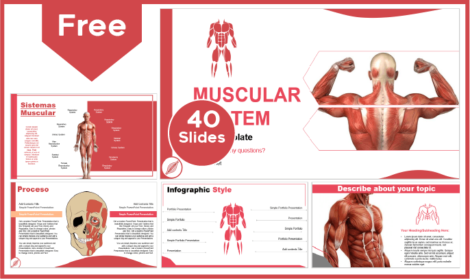
Main features
- 40 slides 100% editable
- 16:9 widescreen format suitable for all screens
- High quality royalty-free images
- Included resources: charts, graphs, timelines and diagrams
- More than 100 icons customizable in color and size
- Main font: Arial
- Predominant color: Red
Download this template
If you want to see more templates like this one, check out the complete list of PowerPoint templates and Google Slides biology themes available on our website. If you are interested in professional, premium, free and highly effective designs, do not hesitate and select the one that best suits your needs.
We use cookies to improve the experience of everyone who browses our website. Cookies Policy
Accept Cookies
Muscular System - PowerPoint and Notes

What educators are saying
Also included in.

Description
Introduce or help your students review the muscular system with this PowerPoint presentation. This resource includes a 20 slide PowerPoint presentation and 2 versions of the student notes pages - full size and interactive notebook format (modified notes also included). This resource is perfect to use at the beginning of the human body systems unit.
Save 30% by purchasing the Science PowerPoint and Notes Bundle .
ALL text is 100% EDITABLE!
20 PowerPoint slides with student notes pages
This product includes the following:
-Muscular system function
-Muscle classification - voluntary, involuntary, striated, smooth
-Types of muscle - skeletal, smooth, cardiac
-Muscle contraction - isotonic, concentric, isotonic, eccentric, isometric
-How do they work?
-Body movements
-Effects of exercise
-Checkpoint - 5 questions
-2 Versions of Student Notes Pages - Full Size and Interactive Notebook Format
-Modified Student Notes Pages
-Student Notes Pages Answer Key
-100% Editable Text - PowerPoint and Notes Pages
*Clipart included is secured and not editable*
Other products you may like:
Science Interactive Notebook Bundle
Science Card Sort MEGA Bundle
Science Task Card Bundle
Around the Room-Science Circuit Bundle
Science Warm-Up Bundle
Terms of Use:
© Copyright The Science Duo. All rights reserved by author. This product is to be used by the original purchaser only. Copying for more than one teacher or classroom, or for an entire department, school, or school system is prohibited. This product may not be distributed or displayed digitally for public view, uploaded to school or district websites, distributed via email, or submitted to file sharing sites such as Amazon Inspire. Failure to comply is a copyright infringement and a violation of the Digital Millennium Copyright Act (DMCA). Intended for single classroom and personal use only.
Questions & Answers
The science duo.
- We're hiring
- Help & FAQ
- Privacy policy
- Student privacy
- Terms of service
- Tell us what you think
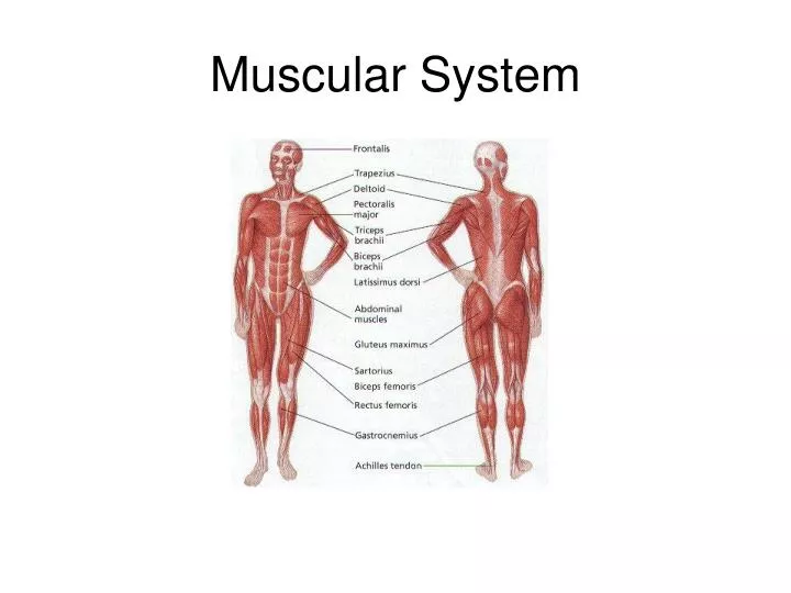
Muscular System
Jul 15, 2014
510 likes | 747 Views
Muscular System. The Skeleton and its joints support, protect, and provide flexibility for the body, but the Skeleton CANNOT Move Itself. 2. That job is performed by the Muscle Tissue that makes up the MUSCULAR SYSTEM .
Share Presentation
- protein filaments thick
- skeleton muscle cells
- dense bundles
- actin filaments
- connective tissue

Presentation Transcript
The Skeleton and its joints support, protect, and provide flexibility for the body, but the Skeleton CANNOT Move Itself. 2. That job is performed by the Muscle Tissue that makes up the MUSCULAR SYSTEM. 3. A MUSCLE TISSUE IS TISSUE THAT CAN CONTRACT IN A COORDINATED FASHION AND INCLUDES MUSCLES TISSUE, BLOOD VESSELS, NERVES, AND CONNECTIVE TISSUE. 4. Approximately 40 to 50 percent of the MASS of the Human Body is composed of Muscle Tissue. 5. THE MUSCULAR SYSTEM IS COMPOSED OF MUSCLE TISSUE (MUSCLE FIBER or cells) THAT IS HIGHLY SPECIALIZED TO CONTRACT TO PRODUCE MOVEMENT WHEN STIMULATED. 6. The Word Muscle is derived from the Latin word "MUS", meaning mouse. 7. Muscle tissue is found everywhere within the body, not only beneath the skin but deep within the body, surrounding many internal organs and blood vessels. 8. The size and location of muscle tissue helps determine the shape of our bodies and the way we move. Muscular System
TYPES OF MUSCLE TISSUE (THREE TYPES) EACH TYPE HAS A DIFFERENT STRUCTURE AND PLAYS A DIFFERENT ROLE IN THE BODY.
SKELETAL MUSCLE
Skeletal Muscle • Skeletal Muscle is Responsible for moving parts of the body, such as the limbs, trunk, and face. 2. SKELETAL MUSCLES ARE GENERALLY ATTACHED TO BONES AND ARE AT WORK EVERY TIME WE MAKE A MOVE. 3. SKELETAL MUSCLES ARE RESPONSIBLE FOR VOLUNTARY (CONSCIOUS) MOVEMENT. 4. A Skeletal Muscle is made of Elongated Cells called MUSCLE FIBERS. Varying movements require Contraction of variable numbers of Muscles Fibers in a Muscle. 5. Skeletal Muscle fibers are grouped into dense bundles called FASCICLES. A group of Fascicles are bound together by Connective Tissue to form a MUSCLE.
Skeletal Muscle • 6. When viewed under a microscope, Skeletal Muscles appear to have STRIATIONS (BANDS OR STRIPES). This gives Skeleton Muscle the name of VOLUNTARY OR STRIATED MUSCLE. • 7. MOST SKELETAL MUSCLES ARE CONSCIOUSLY CONTROLLED BY THE CENTRAL NERVOUS SYSTEM (CNS). • 8. Skeleton Muscle Cells are LARGE and have MORE than ONE NUCLEUS. They vary in length from 1mm to 30 to 60 cm. • 9. Because they are so long and slender, they are often called MUSCLE FIBERS rather than Muscle Cells. • 10. Muscle Fiber together with the Connective Tissue, Blood Vessels, and Nerves form a Skeletal Muscle
Smooth Muscles
Smooth Muscle • SMOOTH MUSCLES ARE USUALLY NOT UNDER VOLUNTARY CONTROL. 2. SMOOTH MUSCLE CELLS ARE SPINDLE-SHAPED AND HAVE A SINGLE NUCLEUS, ARE NOT STRIATED and Interlace to form Sheets of smooth Muscle Tissue. 3. SMOOTH MUSCLES ARE FOUND IN MANY INTERNAL ORGANS, STOMACH, INTESTINES, AND IN THE WALLS OF BLOOD VESSELS. 4. Smooth muscle fibers are surrounded by connective tissue, but the connective tissue Does Not unite to form TENDONS as it does in Skeletal Muscles. 5. Most Smooth Muscle Cells can CONTRACT WITHOUT Nervous Stimulation. Because most of its movements Cannot be consciously controlled, Smooth Muscle is referred to as Involuntary Muscle. 6. The contractions in Smooth Muscles move food through our digestive tract, control the way blood flows through the circulatory system, and increases the size of the pupils of our eyes in bright light.
Cardiac Muscle
Cardiac Muscle • THE ONLY PLACE IN THE BODY WHERE CARDIAC MUSCLE IS FOUND IS IN THE HEART. 2. Cardiac Cells are Striated, but they are NOT under Voluntary Control. 3. Cardiac Muscle Contract Without Direct stimulation by the Nervous System. A bundle of specialized muscle cells in the upper part of the heart sends electrical signals through cardiac muscle tissue, causing the heart to rhythmically contract and pump blood through the body. 4. The Cardiac Muscle Cell contains ONE Nucleus located near the center, adjacent cells form branching fibers that allow Nerve Impulses to pass from cell to cell.
Muscle Structure • A Muscle Fiber is a single, Multinucleated Muscle Cell. 2. A Muscle may be made up of hundreds or even thousands of Muscle Fibers, depending on the Muscles Size. 3. Although Muscle Fiber makes up most of the Muscle Tissue, a large amount of Connective Tissue, Blood Vessels, and Nerves are also present. 4. Connective Tissue Covers and Supports each Muscle Fiber and reinforces the Muscle as a whole. 5. The health of Muscle depends on a sufficient Nerve and Blood Supply. Each Skeletal Muscle has a Nerve Ending that controls its activity.
6. Active Muscles use a lot of Energy and require a continuous supply of Oxygen and Nutrients, which are supplied by Arteries. Muscles produce large amounts of Metabolic Waste that must be removed by Veins. 7. Muscle Fibers consist of Bundles of threadlike structures called MYOFIBRILS. 8. Each Myofibril is made up of TWO Types of Protein Filaments- Thick ones and Thin ones. 9. The THICK FILAMENTS are made up of a PROTEIN called MYOSIN. 10. The THIN FILAMENTS are made of a PROTEIN called ACTIN. 11. Myosin and Actin Filaments are arranged to form overlapping patterns, which are responsible for the Light and Dark Bands that can be seen in Skeletal (Striated Appearance) Muscle. 12. Thin Actin Filaments are Anchored at their Midpoints to a structure called the Z-LINE. 13. The Region From one Z-line to the next is called a SARCOMERE the Functional Unit of Muscle Contractions
MECHANISM OF MUSCLE CONTRACTIONS 1. The Sarcomere is the functional unit of Muscle contractions. 2. When Muscle Cells Contract, the light and dark bands contained in Muscle Cells get closer together. 3. This happens because when a Muscle Contracts, Myosin Filaments and Actin filaments interact to shorten the length of a Sarcomere. 4. When Myosin Filaments and Actin Filaments come near each other, many knob (heads) like projections in each Myosin Filament form CROSS-BRIDGES with an Actin Filament. 5. When the Muscle is Stimulated to Contract, the Cross-bridges MOVE, PULLING the Two Filaments past each other.
MECHANISM OF MUSCLE CONTRACTIONS • 6. After each Cross-bridge has moved as far as it can, it releases the Actin Filament and returns to its original position. The Cross-bridge then attaches to the Actin Filament at another place and the cycle is repeated. This action Shortens the Length of the Sarcomere. • 7. The synchronized shortening of Sarcomeres along the full length of a Muscle Fiber causes the Whole Fiber, and hence the Muscle, to Contract. • 8. WHEN THOUSANDS OF ACTIN AND MYOSIN FILAMENTS INTERACT IN THIS WAY, THE ENTIRE MUSCLE CELL SHORTENS. • 9. THIS CONCEPT IS THE SLIDING FILAMENT THEORY. • 10. Muscle Contractions require Energy, which is supplied by ATP. This Energy is used to Detach the Myosin Heads from the Actin Filaments.
MECHANISM OF MUSCLE CONTRACTIONS 11. Because Myosin Heads must Attach and Detach a number of times during a Single Muscle Contraction, Muscle Cells must have a Continuous Supply of ATP. 12. Without ATP the Myosin Heads would stay Attached to the Actin Filaments, keeping Muscles Permanently Contracted. 13. A Muscle Contraction, like a Nerve Impulse, is an All-or-None Response- either Fibers Contract or they Remain Relaxed. 14. The force of a Muscle Contraction is determined by the number of Muscle fibers that are Stimulated. As more fibers are activated, the force of the contraction Increases. 15. Some Muscles, such as the muscles that hold the body in an upright position and maintain posture, are nearly always at least Partially Contracted.
CONTROL OF MUSCLE CONTRACTION • Muscles are useful only if they Contract in a Controlled fashion. 2. Motor Neurons connect the CNS to Skeleton Muscle Cells (EFFECTORS); Impulses (ACTION POTENTIALS) from Motor Neurons Control the Contraction of Skeleton Muscle Cells. 3. The point of contact between a Motor Neuron and a Muscle Cell is called the NEUROMUSCULAR JUNCTION. 4. Vesicles, or pockets, in the AXON TERMINALS of the Motor Neuron release molecules of the NEUROTRANSMITTER ACETYLCHOLINE. 5. These molecules Diffuse across the SYNAPSE, producing and IMPULSE in the Cell Membrane of the Muscle Cell. 6. The impulse causes the release of Calcium ions within the cell. The Calcium Ions affect regulatory proteins that allow Actin and Myosin Filaments to interact and form cross-bridges.
CONTROL OF MUSCLE CONTRACTION 7. A Muscle Cell WILL remain in a state of CONTRACTION until the production of Acetylcholine STOPS. 8. An ENZYME called ACETYLCHOLINESTERASE, also produced at the Neuromuscular Junction, DESTROYS ACETYLCHOLINE, permits the reabsorption of Calcium Ions into the Muscle Cell, and Terminates the Contraction. 9. You can have a Weak or Strong Contraction depending on what you are trying to accomplish. The BRAIN (frontal lobes of the cerebrum) decides what and how many Muscles Cells need to Contract. Blinking your eye would be a Weak Contraction, but lifting heavy weights, the brain would signal most Muscle Cells to Contract. 10. MUSCLE SENSE IS THE BRAINS ABILITY TO KNOW WHERE OUR MUSCLES ARE AND WHAT THEY ARE DOING. Permits us to perform everyday activities without having to concentrate on muscle position.
HOW MUSCLES AND BONES INTERACT • Skeleton Muscles generate Force and produce Movement only by CONTRACTING or PULLING on Body Parts. 2. Individual Muscles can only PULL; they CANNOT PUSH. 3. Skeleton Muscles are joined to bone by TOUGH CONNECTIVE TISSUE CALLED TENDONS. 4. TENDONS ATTACH MUSCLE TO BONE; THE ORIGIN IS THE MORE STATIONARY BONE, THE INSERTION IS THE MORE MOVABLE BONE. 5. Tendons are attached in such a way that they PULL on the Bones and make them work like LEVERS. The movements of the Muscles and Joints enable the Bones to act as LEVERS.
HOW MUSCLES AND BONES INTERACT 6. The Joint functions as a FULCRUM (The fixed point around which the lever moves) and the Muscles provide the FORCE to move the Lever. 7. Usually there several Muscles surrounding each Joint that PULL in DIFFERENT DIRECTIONS. 8. MOST SKELETAL MUSCLES WORK IN PAIRS. 9. When one Muscle or set of Muscles CONTRACTS, the other RELAXES.
HOW MUSCLES AND BONES INTERACT 10. The Muscles of the upper arm are a good example of this dual action: ANTAGONISTIC MUSCLES. FLEXOR, A MUSCLE THAT BENDS A JOINT. EXTENSOR, A MUSCLE THAT STRAIGHTENS A JOINT. A. When the BICEPS Muscle (on the front of the upper arm, FLEXOR) CONTRACTS, it BENDS OR FLEXES THE ELBOW JOINT. B. When the TRICEPS Muscle (on the back of the upper arm, EXTENSOR) CONTRACTS, it opens, or extends, the elbow joint. C. A controlled movement requires contraction by both muscles. 11. ANTAGONISTIC MUSCLES ARE OPPONENTS, MUSCLES WHICH HAVE OPPOSING OR OPPOSITE FUNCTIONS. A muscle pulls when it contracts, but exerts no force when it relaxes and CANNOT PUSH. When one muscle Pulls a bone in one direction, another muscle is needed to PULL the bone in the other direction. 12. SYNERGISTIC MUSCLES ARE THOSE WITH THE SAME FUNCTION, OR THOSE THAT WORK TOGETHER TO PERFORM A PARTICULAR FUNCTION. They also stabilize a joint to make a more precise movement possible.
HOW MUSCLES AND BONES INTERACT 13. A normal characteristic of all Skeleton Muscles is that they remain in a state of PARTIAL CONTRACTION. 14. At any given time, some Muscles are being Stimulated while other are not. This causes a TIGHTENED, or FIRMED, Muscle and is known as MUSCLE TONE. 15. Muscle Tone is responsible for keeping the back and legs straight and the head upright even when you are relaxed. 16. EXERCISE IS THE KEY TO MAINTAINING GOOD MUSCLE TONE WITHIN YOUR BODY. 17. MUSCLES THAT ARE EXERCISED REGULARLY STAY FIRM AND INCREASE IN SIZE BY ADDING MORE MATERIALS TO THE INSIDE OF MUSCLE FIBERS.
HOW MUSCLES AND BONES INTERACT • 18. MUSCLE FATIGUE is a Physiological Inability of a muscle to contract. Muscle fatigue is a result of a relative depletion of ATP. When ATP is absent, a state of continuous contraction occurs. This causes severe muscle cramps. • 19. OXYGEN DEBT is a temporary Lack of Oxygen. When this occurs Muscles will switch from the normal Aerobic Respiration to a form of Anaerobic Respiration called Lactic Acid Fermentation. As the oxygen becomes Depleted, the muscle cells begin to switch. Oxygen debt leads to the accumulation of Metabolic Waste (Lactic Acid) in the muscle fibers, resulting in muscle fatigue, pain, and even cramps. Eventually, the lactic acid diffuses into the blood and is transported to the Liver. So if you ever experienced Soreness after prolong exercise, it may have been caused by Oxygen Debt - Your body could not provide your Muscles the Oxygen they needed to function properly.
http://www.gwc.maricopa.edu/class/bio201/muscle/mustut.htm
Muscle dysmorphia or bigorexia • a disorder in which a person becomes obsessed with the idea that he or she is not muscular enough • hold delusions that they are "skinny" or "too small" but are often above average in musculature • Muscle dysmorphia can cause people to: • Constantly examine themselves in a mirror • Frequently compare themselves with others • Hate their reflections • Become distressed if they miss a workout session or one of their many meals a day • Become distressed if they do not receive enough protein per day in their diet • Take potentially dangerous anabolic steroids • Neglect jobs, relationships, or family because of excessive exercising • Have delusions of being underweight or below average in musculature. • In extreme cases, inject appendages with fluid (e.g. synthol--A site enhancement oil (SEO) used by some bodybuilders to increase the apparent size of some muscles. The effects of SEOs are purely cosmetic and there is no increase in muscular performance.
Effects of SEO • Many doctors advise that the use of SEOs is extremely dangerous, if not potentially fatal, as injection into a major blood vessel can cause embolism, leading to heart attacks, strokes, pulmonary embolism and/or permanent brain damage if SEO traces find their way into cerebral vessels. • In some individuals, the use of SEOs can lead to chronic inflation of the treated muscle, which over time can result in deformation of the muscle.
Anabolic steroid illegal!!! • Between 1 million and 3 million people (1% of the population) are thought to have misused AAS in the United States • drugs which mimic the effects of the male sex hormones testosterone and dihydrotestosterone. • increase protein synthesis within cells, which results in the buildup of cellular tissue (anabolism), especially in muscles • Anabolic steroids also have androgenic and virilizing properties, including the development and maintenance of masculine characteristics such as the growth of the vocal cords and body hair.
Anabolic steroids were first • to stimulate bone growth • To stimulate appetite • induce male puberty • treat chronic wasting conditions, such as cancer and AIDS
These effects include harmful changes in: • cholesterol levels (increased low-density lipoprotein and decreased high-density lipoprotein), • acne, • high blood pressure, • liver damage (mainly with oral steroids), • dangerous changes in the structure of the left ventricle of the heart. • can accelerate the rate of premature baldness for males who are genetically predisposed, but testosterone itself can produce baldness in females
sex-specific side effects of anabolic steroids • Development of breast tissue in males, a condition called gynecomastia • Reduced sexual function and temporary infertility can also occur in males • testicular atrophy • Female-specific side effects include • increases in body hair, • deepening of the voice, • enlarged clitoris, • temporary decreases in menstrual cycles. • When taken during pregnancy, anabolic steroids can affect fetal development by causing the development of male features in the female fetus and female features in the male fetus.
A number of severe side effects can occur if adolescents use anabolic steroids. • the steroids may prematurely stop the lengthening of bones resulting in stunted growth. • accelerated bone maturation, • increased frequency and duration of erections, and premature sexual development. • Anabolic steroid use in adolescence is also correlated with poorer attitudes related to health. • alterations in the structure of the heart, such as enlargement and thickening of the left ventricle, which impairs its contraction and relaxation.Possible effects of these alterations in the heart are hypertension, cardiac arrhythmias, congestive heart failure, heart attacks, and sudden cardiac death.
significant psychiatric symptoms including aggression and violence, mania, and less frequently psychosis and suicide have been associated with steroid abuse • "roid rage”
Muscular Dystrophy • Muscular Dystrophy (MD) is a group of genetic diseases that cause the muscle fibers to become easily damaged. • There are over one dozen different types, but the most common general symptoms are muscle weakness, lack of coordination and loss of mobility. • The most severe type, Duchenne's muscular dystrophy, can cause mental retardation and primarily affects young boys. Often fatal • MD is diagnosed through genetic tests, muscle biopsies, blood tests that measure for high levels of creatine kinase and ultrasounds. • There is no cure for MD but it can be treated to reduce the severity with physical therapy, medication, surgery and special braces.
Dermatomyositis • This uncommon autoimmune disease causes muscle weakness accompanied by a skin rash. • It can affect anyone but is most commonly seen in adults ages 40 to 60 and children ages five to 15. • Symptoms include a light purple or red rash on the face, hands, knees, chest and back and progressive muscle weakness. It may also cause difficulty swallowing, muscle pain, ulcers, fever, fatigue and weight loss. • Doctors are uncertain of the cause but believe it may be genetic. • Treatment includes pain management, corticosteroids and immunosuppressive drugs. Although dermatomyositis has no cure, it can go into remission.
Compartment Syndrome • Compartment syndrome occurs when too much pressure builds up in and around the muscles. • It can result from crushing injuries, extended pressure on a blood vessel, swelling inside a cast or complications from surgery. • Symptoms include severe pain, a feeling of fullness or tightness in the muscle and a tingling sensation. Numbness indicates cellular death and it may be difficult to restore full function once it reaches that point. Surgery to relieve the pressure is usually required.
Rhabdomyolysis • Rhabdomylosis damages both the muscles and the kidneys by causing the muscle fibers to breakdown and be released into the blood stream. • The fibers erode into a substance called myoglobin, which blocks the kidney structures and can lead to kidney failure. • Alcoholism, heatstroke, cocaine and heroin overdoses, seizures and severe exertion are possible causes. • If caught early, intravenous fluids are given to restore hydration. Once kidney damage occurs, treatment focuses on restoring renal functions and preventing further damage. • Signs of rhabdomyolysis include weakness, muscle stiffness and pain, joint pain and weight gain.
Fibrodysplasia Ossificans Progressiva • Fibrodysplasia Ossificans Progressiva (FOP) is a rare congenital disease that affects approximately one in two million people worldwide and causes muscles, tendons and ligaments to be replaced with bone tissue. • Since it is a congenital disease, it begins before birth but is generally diagnosed in childhood. • The earliest sign is malformed big toes at birth. • The disease generally affects the neck and shoulders during childhood and continues downward throughout adolescence and adulthood. • Any type of trauma, including falls and medical procedures, can trigger an episode. There are no effective treatments for FOP. Medication is usually given to treat the pain associated with the new bone formation.
Myasthenia gravis • a neuromuscular disorder characterized by variable weakness of voluntary muscles, which often improves with rest and worsens with activity. • The condition is caused by an abnormal immune response-immune cells target and attack the body's own cells (an autoimmune response). This immune response produces antibodies that attach to affected areas, preventing muscle cells from receiving chemical messages (neurotransmitters) from the nerve cell. • Cause is unknown--may be associated with tumors of the thymus (an organ of the immune system). • affects about 3 of every 10,000 people and can affect people at any age. It is most common in young women and older men.
Symptoms Muscle weakness, including: • Swallowing difficulty, frequent gagging, or choking • Paralysis • Muscles that function best after rest • Drooping head • Difficulty climbing stairs • Difficulty lifting objects • Need to use hands to rise from sitting positions • Difficulty talking • Difficulty chewing • Vision problems: • Double vision • Difficulty maintaining steady gaze • Eyelid drooping • Additional symptoms that may be associated with this disease: • Hoarseness or changing voice • Fatigue • Facial paralysis • Drooling • Breathing difficulty
Multiple Sclerosis • Multiple sclerosis (or MS) is a chronic, often disabling disease that attacks the central nervous system (CNS) • Symptoms may be mild, such as numbness in the limbs, or severe, such as paralysis or loss of vision. • the cause (etiology) of MS is still not known, scientists believe that a combination of several factors may be involved • MS occurs in most ethnic groups, including African-Americans, Asians and Hispanics/Latinos, but is more common in Caucasians of northern European ancestry
Symptoms (most common) • Fatigue • Numbness • Numbness of the face, body, or extremities (arms and legs) is one of the most common symptoms of MS, and is often the first symptom experienced by those eventually diagnosed as having MS. • Walking (Gait), Balance, & Coordination Problems • Bladder Dysfunction • Bowel Dysfunction • Constipation is a particular concern among people living with MS, as is loss of control of the bowels. Diarrhea and other problems of the stomach and bowels also can occur. • Vision Problems • The sudden onset of double vision, poor contrast, eye pain, or heavy blurring • Dizziness and Vertigo
Sexual Dysfunction • Sexual arousal begins in the central nervous system, as the brain sends messages to the sexual organs along nerves running through the spinal cord. If MS damages these nerve pathways, sexual respons can be directly affected. • Pain • Cognitive Function • Cognition refers to a range of high-level brain functions, including the ability to learn and remember information: organize, plan, and problem-solve; focus, maintain, and shift attention as necessary; understand and use language; accurately perceive the environment, and perform calculations. • Emotional Changes • Bouts of severe depression (which is different from the healthy grieving that needs to occur in the face of losses and changes caused by MS), mood swings, irritability, and episodes of uncontrollable laughing and crying (called pseudobulbar affect) • Depression • Spasticity: the involuntary movement (jerking) of muscles
Tetanus • infection of the nervous system with the potentially deadly bacteria Clostridium tetani (C. tetani). • begins with mild spasms in the jaw muscles (lockjaw). The spasms can also affect the chest, neck, back, and abdominal muscles. Back muscle spasms often cause arching, called opisthotonos. • prolonged muscular action causes sudden, powerful, and painful contractions of muscle groups. This is called tetany. These episodes can cause fractures and muscle tears. • Other symptoms include: • Drooling • Excessive sweating • Fever • Hand or foot spasms • Irritability • Swallowing difficulty • Uncontrolled urination or defecation
- More by User
Muscular System. By: Emily Brosten Stephanie Elhard JAMES VALLEY VOCATIONAL TECHNICAL CENTER. Introduction. 1. 600 muscles in the body 2. Muscles are ~ made of bundles of muscle fibers which are held together by connective tissue.
1.11k views • 36 slides

Muscular System. What is the purpose of muscle?. Movement Posture Stabilize joints Generate heat. Types of Muscle. Compare all three muscle types. Look at: Location/number of nuclei Associated structures What controls the muscle contraction?
801 views • 65 slides

Muscular System. Muscles and Muscle Tissue. Muscle Review Questions . ADD THESE TO YOUR PACKET ON BACK! 1. What are the three types of muscle tissue? What are their functions? Where are they located in the body? Voluntary ? Striated? 2. What are the four functions of muscle?
377 views • 19 slides

Muscular system
Muscular system . The horse has a large muscular system which allows movement of the horse, gives it definitions as well as allowing posture like standing by contracting both muscle groups at the same time. 3 Types of Muscles. Smooth Cardiac Skeletal. Smooth muscle . Smooth Muscle.
494 views • 22 slides

Muscular System. By: Jasmine Jones. Functions. 1.Body movement due to the contraction of skeletal muscles 2.Maintence of posture also due to skeletal muscles 3. Respiration due to movements of the muscles of the thorax
228 views • 5 slides
Parker Andrews. Muscular System. function. Generate force Movement In some ways communicate (physical expressions) The ability to use chemical energy to produce force and energy is present in all cells but is a dominate characteristic of muscle cells . 3 Types of muscle tissue. Skeletal
267 views • 11 slides

Muscular System. It permits movement of the body, maintains posture, and circulates blood throughout the body. 1 . Cardiac: A type of muscle tissue that is found only in the heart 2. Smooth: not under voluntary control Examples: 3 Skeletal: moves the limbs Examples.
118 views • 4 slides

Muscular System. Chapter 6. Functions of the Muscular System. Produces Movement-skeletal muscles are responsible for all locomotion and manipulation of the skeleton. Their speed and power help us stay safe.
698 views • 42 slides

MUSCULAR SYSTEM
MUSCULAR SYSTEM. anatomical terminology. ? Assume the anatomical position, what do these words mean? Inferior; superior Proximal; distal Medial; lateral Posterior; anterior. TYPES OF MUSCLES. SKELETAL : voluntary control striated appearance (alternating dark & light bands)
470 views • 20 slides

Muscular System. Muscular System Functions:. 1. produce movement 2. Maintain posture 3. Stabilize joints 4. Generate heat 5. Move substances (fluid, food etc). Properties of Muscles: . Excitability : capacity of muscle to respond to a stimulus
2.25k views • 32 slides

MUSCULAR SYSTEM. WHAT DOES THE MUSCULAR SYSTEM DO?. ACCOUNTS FOR ALL OF THE WAYS THAT THE PARTS OF THE BODY MOVE. ACTIONS SUCH AS RUNNING,EATING,BREATHING,DIGESTING FOOD,PUMPING BLOOD. HELPS PROTECT YOUR JOINTS AND HELPS CREATE HEAT THAT KEEPS YOUR BODY WARM. WHAT ARE MUSCLES MADE OF?.
255 views • 10 slides

Muscular System. Section 9.3 Energy Sources for Muscle Contraction. Creatine Phosphate. 4-6x more CP in muscles than ATP Storage form of PO 4 Made when excess ATP is available. There is enough ATP and CP for 10 seconds of contraction
140 views • 6 slides

Muscular System. BY: Slyder Welch. Made up of?. Muscles & Tendons. Important because?. Holds organs in place. Holds the bones together, so that you can move. Helps you chew food. Pumps blood throughout the body. There are even MORE !!!. Functions?.
245 views • 11 slides

Muscular System. Ms. Phayre & Ms. Cauley. “Muscle Madness” Activity. Students will split up into 6 groups. Each group will have 2-3 students. Find a station around the room. There should only be one group per station.
505 views • 19 slides

Muscular System. Key Concepts and Vocabulary Words. The muscular system produces movement and maintains posture. There are three kinds of muscles: skeletal , cardiac , and smooth . Muscles are excitable , contractile , extensible , and elastic .
369 views • 17 slides

Muscular System. Chapter 11 Part 2. Gross Structure of Skeletal Muscle. Skeletal muscles is encased by epi mysium : fascia of fibrous connective tissue. Fasciculus , bundle of cylindrical muscle fibers surrounded by peri mysium .
226 views • 21 slides

MUSCULAR SYSTEM. Structure of the Muscles. Muscles. Comprise a large part of the human body Nearly half our body weight comes from muscle tissue If you weigh 140 lbs, 60 lbs is from muscle attached to bones Over 650 different muscles. Responsibilities of Muscle System. 3 main:
248 views • 19 slides

Muscular system. What is the muscular system?. The muscular system consists of all the muscles of the body. These make up approximately 42% of total body weight, and are composed of long, slender cells known as fibers. The fibers are different lengths and vary in color from white to deep red.
371 views • 14 slides

Muscular System. Vocabulary. bi- two -ia condition of -lysis destruction, dissolve myo- muscle -plegia paralysis tri- three tendo- tendon para- lower half fasci- fibrous band Carp – wrist. ad- to, toward or near ab- from, away circum- around
275 views • 25 slides

Muscular System. Function of the Muscular System. Provides movement for the body and its parts, maintains posture, generates heat and stabilizes joints. Essential function is to shorten. Types of Muscles. 1. Skeletal 2. Smooth 3. Cardiac. Skeletal Muscle.
568 views • 52 slides

Muscular System. Three types of Muscles Found in the Body. Muscle Structure Functions • Movement (constriction of organs & vessels, heat beating, respiration) • Support (posture) • Heat production Communication Properties of Skeletal (striated) muscle (40% of body weight)
466 views • 36 slides

Muscular System. Agriculture, Food, and, Natural Resource Standards Addressed. AS.01.01. Evaluate the development and implications of animal origin, domestication and distribution on production practices and the environment.
765 views • 40 slides

Got any suggestions?
We want to hear from you! Send us a message and help improve Slidesgo
Top searches
Trending searches

memorial day
12 templates

151 templates

15 templates

11 templates

39 templates

christian church
29 templates
Musculoskeletal System
It seems that you like this template, musculoskeletal system presentation, free google slides theme and powerpoint template.
Download the "Musculoskeletal System" presentation for PowerPoint or Google Slides. The education sector constantly demands dynamic and effective ways to present information. This template is created with that very purpose in mind. Offering the best resources, it allows educators or students to efficiently manage their presentations and engage audiences. With its user-friendly and useful features, everyone will find it easy to customize and adapt according to their needs. Whether for a lesson presentation, student report, or administrative purposes, this template offers a unique solution for any case!
"### Features of this template
- 100% editable and easy to modify
- Different slides to impress your audience
- Contains easy-to-edit graphics such as graphs, maps, tables, timelines and mockups
- Includes 500+ icons and Flaticon’s extension for customizing your slides
- Designed to be used in Google Slides and Microsoft PowerPoint
- Includes information about fonts, colors, and credits of the resources used"
How can I use the template?
Am I free to use the templates?
How to attribute?
Attribution required If you are a free user, you must attribute Slidesgo by keeping the slide where the credits appear. How to attribute?
Related posts on our blog.

How to Add, Duplicate, Move, Delete or Hide Slides in Google Slides

How to Change Layouts in PowerPoint

How to Change the Slide Size in Google Slides
Related presentations.

Premium template
Unlock this template and gain unlimited access
Register for free and start editing online
The musculoskeletal system
A PPT presentation about the musculoskeletal system that I have prepared for my students of 3rd of ESO Read less
Recommended
More related content, what's hot, what's hot ( 20 ), viewers also liked, viewers also liked ( 20 ), similar to the musculoskeletal system, similar to the musculoskeletal system ( 20 ), recently uploaded, recently uploaded ( 20 ).
- 1. THE MUSCULOSKELETAL SYSTEM Bones, joints and muscles Tendons, ligaments and cartilage
- 2. The human skeleton • Contains 206 bones •Initially: flexible cartilage •Ossification
- 3. Process of ossification • Approximately 20 years • Growth plates • Bone building: - Osteoblasts - Osteocytes - Osteoclasts • Video http://health.howstuffworks.com/adam-2001 25.htm
- 4. Structure of bones Compact bone • Outside part of the bone • Extremely strong and hard • Periosteum Spongy bone • Mesh-like network (trabeculae) • Red marrow (blood cells) • Yellow marrow (fat) http://youtu.be/yFJ4iswRiu4
- 5. Micrographs
- 6. Anatomical classification of bones • Bones are characterized anatomically as: – long bones (e.g. humerus, femur) – flat bones (membrane bones) – irregular bones (such as the vertebrae) • All these bone types, regardless of their anatomical form, are composed of both spongy and compact bone.
- 7. Functions of the skeleton • Bone provides the internal support of the body and provides sites of attachment of tendons and muscles, essential for locomotion. • Bone provides protection for the vital organs of the body: the skull protects the brain; the ribs protect the heart and lungs. • The hematopoietic bone marrow is protected by the surrounding bony tissue. • The main store of calcium and phosphate is in bone. Bone has several metabolic functions especially in calcium homeostasis. • http://youtu.be/8d-RBe8JBVs
- 8. Joints • Meeting of two bones • Make the skeleton flexible • Types: - Immovable or fibrous - Partially movable, or cartilaginous - Freely movable, or synovial
- 9. Joints • Types of synovial joints: - Hinge: knees and elbows - Gliding: wrists and ankles - Ball and socket: hips and shoulders - Pivot: Head
- 10. Joints consist of the following: •Cartilage: the bones are covered with cartilage (a connective tissue), which is made up of cells and fibers and is wear- resistant. Cartilage helps reduce the friction of movement. •Synovial membrane: a tissue that lines the joint and seals it into a joint capsule. The synovial membrane secretes synovial fluid (a clear, sticky fluid) around the joint to lubricate it. •Ligaments: strong ligaments (tough, elastic bands of connective tissue) surround the joint to give support and limit the joint's movement.
- 11. Joints consist of the following (II) • Tendons: tendons (another type of tough connective tissue) on each side of a joint attach to muscles that control movement of the joint. • Bursas: fluid-filled sacs, between bones, ligaments, or other adjacent structures help cushion the friction in a joint. • Synovial fluid: a clear, sticky fluid secreted by the synovial membrane. • Meniscus: a curved part of cartilage in the knees and other joints.
- 12. Synovial joints
- 13. The Muscles • Pull on the joints, allowing us to move. • Help the body perform other functions. • More than 650 muscles ( half of a person's body weight) • Tendons: tough, cord-like tissues • 3 different kinds of muscle
- 14. How do muscles move? • Contracting and relaxing. • Work in pairs of flexors and extensors. • The flexor contracts to bend a limb at a joint. • The extensor contracts to extend or straighten the limb at the same joint.
- 15. How do skeletal muscles work? http://youtu.be/XoP1diaXVCI

IMAGES
COMMENTS
Muscular System Functions. Body movement. Via muscle contraction; create overall body movements. Posture maintenance. Constant skeletal muscle tone. Respiration. Thoracic muscles. Body heat production. Heat released as byproduct of skeletal muscles.
This PowerPoint presentation covers the anatomy, physiology and common disorders of the muscular system.
The Muscular System. 1. T- 1-855-694-8886 Email- [email protected] By iTutor.com. 2. The muscular system is a complex collection of tissues, each with a different purpose. Understanding the components of the muscular system, including the various types of connective tissues, is a good way to understand how bodies and physical movement work ...
In the muscular system, skeletal muscles are connected to the skeleton, either to bone or to connective tissues such as ligaments. Muscles are always attached at two or more places. When the muscle contracts, the attachment points are pulled closer together; when it relaxes, the attachment points move apart .
The version of the browser you are using is no longer supported. Please upgrade to a supported browser. Dismiss
Muscular System. The muscular system consists of three types of muscles - skeletal, smooth, and cardiac. Skeletal muscles are voluntary and attach to bones to enable movement. Smooth muscles line organs and blood vessels to regulate movement within the body. Cardiac muscle is only found in the heart to pump blood throughout the body.
It's time to talk about the anatomy and physiology of the muscular system! This template perfect for high school anatomy classes will allow you to explain different muscles of the body.
Exercise increases muscle size, strength, and endurance. Aerobic (endurance) exercise (biking, jogging) results in stronger, more flexible muscles with greater resistance to fatigue. Increases blood supply, increase in number of mitochondria, & ability to store O2. Makes body metabolism more efficient.
Anatomy of the Muscular System. Anatomy & Physiology. Skeletal Muscle Structure. Endomysium : connective tissue membrane that surrounds individual muscle fibers or cells Fascicles : groups of skeletal muscle fibers Perimysium : connective tissue membrane surrounding fascicles
PowerPoint Presentation. Muscular System. Poudre High School. Human Anatomy/Physiology. Mr. Bradley. The Muscular System Characteristics of Muscles Skeletal Muscle striated, voluntary Multi-nucleated - fibers in bundles 1-40 mm long, 10-100 microns thick 42% of male body weight, 36% in females General 1. sarcoplasm - cytoplasm of muscle ...
The muscular system is the one that allows us to be mobile, active and healthy. Explain what this important system of the human body consists of with this template that contains 30 different infographics that you can customize to use as you need. They are designed in red, black and white colors and include all the necessary resources to describe the muscular system and make its understanding ...
Muscular System PPT. Chapter 10 ppt.ppt — Microsoft PowerPoint presentation, 23526 kB (24090624 bytes) Print this.
Presentation Transcript. Muscular System Chapter 16 (pgs 310-324) Muscular System • Muscles responsible for all types of body movement • 3 basic muscle types found in body: • Cardiac muscle • Smooth muscle • Skeletal muscle. Muscle Types. Cardiac Muscle • Only found in heart • Striated and branched • Involuntary • Membranes of ...
The Muscular System Powerpoint - Download as a PDF or view online for free
Presentation Transcript. Chapter 6: The Muscular System Anatomy & Physiology. Five Golden Rules of Skeletal Muscle Activity. Muscles and Body Movements • Movement can occur because muscles attach to our bones • Muscles are attached to at least two points known as: • Origin • Attachment to a moveable bone • Insertion • Attachment to ...
This template provides visuals and simple explanations to help students understand the various muscles, their muscle actions, and major injuries associated with them. Organize muscles in different groups and explain all about them with this soft and clear template with many visual illustrations. With this template, your physiotherapy classes ...
The human muscular system template for PowerPoint and Google Slides has 40 100% editable slides that you can have for free.
Introduce or help your students review the muscular system with this PowerPoint presentation. This resource includes a 20 slide PowerPoint presentation and 2 versions of the student notes pages - full size and interactive notebook format (modified notes also included). This resource is perfect to ...
Muscular System. The Skeleton and its joints support, protect, and provide flexibility for the body, but the Skeleton CANNOT Move Itself. 2. That job is performed by the Muscle Tissue that makes up the MUSCULAR SYSTEM .
Introduction to Anatomy (Muscular System) Jan 7, 2014 • Download as PPTX, PDF •. 28 likes • 13,011 views. Dr. Seyed Morteza Mahmoudi. Introduction to Muscular System. Health & Medicine Technology. 1 of 36. Download now.
Present a clinical case on the muscular system with this medical template, complete with illustrations and other useful resources! For Google Slides and PPT
Download the "Musculoskeletal System" presentation for PowerPoint or Google Slides. The education sector constantly demands dynamic and effective ways to present information.
37 likes • 33,283 views. M. Maria Casadevall. A PPT presentation about the musculoskeletal system that I have prepared for my students of 3rd of ESO. Business Technology. 1 of 15. Download now. The musculoskeletal system - Download as a PDF or view online for free.