- Bipolar Disorder
- Therapy Center
- When To See a Therapist
- Types of Therapy
- Best Online Therapy
- Best Couples Therapy
- Best Family Therapy
- Managing Stress
- Sleep and Dreaming
- Understanding Emotions
- Self-Improvement
- Healthy Relationships
- Student Resources
- Personality Types
- Guided Meditations
- Verywell Mind Insights
- 2024 Verywell Mind 25
- Mental Health in the Classroom
- Editorial Process
- Meet Our Review Board
- Crisis Support

How to Write a Great Hypothesis
Hypothesis Definition, Format, Examples, and Tips
Kendra Cherry, MS, is a psychosocial rehabilitation specialist, psychology educator, and author of the "Everything Psychology Book."
:max_bytes(150000):strip_icc():format(webp)/IMG_9791-89504ab694d54b66bbd72cb84ffb860e.jpg)
Amy Morin, LCSW, is a psychotherapist and international bestselling author. Her books, including "13 Things Mentally Strong People Don't Do," have been translated into more than 40 languages. Her TEDx talk, "The Secret of Becoming Mentally Strong," is one of the most viewed talks of all time.
:max_bytes(150000):strip_icc():format(webp)/VW-MIND-Amy-2b338105f1ee493f94d7e333e410fa76.jpg)
Verywell / Alex Dos Diaz
- The Scientific Method
Hypothesis Format
Falsifiability of a hypothesis.
- Operationalization
Hypothesis Types
Hypotheses examples.
- Collecting Data
A hypothesis is a tentative statement about the relationship between two or more variables. It is a specific, testable prediction about what you expect to happen in a study. It is a preliminary answer to your question that helps guide the research process.
Consider a study designed to examine the relationship between sleep deprivation and test performance. The hypothesis might be: "This study is designed to assess the hypothesis that sleep-deprived people will perform worse on a test than individuals who are not sleep-deprived."
At a Glance
A hypothesis is crucial to scientific research because it offers a clear direction for what the researchers are looking to find. This allows them to design experiments to test their predictions and add to our scientific knowledge about the world. This article explores how a hypothesis is used in psychology research, how to write a good hypothesis, and the different types of hypotheses you might use.
The Hypothesis in the Scientific Method
In the scientific method , whether it involves research in psychology, biology, or some other area, a hypothesis represents what the researchers think will happen in an experiment. The scientific method involves the following steps:
- Forming a question
- Performing background research
- Creating a hypothesis
- Designing an experiment
- Collecting data
- Analyzing the results
- Drawing conclusions
- Communicating the results
The hypothesis is a prediction, but it involves more than a guess. Most of the time, the hypothesis begins with a question which is then explored through background research. At this point, researchers then begin to develop a testable hypothesis.
Unless you are creating an exploratory study, your hypothesis should always explain what you expect to happen.
In a study exploring the effects of a particular drug, the hypothesis might be that researchers expect the drug to have some type of effect on the symptoms of a specific illness. In psychology, the hypothesis might focus on how a certain aspect of the environment might influence a particular behavior.
Remember, a hypothesis does not have to be correct. While the hypothesis predicts what the researchers expect to see, the goal of the research is to determine whether this guess is right or wrong. When conducting an experiment, researchers might explore numerous factors to determine which ones might contribute to the ultimate outcome.
In many cases, researchers may find that the results of an experiment do not support the original hypothesis. When writing up these results, the researchers might suggest other options that should be explored in future studies.
In many cases, researchers might draw a hypothesis from a specific theory or build on previous research. For example, prior research has shown that stress can impact the immune system. So a researcher might hypothesize: "People with high-stress levels will be more likely to contract a common cold after being exposed to the virus than people who have low-stress levels."
In other instances, researchers might look at commonly held beliefs or folk wisdom. "Birds of a feather flock together" is one example of folk adage that a psychologist might try to investigate. The researcher might pose a specific hypothesis that "People tend to select romantic partners who are similar to them in interests and educational level."
Elements of a Good Hypothesis
So how do you write a good hypothesis? When trying to come up with a hypothesis for your research or experiments, ask yourself the following questions:
- Is your hypothesis based on your research on a topic?
- Can your hypothesis be tested?
- Does your hypothesis include independent and dependent variables?
Before you come up with a specific hypothesis, spend some time doing background research. Once you have completed a literature review, start thinking about potential questions you still have. Pay attention to the discussion section in the journal articles you read . Many authors will suggest questions that still need to be explored.
How to Formulate a Good Hypothesis
To form a hypothesis, you should take these steps:
- Collect as many observations about a topic or problem as you can.
- Evaluate these observations and look for possible causes of the problem.
- Create a list of possible explanations that you might want to explore.
- After you have developed some possible hypotheses, think of ways that you could confirm or disprove each hypothesis through experimentation. This is known as falsifiability.
In the scientific method , falsifiability is an important part of any valid hypothesis. In order to test a claim scientifically, it must be possible that the claim could be proven false.
Students sometimes confuse the idea of falsifiability with the idea that it means that something is false, which is not the case. What falsifiability means is that if something was false, then it is possible to demonstrate that it is false.
One of the hallmarks of pseudoscience is that it makes claims that cannot be refuted or proven false.
The Importance of Operational Definitions
A variable is a factor or element that can be changed and manipulated in ways that are observable and measurable. However, the researcher must also define how the variable will be manipulated and measured in the study.
Operational definitions are specific definitions for all relevant factors in a study. This process helps make vague or ambiguous concepts detailed and measurable.
For example, a researcher might operationally define the variable " test anxiety " as the results of a self-report measure of anxiety experienced during an exam. A "study habits" variable might be defined by the amount of studying that actually occurs as measured by time.
These precise descriptions are important because many things can be measured in various ways. Clearly defining these variables and how they are measured helps ensure that other researchers can replicate your results.
Replicability
One of the basic principles of any type of scientific research is that the results must be replicable.
Replication means repeating an experiment in the same way to produce the same results. By clearly detailing the specifics of how the variables were measured and manipulated, other researchers can better understand the results and repeat the study if needed.
Some variables are more difficult than others to define. For example, how would you operationally define a variable such as aggression ? For obvious ethical reasons, researchers cannot create a situation in which a person behaves aggressively toward others.
To measure this variable, the researcher must devise a measurement that assesses aggressive behavior without harming others. The researcher might utilize a simulated task to measure aggressiveness in this situation.
Hypothesis Checklist
- Does your hypothesis focus on something that you can actually test?
- Does your hypothesis include both an independent and dependent variable?
- Can you manipulate the variables?
- Can your hypothesis be tested without violating ethical standards?
The hypothesis you use will depend on what you are investigating and hoping to find. Some of the main types of hypotheses that you might use include:
- Simple hypothesis : This type of hypothesis suggests there is a relationship between one independent variable and one dependent variable.
- Complex hypothesis : This type suggests a relationship between three or more variables, such as two independent and dependent variables.
- Null hypothesis : This hypothesis suggests no relationship exists between two or more variables.
- Alternative hypothesis : This hypothesis states the opposite of the null hypothesis.
- Statistical hypothesis : This hypothesis uses statistical analysis to evaluate a representative population sample and then generalizes the findings to the larger group.
- Logical hypothesis : This hypothesis assumes a relationship between variables without collecting data or evidence.
A hypothesis often follows a basic format of "If {this happens} then {this will happen}." One way to structure your hypothesis is to describe what will happen to the dependent variable if you change the independent variable .
The basic format might be: "If {these changes are made to a certain independent variable}, then we will observe {a change in a specific dependent variable}."
A few examples of simple hypotheses:
- "Students who eat breakfast will perform better on a math exam than students who do not eat breakfast."
- "Students who experience test anxiety before an English exam will get lower scores than students who do not experience test anxiety."
- "Motorists who talk on the phone while driving will be more likely to make errors on a driving course than those who do not talk on the phone."
- "Children who receive a new reading intervention will have higher reading scores than students who do not receive the intervention."
Examples of a complex hypothesis include:
- "People with high-sugar diets and sedentary activity levels are more likely to develop depression."
- "Younger people who are regularly exposed to green, outdoor areas have better subjective well-being than older adults who have limited exposure to green spaces."
Examples of a null hypothesis include:
- "There is no difference in anxiety levels between people who take St. John's wort supplements and those who do not."
- "There is no difference in scores on a memory recall task between children and adults."
- "There is no difference in aggression levels between children who play first-person shooter games and those who do not."
Examples of an alternative hypothesis:
- "People who take St. John's wort supplements will have less anxiety than those who do not."
- "Adults will perform better on a memory task than children."
- "Children who play first-person shooter games will show higher levels of aggression than children who do not."
Collecting Data on Your Hypothesis
Once a researcher has formed a testable hypothesis, the next step is to select a research design and start collecting data. The research method depends largely on exactly what they are studying. There are two basic types of research methods: descriptive research and experimental research.
Descriptive Research Methods
Descriptive research such as case studies , naturalistic observations , and surveys are often used when conducting an experiment is difficult or impossible. These methods are best used to describe different aspects of a behavior or psychological phenomenon.
Once a researcher has collected data using descriptive methods, a correlational study can examine how the variables are related. This research method might be used to investigate a hypothesis that is difficult to test experimentally.
Experimental Research Methods
Experimental methods are used to demonstrate causal relationships between variables. In an experiment, the researcher systematically manipulates a variable of interest (known as the independent variable) and measures the effect on another variable (known as the dependent variable).
Unlike correlational studies, which can only be used to determine if there is a relationship between two variables, experimental methods can be used to determine the actual nature of the relationship—whether changes in one variable actually cause another to change.
The hypothesis is a critical part of any scientific exploration. It represents what researchers expect to find in a study or experiment. In situations where the hypothesis is unsupported by the research, the research still has value. Such research helps us better understand how different aspects of the natural world relate to one another. It also helps us develop new hypotheses that can then be tested in the future.
Thompson WH, Skau S. On the scope of scientific hypotheses . R Soc Open Sci . 2023;10(8):230607. doi:10.1098/rsos.230607
Taran S, Adhikari NKJ, Fan E. Falsifiability in medicine: what clinicians can learn from Karl Popper [published correction appears in Intensive Care Med. 2021 Jun 17;:]. Intensive Care Med . 2021;47(9):1054-1056. doi:10.1007/s00134-021-06432-z
Eyler AA. Research Methods for Public Health . 1st ed. Springer Publishing Company; 2020. doi:10.1891/9780826182067.0004
Nosek BA, Errington TM. What is replication ? PLoS Biol . 2020;18(3):e3000691. doi:10.1371/journal.pbio.3000691
Aggarwal R, Ranganathan P. Study designs: Part 2 - Descriptive studies . Perspect Clin Res . 2019;10(1):34-36. doi:10.4103/picr.PICR_154_18
Nevid J. Psychology: Concepts and Applications. Wadworth, 2013.
By Kendra Cherry, MSEd Kendra Cherry, MS, is a psychosocial rehabilitation specialist, psychology educator, and author of the "Everything Psychology Book."
- Privacy Policy

Home » What is a Hypothesis – Types, Examples and Writing Guide
What is a Hypothesis – Types, Examples and Writing Guide
Table of Contents

Definition:
Hypothesis is an educated guess or proposed explanation for a phenomenon, based on some initial observations or data. It is a tentative statement that can be tested and potentially proven or disproven through further investigation and experimentation.
Hypothesis is often used in scientific research to guide the design of experiments and the collection and analysis of data. It is an essential element of the scientific method, as it allows researchers to make predictions about the outcome of their experiments and to test those predictions to determine their accuracy.
Types of Hypothesis
Types of Hypothesis are as follows:
Research Hypothesis
A research hypothesis is a statement that predicts a relationship between variables. It is usually formulated as a specific statement that can be tested through research, and it is often used in scientific research to guide the design of experiments.
Null Hypothesis
The null hypothesis is a statement that assumes there is no significant difference or relationship between variables. It is often used as a starting point for testing the research hypothesis, and if the results of the study reject the null hypothesis, it suggests that there is a significant difference or relationship between variables.
Alternative Hypothesis
An alternative hypothesis is a statement that assumes there is a significant difference or relationship between variables. It is often used as an alternative to the null hypothesis and is tested against the null hypothesis to determine which statement is more accurate.
Directional Hypothesis
A directional hypothesis is a statement that predicts the direction of the relationship between variables. For example, a researcher might predict that increasing the amount of exercise will result in a decrease in body weight.
Non-directional Hypothesis
A non-directional hypothesis is a statement that predicts the relationship between variables but does not specify the direction. For example, a researcher might predict that there is a relationship between the amount of exercise and body weight, but they do not specify whether increasing or decreasing exercise will affect body weight.
Statistical Hypothesis
A statistical hypothesis is a statement that assumes a particular statistical model or distribution for the data. It is often used in statistical analysis to test the significance of a particular result.
Composite Hypothesis
A composite hypothesis is a statement that assumes more than one condition or outcome. It can be divided into several sub-hypotheses, each of which represents a different possible outcome.
Empirical Hypothesis
An empirical hypothesis is a statement that is based on observed phenomena or data. It is often used in scientific research to develop theories or models that explain the observed phenomena.
Simple Hypothesis
A simple hypothesis is a statement that assumes only one outcome or condition. It is often used in scientific research to test a single variable or factor.
Complex Hypothesis
A complex hypothesis is a statement that assumes multiple outcomes or conditions. It is often used in scientific research to test the effects of multiple variables or factors on a particular outcome.
Applications of Hypothesis
Hypotheses are used in various fields to guide research and make predictions about the outcomes of experiments or observations. Here are some examples of how hypotheses are applied in different fields:
- Science : In scientific research, hypotheses are used to test the validity of theories and models that explain natural phenomena. For example, a hypothesis might be formulated to test the effects of a particular variable on a natural system, such as the effects of climate change on an ecosystem.
- Medicine : In medical research, hypotheses are used to test the effectiveness of treatments and therapies for specific conditions. For example, a hypothesis might be formulated to test the effects of a new drug on a particular disease.
- Psychology : In psychology, hypotheses are used to test theories and models of human behavior and cognition. For example, a hypothesis might be formulated to test the effects of a particular stimulus on the brain or behavior.
- Sociology : In sociology, hypotheses are used to test theories and models of social phenomena, such as the effects of social structures or institutions on human behavior. For example, a hypothesis might be formulated to test the effects of income inequality on crime rates.
- Business : In business research, hypotheses are used to test the validity of theories and models that explain business phenomena, such as consumer behavior or market trends. For example, a hypothesis might be formulated to test the effects of a new marketing campaign on consumer buying behavior.
- Engineering : In engineering, hypotheses are used to test the effectiveness of new technologies or designs. For example, a hypothesis might be formulated to test the efficiency of a new solar panel design.
How to write a Hypothesis
Here are the steps to follow when writing a hypothesis:
Identify the Research Question
The first step is to identify the research question that you want to answer through your study. This question should be clear, specific, and focused. It should be something that can be investigated empirically and that has some relevance or significance in the field.
Conduct a Literature Review
Before writing your hypothesis, it’s essential to conduct a thorough literature review to understand what is already known about the topic. This will help you to identify the research gap and formulate a hypothesis that builds on existing knowledge.
Determine the Variables
The next step is to identify the variables involved in the research question. A variable is any characteristic or factor that can vary or change. There are two types of variables: independent and dependent. The independent variable is the one that is manipulated or changed by the researcher, while the dependent variable is the one that is measured or observed as a result of the independent variable.
Formulate the Hypothesis
Based on the research question and the variables involved, you can now formulate your hypothesis. A hypothesis should be a clear and concise statement that predicts the relationship between the variables. It should be testable through empirical research and based on existing theory or evidence.
Write the Null Hypothesis
The null hypothesis is the opposite of the alternative hypothesis, which is the hypothesis that you are testing. The null hypothesis states that there is no significant difference or relationship between the variables. It is important to write the null hypothesis because it allows you to compare your results with what would be expected by chance.
Refine the Hypothesis
After formulating the hypothesis, it’s important to refine it and make it more precise. This may involve clarifying the variables, specifying the direction of the relationship, or making the hypothesis more testable.
Examples of Hypothesis
Here are a few examples of hypotheses in different fields:
- Psychology : “Increased exposure to violent video games leads to increased aggressive behavior in adolescents.”
- Biology : “Higher levels of carbon dioxide in the atmosphere will lead to increased plant growth.”
- Sociology : “Individuals who grow up in households with higher socioeconomic status will have higher levels of education and income as adults.”
- Education : “Implementing a new teaching method will result in higher student achievement scores.”
- Marketing : “Customers who receive a personalized email will be more likely to make a purchase than those who receive a generic email.”
- Physics : “An increase in temperature will cause an increase in the volume of a gas, assuming all other variables remain constant.”
- Medicine : “Consuming a diet high in saturated fats will increase the risk of developing heart disease.”
Purpose of Hypothesis
The purpose of a hypothesis is to provide a testable explanation for an observed phenomenon or a prediction of a future outcome based on existing knowledge or theories. A hypothesis is an essential part of the scientific method and helps to guide the research process by providing a clear focus for investigation. It enables scientists to design experiments or studies to gather evidence and data that can support or refute the proposed explanation or prediction.
The formulation of a hypothesis is based on existing knowledge, observations, and theories, and it should be specific, testable, and falsifiable. A specific hypothesis helps to define the research question, which is important in the research process as it guides the selection of an appropriate research design and methodology. Testability of the hypothesis means that it can be proven or disproven through empirical data collection and analysis. Falsifiability means that the hypothesis should be formulated in such a way that it can be proven wrong if it is incorrect.
In addition to guiding the research process, the testing of hypotheses can lead to new discoveries and advancements in scientific knowledge. When a hypothesis is supported by the data, it can be used to develop new theories or models to explain the observed phenomenon. When a hypothesis is not supported by the data, it can help to refine existing theories or prompt the development of new hypotheses to explain the phenomenon.
When to use Hypothesis
Here are some common situations in which hypotheses are used:
- In scientific research , hypotheses are used to guide the design of experiments and to help researchers make predictions about the outcomes of those experiments.
- In social science research , hypotheses are used to test theories about human behavior, social relationships, and other phenomena.
- I n business , hypotheses can be used to guide decisions about marketing, product development, and other areas. For example, a hypothesis might be that a new product will sell well in a particular market, and this hypothesis can be tested through market research.
Characteristics of Hypothesis
Here are some common characteristics of a hypothesis:
- Testable : A hypothesis must be able to be tested through observation or experimentation. This means that it must be possible to collect data that will either support or refute the hypothesis.
- Falsifiable : A hypothesis must be able to be proven false if it is not supported by the data. If a hypothesis cannot be falsified, then it is not a scientific hypothesis.
- Clear and concise : A hypothesis should be stated in a clear and concise manner so that it can be easily understood and tested.
- Based on existing knowledge : A hypothesis should be based on existing knowledge and research in the field. It should not be based on personal beliefs or opinions.
- Specific : A hypothesis should be specific in terms of the variables being tested and the predicted outcome. This will help to ensure that the research is focused and well-designed.
- Tentative: A hypothesis is a tentative statement or assumption that requires further testing and evidence to be confirmed or refuted. It is not a final conclusion or assertion.
- Relevant : A hypothesis should be relevant to the research question or problem being studied. It should address a gap in knowledge or provide a new perspective on the issue.
Advantages of Hypothesis
Hypotheses have several advantages in scientific research and experimentation:
- Guides research: A hypothesis provides a clear and specific direction for research. It helps to focus the research question, select appropriate methods and variables, and interpret the results.
- Predictive powe r: A hypothesis makes predictions about the outcome of research, which can be tested through experimentation. This allows researchers to evaluate the validity of the hypothesis and make new discoveries.
- Facilitates communication: A hypothesis provides a common language and framework for scientists to communicate with one another about their research. This helps to facilitate the exchange of ideas and promotes collaboration.
- Efficient use of resources: A hypothesis helps researchers to use their time, resources, and funding efficiently by directing them towards specific research questions and methods that are most likely to yield results.
- Provides a basis for further research: A hypothesis that is supported by data provides a basis for further research and exploration. It can lead to new hypotheses, theories, and discoveries.
- Increases objectivity: A hypothesis can help to increase objectivity in research by providing a clear and specific framework for testing and interpreting results. This can reduce bias and increase the reliability of research findings.
Limitations of Hypothesis
Some Limitations of the Hypothesis are as follows:
- Limited to observable phenomena: Hypotheses are limited to observable phenomena and cannot account for unobservable or intangible factors. This means that some research questions may not be amenable to hypothesis testing.
- May be inaccurate or incomplete: Hypotheses are based on existing knowledge and research, which may be incomplete or inaccurate. This can lead to flawed hypotheses and erroneous conclusions.
- May be biased: Hypotheses may be biased by the researcher’s own beliefs, values, or assumptions. This can lead to selective interpretation of data and a lack of objectivity in research.
- Cannot prove causation: A hypothesis can only show a correlation between variables, but it cannot prove causation. This requires further experimentation and analysis.
- Limited to specific contexts: Hypotheses are limited to specific contexts and may not be generalizable to other situations or populations. This means that results may not be applicable in other contexts or may require further testing.
- May be affected by chance : Hypotheses may be affected by chance or random variation, which can obscure or distort the true relationship between variables.
About the author
Muhammad Hassan
Researcher, Academic Writer, Web developer
You may also like

Data Collection – Methods Types and Examples

Delimitations in Research – Types, Examples and...

Research Process – Steps, Examples and Tips

Research Design – Types, Methods and Examples

Institutional Review Board – Application Sample...

Evaluating Research – Process, Examples and...
What Is a Hypothesis? (Science)
If...,Then...
Angela Lumsden/Getty Images
- Scientific Method
- Chemical Laws
- Periodic Table
- Projects & Experiments
- Biochemistry
- Physical Chemistry
- Medical Chemistry
- Chemistry In Everyday Life
- Famous Chemists
- Activities for Kids
- Abbreviations & Acronyms
- Weather & Climate
- Ph.D., Biomedical Sciences, University of Tennessee at Knoxville
- B.A., Physics and Mathematics, Hastings College
A hypothesis (plural hypotheses) is a proposed explanation for an observation. The definition depends on the subject.
In science, a hypothesis is part of the scientific method. It is a prediction or explanation that is tested by an experiment. Observations and experiments may disprove a scientific hypothesis, but can never entirely prove one.
In the study of logic, a hypothesis is an if-then proposition, typically written in the form, "If X , then Y ."
In common usage, a hypothesis is simply a proposed explanation or prediction, which may or may not be tested.
Writing a Hypothesis
Most scientific hypotheses are proposed in the if-then format because it's easy to design an experiment to see whether or not a cause and effect relationship exists between the independent variable and the dependent variable . The hypothesis is written as a prediction of the outcome of the experiment.
Null Hypothesis and Alternative Hypothesis
Statistically, it's easier to show there is no relationship between two variables than to support their connection. So, scientists often propose the null hypothesis . The null hypothesis assumes changing the independent variable will have no effect on the dependent variable.
In contrast, the alternative hypothesis suggests changing the independent variable will have an effect on the dependent variable. Designing an experiment to test this hypothesis can be trickier because there are many ways to state an alternative hypothesis.
For example, consider a possible relationship between getting a good night's sleep and getting good grades. The null hypothesis might be stated: "The number of hours of sleep students get is unrelated to their grades" or "There is no correlation between hours of sleep and grades."
An experiment to test this hypothesis might involve collecting data, recording average hours of sleep for each student and grades. If a student who gets eight hours of sleep generally does better than students who get four hours of sleep or 10 hours of sleep, the hypothesis might be rejected.
But the alternative hypothesis is harder to propose and test. The most general statement would be: "The amount of sleep students get affects their grades." The hypothesis might also be stated as "If you get more sleep, your grades will improve" or "Students who get nine hours of sleep have better grades than those who get more or less sleep."
In an experiment, you can collect the same data, but the statistical analysis is less likely to give you a high confidence limit.
Usually, a scientist starts out with the null hypothesis. From there, it may be possible to propose and test an alternative hypothesis, to narrow down the relationship between the variables.
Example of a Hypothesis
Examples of a hypothesis include:
- If you drop a rock and a feather, (then) they will fall at the same rate.
- Plants need sunlight in order to live. (if sunlight, then life)
- Eating sugar gives you energy. (if sugar, then energy)
- White, Jay D. Research in Public Administration . Conn., 1998.
- Schick, Theodore, and Lewis Vaughn. How to Think about Weird Things: Critical Thinking for a New Age . McGraw-Hill Higher Education, 2002.
- Null Hypothesis Examples
- Examples of Independent and Dependent Variables
- Difference Between Independent and Dependent Variables
- Definition of a Hypothesis
- Null Hypothesis Definition and Examples
- What Are the Elements of a Good Hypothesis?
- Six Steps of the Scientific Method
- What Are Examples of a Hypothesis?
- Independent Variable Definition and Examples
- Understanding Simple vs Controlled Experiments
- Scientific Method Flow Chart
- What Is a Testable Hypothesis?
- Scientific Method Vocabulary Terms
- What 'Fail to Reject' Means in a Hypothesis Test
- How To Design a Science Fair Experiment
- What Is an Experiment? Definition and Design
Research Hypothesis In Psychology: Types, & Examples
Saul Mcleod, PhD
Editor-in-Chief for Simply Psychology
BSc (Hons) Psychology, MRes, PhD, University of Manchester
Saul Mcleod, PhD., is a qualified psychology teacher with over 18 years of experience in further and higher education. He has been published in peer-reviewed journals, including the Journal of Clinical Psychology.
Learn about our Editorial Process
Olivia Guy-Evans, MSc
Associate Editor for Simply Psychology
BSc (Hons) Psychology, MSc Psychology of Education
Olivia Guy-Evans is a writer and associate editor for Simply Psychology. She has previously worked in healthcare and educational sectors.
On This Page:
A research hypothesis, in its plural form “hypotheses,” is a specific, testable prediction about the anticipated results of a study, established at its outset. It is a key component of the scientific method .
Hypotheses connect theory to data and guide the research process towards expanding scientific understanding
Some key points about hypotheses:
- A hypothesis expresses an expected pattern or relationship. It connects the variables under investigation.
- It is stated in clear, precise terms before any data collection or analysis occurs. This makes the hypothesis testable.
- A hypothesis must be falsifiable. It should be possible, even if unlikely in practice, to collect data that disconfirms rather than supports the hypothesis.
- Hypotheses guide research. Scientists design studies to explicitly evaluate hypotheses about how nature works.
- For a hypothesis to be valid, it must be testable against empirical evidence. The evidence can then confirm or disprove the testable predictions.
- Hypotheses are informed by background knowledge and observation, but go beyond what is already known to propose an explanation of how or why something occurs.
Predictions typically arise from a thorough knowledge of the research literature, curiosity about real-world problems or implications, and integrating this to advance theory. They build on existing literature while providing new insight.
Types of Research Hypotheses
Alternative hypothesis.
The research hypothesis is often called the alternative or experimental hypothesis in experimental research.
It typically suggests a potential relationship between two key variables: the independent variable, which the researcher manipulates, and the dependent variable, which is measured based on those changes.
The alternative hypothesis states a relationship exists between the two variables being studied (one variable affects the other).
A hypothesis is a testable statement or prediction about the relationship between two or more variables. It is a key component of the scientific method. Some key points about hypotheses:
- Important hypotheses lead to predictions that can be tested empirically. The evidence can then confirm or disprove the testable predictions.
In summary, a hypothesis is a precise, testable statement of what researchers expect to happen in a study and why. Hypotheses connect theory to data and guide the research process towards expanding scientific understanding.
An experimental hypothesis predicts what change(s) will occur in the dependent variable when the independent variable is manipulated.
It states that the results are not due to chance and are significant in supporting the theory being investigated.
The alternative hypothesis can be directional, indicating a specific direction of the effect, or non-directional, suggesting a difference without specifying its nature. It’s what researchers aim to support or demonstrate through their study.
Null Hypothesis
The null hypothesis states no relationship exists between the two variables being studied (one variable does not affect the other). There will be no changes in the dependent variable due to manipulating the independent variable.
It states results are due to chance and are not significant in supporting the idea being investigated.
The null hypothesis, positing no effect or relationship, is a foundational contrast to the research hypothesis in scientific inquiry. It establishes a baseline for statistical testing, promoting objectivity by initiating research from a neutral stance.
Many statistical methods are tailored to test the null hypothesis, determining the likelihood of observed results if no true effect exists.
This dual-hypothesis approach provides clarity, ensuring that research intentions are explicit, and fosters consistency across scientific studies, enhancing the standardization and interpretability of research outcomes.
Nondirectional Hypothesis
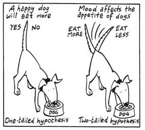
A non-directional hypothesis, also known as a two-tailed hypothesis, predicts that there is a difference or relationship between two variables but does not specify the direction of this relationship.
It merely indicates that a change or effect will occur without predicting which group will have higher or lower values.
For example, “There is a difference in performance between Group A and Group B” is a non-directional hypothesis.
Directional Hypothesis
A directional (one-tailed) hypothesis predicts the nature of the effect of the independent variable on the dependent variable. It predicts in which direction the change will take place. (i.e., greater, smaller, less, more)
It specifies whether one variable is greater, lesser, or different from another, rather than just indicating that there’s a difference without specifying its nature.
For example, “Exercise increases weight loss” is a directional hypothesis.
Falsifiability
The Falsification Principle, proposed by Karl Popper , is a way of demarcating science from non-science. It suggests that for a theory or hypothesis to be considered scientific, it must be testable and irrefutable.
Falsifiability emphasizes that scientific claims shouldn’t just be confirmable but should also have the potential to be proven wrong.
It means that there should exist some potential evidence or experiment that could prove the proposition false.
However many confirming instances exist for a theory, it only takes one counter observation to falsify it. For example, the hypothesis that “all swans are white,” can be falsified by observing a black swan.
For Popper, science should attempt to disprove a theory rather than attempt to continually provide evidence to support a research hypothesis.
Can a Hypothesis be Proven?
Hypotheses make probabilistic predictions. They state the expected outcome if a particular relationship exists. However, a study result supporting a hypothesis does not definitively prove it is true.
All studies have limitations. There may be unknown confounding factors or issues that limit the certainty of conclusions. Additional studies may yield different results.
In science, hypotheses can realistically only be supported with some degree of confidence, not proven. The process of science is to incrementally accumulate evidence for and against hypothesized relationships in an ongoing pursuit of better models and explanations that best fit the empirical data. But hypotheses remain open to revision and rejection if that is where the evidence leads.
- Disproving a hypothesis is definitive. Solid disconfirmatory evidence will falsify a hypothesis and require altering or discarding it based on the evidence.
- However, confirming evidence is always open to revision. Other explanations may account for the same results, and additional or contradictory evidence may emerge over time.
We can never 100% prove the alternative hypothesis. Instead, we see if we can disprove, or reject the null hypothesis.
If we reject the null hypothesis, this doesn’t mean that our alternative hypothesis is correct but does support the alternative/experimental hypothesis.
Upon analysis of the results, an alternative hypothesis can be rejected or supported, but it can never be proven to be correct. We must avoid any reference to results proving a theory as this implies 100% certainty, and there is always a chance that evidence may exist which could refute a theory.
How to Write a Hypothesis
- Identify variables . The researcher manipulates the independent variable and the dependent variable is the measured outcome.
- Operationalized the variables being investigated . Operationalization of a hypothesis refers to the process of making the variables physically measurable or testable, e.g. if you are about to study aggression, you might count the number of punches given by participants.
- Decide on a direction for your prediction . If there is evidence in the literature to support a specific effect of the independent variable on the dependent variable, write a directional (one-tailed) hypothesis. If there are limited or ambiguous findings in the literature regarding the effect of the independent variable on the dependent variable, write a non-directional (two-tailed) hypothesis.
- Make it Testable : Ensure your hypothesis can be tested through experimentation or observation. It should be possible to prove it false (principle of falsifiability).
- Clear & concise language . A strong hypothesis is concise (typically one to two sentences long), and formulated using clear and straightforward language, ensuring it’s easily understood and testable.
Consider a hypothesis many teachers might subscribe to: students work better on Monday morning than on Friday afternoon (IV=Day, DV= Standard of work).
Now, if we decide to study this by giving the same group of students a lesson on a Monday morning and a Friday afternoon and then measuring their immediate recall of the material covered in each session, we would end up with the following:
- The alternative hypothesis states that students will recall significantly more information on a Monday morning than on a Friday afternoon.
- The null hypothesis states that there will be no significant difference in the amount recalled on a Monday morning compared to a Friday afternoon. Any difference will be due to chance or confounding factors.
More Examples
- Memory : Participants exposed to classical music during study sessions will recall more items from a list than those who studied in silence.
- Social Psychology : Individuals who frequently engage in social media use will report higher levels of perceived social isolation compared to those who use it infrequently.
- Developmental Psychology : Children who engage in regular imaginative play have better problem-solving skills than those who don’t.
- Clinical Psychology : Cognitive-behavioral therapy will be more effective in reducing symptoms of anxiety over a 6-month period compared to traditional talk therapy.
- Cognitive Psychology : Individuals who multitask between various electronic devices will have shorter attention spans on focused tasks than those who single-task.
- Health Psychology : Patients who practice mindfulness meditation will experience lower levels of chronic pain compared to those who don’t meditate.
- Organizational Psychology : Employees in open-plan offices will report higher levels of stress than those in private offices.
- Behavioral Psychology : Rats rewarded with food after pressing a lever will press it more frequently than rats who receive no reward.
Related Articles
Research Methodology
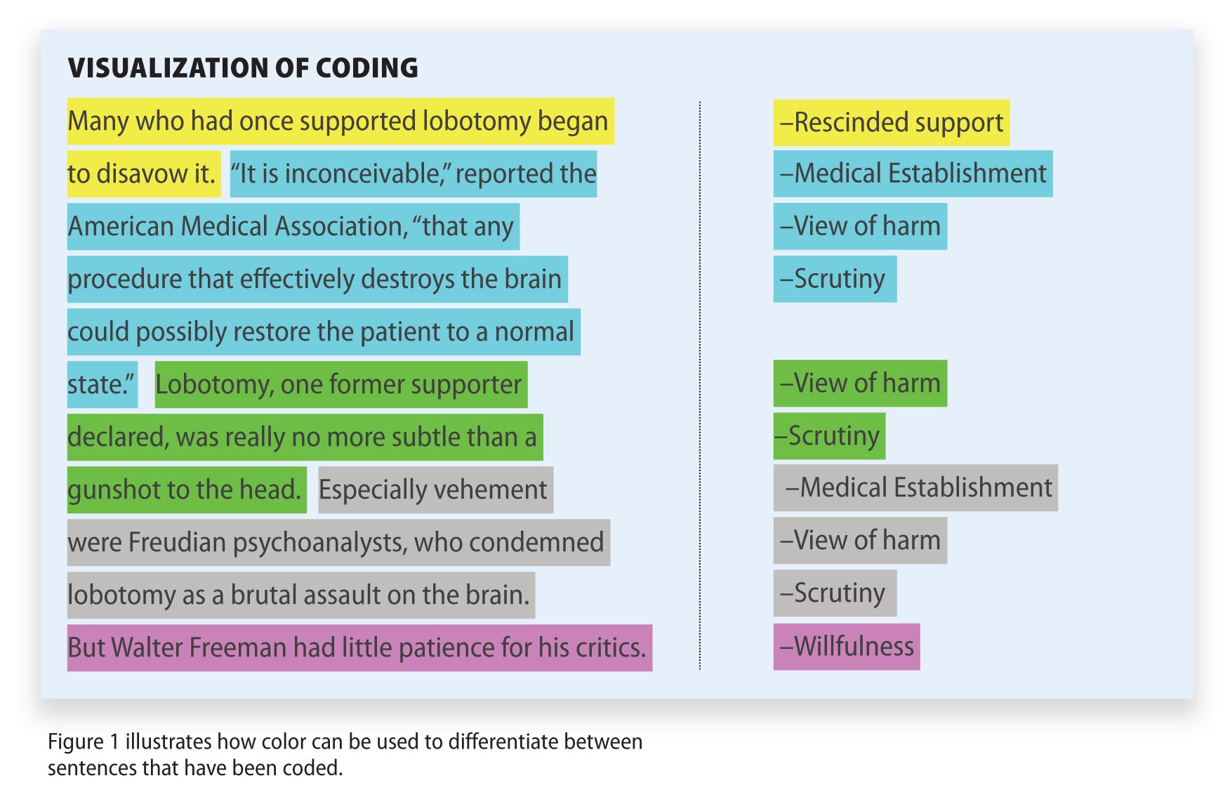
Qualitative Data Coding

What Is a Focus Group?

Cross-Cultural Research Methodology In Psychology

What Is Internal Validity In Research?

Research Methodology , Statistics
What Is Face Validity In Research? Importance & How To Measure

Criterion Validity: Definition & Examples
Collaborate with anyone, anywhere .
Use Hypothesis to annotate anything online with classmates, colleagues, or friends. Create a free personal account , or talk to our sales team about Education solutions.
Get Started
Bringing content and conversation together.
Hypothesis adds a collaborative layer over any online content. Through the power of social annotation, we can make online discussions more meaningful, productive, and engaging.
Hypothesis is for
Keep students engaged and connected with a seamless learning experience.
Create a new method of peer review and engagement for researchers and publications.
Share and discuss intriguing content with family, friends, and like-minded groups.
Comment on and highlight what interests you.
Annotate articles, websites, videos, documents, apps, and more – without clicking away or posting elsewhere. Hypothesis is easy to use and based on open web standards, so it works across the entire internet.
Turn annotations into conversations.
Classrooms, coworkers, and communities can share ideas, ask questions, and respond to annotations. Set groups to public or private to create your ideal discussion.
Build durable, shared knowledge.
No more losing valuable knowledge across email, forums, and apps. Annotations are saved at the source and accessible to everyone, preserving commentary in context.
- 01. Comment & Highlight
- 02. Conversations
- 03. Knowledge
Changing online discussions for good.
Hypothesis is a mission-driven organization building universal collaboration into the internet with open-source technology.
Vision and Values
Start the conversation.
Create a free account and install the Hypothesis browser extension to start annotating. For Education solutions, contact our sales team.
Create A Free Account Contact Sales Team
What Is A Research (Scientific) Hypothesis? A plain-language explainer + examples
By: Derek Jansen (MBA) | Reviewed By: Dr Eunice Rautenbach | June 2020
If you’re new to the world of research, or it’s your first time writing a dissertation or thesis, you’re probably noticing that the words “research hypothesis” and “scientific hypothesis” are used quite a bit, and you’re wondering what they mean in a research context .
“Hypothesis” is one of those words that people use loosely, thinking they understand what it means. However, it has a very specific meaning within academic research. So, it’s important to understand the exact meaning before you start hypothesizing.
Research Hypothesis 101
- What is a hypothesis ?
- What is a research hypothesis (scientific hypothesis)?
- Requirements for a research hypothesis
- Definition of a research hypothesis
- The null hypothesis
What is a hypothesis?
Let’s start with the general definition of a hypothesis (not a research hypothesis or scientific hypothesis), according to the Cambridge Dictionary:
Hypothesis: an idea or explanation for something that is based on known facts but has not yet been proved.
In other words, it’s a statement that provides an explanation for why or how something works, based on facts (or some reasonable assumptions), but that has not yet been specifically tested . For example, a hypothesis might look something like this:
Hypothesis: sleep impacts academic performance.
This statement predicts that academic performance will be influenced by the amount and/or quality of sleep a student engages in – sounds reasonable, right? It’s based on reasonable assumptions , underpinned by what we currently know about sleep and health (from the existing literature). So, loosely speaking, we could call it a hypothesis, at least by the dictionary definition.
But that’s not good enough…
Unfortunately, that’s not quite sophisticated enough to describe a research hypothesis (also sometimes called a scientific hypothesis), and it wouldn’t be acceptable in a dissertation, thesis or research paper . In the world of academic research, a statement needs a few more criteria to constitute a true research hypothesis .
What is a research hypothesis?
A research hypothesis (also called a scientific hypothesis) is a statement about the expected outcome of a study (for example, a dissertation or thesis). To constitute a quality hypothesis, the statement needs to have three attributes – specificity , clarity and testability .
Let’s take a look at these more closely.
Need a helping hand?
Hypothesis Essential #1: Specificity & Clarity
A good research hypothesis needs to be extremely clear and articulate about both what’ s being assessed (who or what variables are involved ) and the expected outcome (for example, a difference between groups, a relationship between variables, etc.).
Let’s stick with our sleepy students example and look at how this statement could be more specific and clear.
Hypothesis: Students who sleep at least 8 hours per night will, on average, achieve higher grades in standardised tests than students who sleep less than 8 hours a night.
As you can see, the statement is very specific as it identifies the variables involved (sleep hours and test grades), the parties involved (two groups of students), as well as the predicted relationship type (a positive relationship). There’s no ambiguity or uncertainty about who or what is involved in the statement, and the expected outcome is clear.
Contrast that to the original hypothesis we looked at – “Sleep impacts academic performance” – and you can see the difference. “Sleep” and “academic performance” are both comparatively vague , and there’s no indication of what the expected relationship direction is (more sleep or less sleep). As you can see, specificity and clarity are key.

Hypothesis Essential #2: Testability (Provability)
A statement must be testable to qualify as a research hypothesis. In other words, there needs to be a way to prove (or disprove) the statement. If it’s not testable, it’s not a hypothesis – simple as that.
For example, consider the hypothesis we mentioned earlier:
Hypothesis: Students who sleep at least 8 hours per night will, on average, achieve higher grades in standardised tests than students who sleep less than 8 hours a night.
We could test this statement by undertaking a quantitative study involving two groups of students, one that gets 8 or more hours of sleep per night for a fixed period, and one that gets less. We could then compare the standardised test results for both groups to see if there’s a statistically significant difference.
Again, if you compare this to the original hypothesis we looked at – “Sleep impacts academic performance” – you can see that it would be quite difficult to test that statement, primarily because it isn’t specific enough. How much sleep? By who? What type of academic performance?
So, remember the mantra – if you can’t test it, it’s not a hypothesis 🙂

Defining A Research Hypothesis
You’re still with us? Great! Let’s recap and pin down a clear definition of a hypothesis.
A research hypothesis (or scientific hypothesis) is a statement about an expected relationship between variables, or explanation of an occurrence, that is clear, specific and testable.
So, when you write up hypotheses for your dissertation or thesis, make sure that they meet all these criteria. If you do, you’ll not only have rock-solid hypotheses but you’ll also ensure a clear focus for your entire research project.
What about the null hypothesis?
You may have also heard the terms null hypothesis , alternative hypothesis, or H-zero thrown around. At a simple level, the null hypothesis is the counter-proposal to the original hypothesis.
For example, if the hypothesis predicts that there is a relationship between two variables (for example, sleep and academic performance), the null hypothesis would predict that there is no relationship between those variables.
At a more technical level, the null hypothesis proposes that no statistical significance exists in a set of given observations and that any differences are due to chance alone.
And there you have it – hypotheses in a nutshell.
If you have any questions, be sure to leave a comment below and we’ll do our best to help you. If you need hands-on help developing and testing your hypotheses, consider our private coaching service , where we hold your hand through the research journey.

Psst... there’s more!
This post was based on one of our popular Research Bootcamps . If you're working on a research project, you'll definitely want to check this out ...
You Might Also Like:

16 Comments
Very useful information. I benefit more from getting more information in this regard.
Very great insight,educative and informative. Please give meet deep critics on many research data of public international Law like human rights, environment, natural resources, law of the sea etc
In a book I read a distinction is made between null, research, and alternative hypothesis. As far as I understand, alternative and research hypotheses are the same. Can you please elaborate? Best Afshin
This is a self explanatory, easy going site. I will recommend this to my friends and colleagues.
Very good definition. How can I cite your definition in my thesis? Thank you. Is nul hypothesis compulsory in a research?
It’s a counter-proposal to be proven as a rejection
Please what is the difference between alternate hypothesis and research hypothesis?
It is a very good explanation. However, it limits hypotheses to statistically tasteable ideas. What about for qualitative researches or other researches that involve quantitative data that don’t need statistical tests?
In qualitative research, one typically uses propositions, not hypotheses.
could you please elaborate it more
I’ve benefited greatly from these notes, thank you.
This is very helpful
well articulated ideas are presented here, thank you for being reliable sources of information
Excellent. Thanks for being clear and sound about the research methodology and hypothesis (quantitative research)
I have only a simple question regarding the null hypothesis. – Is the null hypothesis (Ho) known as the reversible hypothesis of the alternative hypothesis (H1? – How to test it in academic research?
this is very important note help me much more
Trackbacks/Pingbacks
- What Is Research Methodology? Simple Definition (With Examples) - Grad Coach - […] Contrasted to this, a quantitative methodology is typically used when the research aims and objectives are confirmatory in nature. For example,…
Submit a Comment Cancel reply
Your email address will not be published. Required fields are marked *
Save my name, email, and website in this browser for the next time I comment.
- Print Friendly

- Manuscript Preparation
What is and How to Write a Good Hypothesis in Research?
- 4 minute read
- 318.5K views
Table of Contents
One of the most important aspects of conducting research is constructing a strong hypothesis. But what makes a hypothesis in research effective? In this article, we’ll look at the difference between a hypothesis and a research question, as well as the elements of a good hypothesis in research. We’ll also include some examples of effective hypotheses, and what pitfalls to avoid.
What is a Hypothesis in Research?
Simply put, a hypothesis is a research question that also includes the predicted or expected result of the research. Without a hypothesis, there can be no basis for a scientific or research experiment. As such, it is critical that you carefully construct your hypothesis by being deliberate and thorough, even before you set pen to paper. Unless your hypothesis is clearly and carefully constructed, any flaw can have an adverse, and even grave, effect on the quality of your experiment and its subsequent results.
Research Question vs Hypothesis
It’s easy to confuse research questions with hypotheses, and vice versa. While they’re both critical to the Scientific Method, they have very specific differences. Primarily, a research question, just like a hypothesis, is focused and concise. But a hypothesis includes a prediction based on the proposed research, and is designed to forecast the relationship of and between two (or more) variables. Research questions are open-ended, and invite debate and discussion, while hypotheses are closed, e.g. “The relationship between A and B will be C.”
A hypothesis is generally used if your research topic is fairly well established, and you are relatively certain about the relationship between the variables that will be presented in your research. Since a hypothesis is ideally suited for experimental studies, it will, by its very existence, affect the design of your experiment. The research question is typically used for new topics that have not yet been researched extensively. Here, the relationship between different variables is less known. There is no prediction made, but there may be variables explored. The research question can be casual in nature, simply trying to understand if a relationship even exists, descriptive or comparative.
How to Write Hypothesis in Research
Writing an effective hypothesis starts before you even begin to type. Like any task, preparation is key, so you start first by conducting research yourself, and reading all you can about the topic that you plan to research. From there, you’ll gain the knowledge you need to understand where your focus within the topic will lie.
Remember that a hypothesis is a prediction of the relationship that exists between two or more variables. Your job is to write a hypothesis, and design the research, to “prove” whether or not your prediction is correct. A common pitfall is to use judgments that are subjective and inappropriate for the construction of a hypothesis. It’s important to keep the focus and language of your hypothesis objective.
An effective hypothesis in research is clearly and concisely written, and any terms or definitions clarified and defined. Specific language must also be used to avoid any generalities or assumptions.
Use the following points as a checklist to evaluate the effectiveness of your research hypothesis:
- Predicts the relationship and outcome
- Simple and concise – avoid wordiness
- Clear with no ambiguity or assumptions about the readers’ knowledge
- Observable and testable results
- Relevant and specific to the research question or problem
Research Hypothesis Example
Perhaps the best way to evaluate whether or not your hypothesis is effective is to compare it to those of your colleagues in the field. There is no need to reinvent the wheel when it comes to writing a powerful research hypothesis. As you’re reading and preparing your hypothesis, you’ll also read other hypotheses. These can help guide you on what works, and what doesn’t, when it comes to writing a strong research hypothesis.
Here are a few generic examples to get you started.
Eating an apple each day, after the age of 60, will result in a reduction of frequency of physician visits.
Budget airlines are more likely to receive more customer complaints. A budget airline is defined as an airline that offers lower fares and fewer amenities than a traditional full-service airline. (Note that the term “budget airline” is included in the hypothesis.
Workplaces that offer flexible working hours report higher levels of employee job satisfaction than workplaces with fixed hours.
Each of the above examples are specific, observable and measurable, and the statement of prediction can be verified or shown to be false by utilizing standard experimental practices. It should be noted, however, that often your hypothesis will change as your research progresses.
Language Editing Plus
Elsevier’s Language Editing Plus service can help ensure that your research hypothesis is well-designed, and articulates your research and conclusions. Our most comprehensive editing package, you can count on a thorough language review by native-English speakers who are PhDs or PhD candidates. We’ll check for effective logic and flow of your manuscript, as well as document formatting for your chosen journal, reference checks, and much more.

- Research Process
Systematic Literature Review or Literature Review?

What is a Problem Statement? [with examples]
You may also like.

Page-Turner Articles are More Than Just Good Arguments: Be Mindful of Tone and Structure!

A Must-see for Researchers! How to Ensure Inclusivity in Your Scientific Writing

Make Hook, Line, and Sinker: The Art of Crafting Engaging Introductions

Can Describing Study Limitations Improve the Quality of Your Paper?

A Guide to Crafting Shorter, Impactful Sentences in Academic Writing

6 Steps to Write an Excellent Discussion in Your Manuscript

How to Write Clear and Crisp Civil Engineering Papers? Here are 5 Key Tips to Consider

The Clear Path to An Impactful Paper: ②
Input your search keywords and press Enter.
How to Write a Research Hypothesis
- Research Process
- Peer Review
Since grade school, we've all been familiar with hypotheses. The hypothesis is an essential step of the scientific method. But what makes an effective research hypothesis, how do you create one, and what types of hypotheses are there? We answer these questions and more.
Updated on April 27, 2022

What is a research hypothesis?
General hypothesis.
Since grade school, we've all been familiar with the term “hypothesis.” A hypothesis is a fact-based guess or prediction that has not been proven. It is an essential step of the scientific method. The hypothesis of a study is a drive for experimentation to either prove the hypothesis or dispute it.
Research Hypothesis
A research hypothesis is more specific than a general hypothesis. It is an educated, expected prediction of the outcome of a study that is testable.
What makes an effective research hypothesis?
A good research hypothesis is a clear statement of the relationship between a dependent variable(s) and independent variable(s) relevant to the study that can be disproven.
Research hypothesis checklist
Once you've written a possible hypothesis, make sure it checks the following boxes:
- It must be testable: You need a means to prove your hypothesis. If you can't test it, it's not a hypothesis.
- It must include a dependent and independent variable: At least one independent variable ( cause ) and one dependent variable ( effect ) must be included.
- The language must be easy to understand: Be as clear and concise as possible. Nothing should be left to interpretation.
- It must be relevant to your research topic: You probably shouldn't be talking about cats and dogs if your research topic is outer space. Stay relevant to your topic.
How to create an effective research hypothesis
Pose it as a question first.
Start your research hypothesis from a journalistic approach. Ask one of the five W's: Who, what, when, where, or why.
A possible initial question could be: Why is the sky blue?
Do the preliminary research
Once you have a question in mind, read research around your topic. Collect research from academic journals.
If you're looking for information about the sky and why it is blue, research information about the atmosphere, weather, space, the sun, etc.
Write a draft hypothesis
Once you're comfortable with your subject and have preliminary knowledge, create a working hypothesis. Don't stress much over this. Your first hypothesis is not permanent. Look at it as a draft.
Your first draft of a hypothesis could be: Certain molecules in the Earth's atmosphere are responsive to the sky being the color blue.
Make your working draft perfect
Take your working hypothesis and make it perfect. Narrow it down to include only the information listed in the “Research hypothesis checklist” above.
Now that you've written your working hypothesis, narrow it down. Your new hypothesis could be: Light from the sun hitting oxygen molecules in the sky makes the color of the sky appear blue.
Write a null hypothesis
Your null hypothesis should be the opposite of your research hypothesis. It should be able to be disproven by your research.
In this example, your null hypothesis would be: Light from the sun hitting oxygen molecules in the sky does not make the color of the sky appear blue.
Why is it important to have a clear, testable hypothesis?
One of the main reasons a manuscript can be rejected from a journal is because of a weak hypothesis. “Poor hypothesis, study design, methodology, and improper use of statistics are other reasons for rejection of a manuscript,” says Dr. Ish Kumar Dhammi and Dr. Rehan-Ul-Haq in Indian Journal of Orthopaedics.
According to Dr. James M. Provenzale in American Journal of Roentgenology , “The clear declaration of a research question (or hypothesis) in the Introduction is critical for reviewers to understand the intent of the research study. It is best to clearly state the study goal in plain language (for example, “We set out to determine whether condition x produces condition y.”) An insufficient problem statement is one of the more common reasons for manuscript rejection.”
Characteristics that make a hypothesis weak include:
- Unclear variables
- Unoriginality
- Too general
- Too specific
A weak hypothesis leads to weak research and methods . The goal of a paper is to prove or disprove a hypothesis - or to prove or disprove a null hypothesis. If the hypothesis is not a dependent variable of what is being studied, the paper's methods should come into question.
A strong hypothesis is essential to the scientific method. A hypothesis states an assumed relationship between at least two variables and the experiment then proves or disproves that relationship with statistical significance. Without a proven and reproducible relationship, the paper feeds into the reproducibility crisis. Learn more about writing for reproducibility .
In a study published in The Journal of Obstetrics and Gynecology of India by Dr. Suvarna Satish Khadilkar, she reviewed 400 rejected manuscripts to see why they were rejected. Her studies revealed that poor methodology was a top reason for the submission having a final disposition of rejection.
Aside from publication chances, Dr. Gareth Dyke believes a clear hypothesis helps efficiency.
“Developing a clear and testable hypothesis for your research project means that you will not waste time, energy, and money with your work,” said Dyke. “Refining a hypothesis that is both meaningful, interesting, attainable, and testable is the goal of all effective research.”
Types of research hypotheses
There can be overlap in these types of hypotheses.
Simple hypothesis
A simple hypothesis is a hypothesis at its most basic form. It shows the relationship of one independent and one independent variable.
Example: Drinking soda (independent variable) every day leads to obesity (dependent variable).
Complex hypothesis
A complex hypothesis shows the relationship of two or more independent and dependent variables.
Example: Drinking soda (independent variable) every day leads to obesity (dependent variable) and heart disease (dependent variable).
Directional hypothesis
A directional hypothesis guesses which way the results of an experiment will go. It uses words like increase, decrease, higher, lower, positive, negative, more, or less. It is also frequently used in statistics.
Example: Humans exposed to radiation have a higher risk of cancer than humans not exposed to radiation.
Non-directional hypothesis
A non-directional hypothesis says there will be an effect on the dependent variable, but it does not say which direction.
Associative hypothesis
An associative hypothesis says that when one variable changes, so does the other variable.
Alternative hypothesis
An alternative hypothesis states that the variables have a relationship.
- The opposite of a null hypothesis
Example: An apple a day keeps the doctor away.

Null hypothesis
A null hypothesis states that there is no relationship between the two variables. It is posed as the opposite of what the alternative hypothesis states.
Researchers use a null hypothesis to work to be able to reject it. A null hypothesis:
- Can never be proven
- Can only be rejected
- Is the opposite of an alternative hypothesis
Example: An apple a day does not keep the doctor away.
Logical hypothesis
A logical hypothesis is a suggested explanation while using limited evidence.
Example: Bats can navigate in the dark better than tigers.
In this hypothesis, the researcher knows that tigers cannot see in the dark, and bats mostly live in darkness.
Empirical hypothesis
An empirical hypothesis is also called a “working hypothesis.” It uses the trial and error method and changes around the independent variables.
- An apple a day keeps the doctor away.
- Two apples a day keep the doctor away.
- Three apples a day keep the doctor away.
In this case, the research changes the hypothesis as the researcher learns more about his/her research.
Statistical hypothesis
A statistical hypothesis is a look of a part of a population or statistical model. This type of hypothesis is especially useful if you are making a statement about a large population. Instead of having to test the entire population of Illinois, you could just use a smaller sample of people who live there.
Example: 70% of people who live in Illinois are iron deficient.
Causal hypothesis
A causal hypothesis states that the independent variable will have an effect on the dependent variable.
Example: Using tobacco products causes cancer.
Final thoughts
Make sure your research is error-free before you send it to your preferred journal . Check our our English Editing services to avoid your chances of desk rejection.

Jonny Rhein, BA
See our "Privacy Policy"
Popular searches
- How to Get Participants For Your Study
- How to Do Segmentation?
- Conjoint Preference Share Simulator
- MaxDiff Analysis
- Likert Scales
- Reliability & Validity
Request consultation
Do you need support in running a pricing or product study? We can help you with agile consumer research and conjoint analysis.
Looking for an online survey platform?
Conjointly offers a great survey tool with multiple question types, randomisation blocks, and multilingual support. The Basic tier is always free.
Research Methods Knowledge Base
- Navigating the Knowledge Base
- Five Big Words
- Types of Research Questions
- Time in Research
- Types of Relationships
- Types of Data
- Unit of Analysis
- Two Research Fallacies
- Philosophy of Research
- Ethics in Research
- Conceptualizing
- Evaluation Research
- Measurement
- Research Design
- Table of Contents
Fully-functional online survey tool with various question types, logic, randomisation, and reporting for unlimited number of surveys.
Completely free for academics and students .
An hypothesis is a specific statement of prediction. It describes in concrete (rather than theoretical) terms what you expect will happen in your study. Not all studies have hypotheses. Sometimes a study is designed to be exploratory (see inductive research ). There is no formal hypothesis, and perhaps the purpose of the study is to explore some area more thoroughly in order to develop some specific hypothesis or prediction that can be tested in future research. A single study may have one or many hypotheses.
Actually, whenever I talk about an hypothesis, I am really thinking simultaneously about two hypotheses. Let’s say that you predict that there will be a relationship between two variables in your study. The way we would formally set up the hypothesis test is to formulate two hypothesis statements, one that describes your prediction and one that describes all the other possible outcomes with respect to the hypothesized relationship. Your prediction is that variable A and variable B will be related (you don’t care whether it’s a positive or negative relationship). Then the only other possible outcome would be that variable A and variable B are not related. Usually, we call the hypothesis that you support (your prediction) the alternative hypothesis, and we call the hypothesis that describes the remaining possible outcomes the null hypothesis. Sometimes we use a notation like HA or H1 to represent the alternative hypothesis or your prediction, and HO or H0 to represent the null case. You have to be careful here, though. In some studies, your prediction might very well be that there will be no difference or change. In this case, you are essentially trying to find support for the null hypothesis and you are opposed to the alternative.
If your prediction specifies a direction, and the null therefore is the no difference prediction and the prediction of the opposite direction, we call this a one-tailed hypothesis . For instance, let’s imagine that you are investigating the effects of a new employee training program and that you believe one of the outcomes will be that there will be less employee absenteeism. Your two hypotheses might be stated something like this:
The null hypothesis for this study is:
HO: As a result of the XYZ company employee training program, there will either be no significant difference in employee absenteeism or there will be a significant increase .
which is tested against the alternative hypothesis:
HA: As a result of the XYZ company employee training program, there will be a significant decrease in employee absenteeism.
In the figure on the left, we see this situation illustrated graphically. The alternative hypothesis – your prediction that the program will decrease absenteeism – is shown there. The null must account for the other two possible conditions: no difference, or an increase in absenteeism. The figure shows a hypothetical distribution of absenteeism differences. We can see that the term “one-tailed” refers to the tail of the distribution on the outcome variable.
When your prediction does not specify a direction, we say you have a two-tailed hypothesis . For instance, let’s assume you are studying a new drug treatment for depression. The drug has gone through some initial animal trials, but has not yet been tested on humans. You believe (based on theory and the previous research) that the drug will have an effect, but you are not confident enough to hypothesize a direction and say the drug will reduce depression (after all, you’ve seen more than enough promising drug treatments come along that eventually were shown to have severe side effects that actually worsened symptoms). In this case, you might state the two hypotheses like this:
HO: As a result of 300mg./day of the ABC drug, there will be no significant difference in depression.
HA: As a result of 300mg./day of the ABC drug, there will be a significant difference in depression.
The figure on the right illustrates this two-tailed prediction for this case. Again, notice that the term “two-tailed” refers to the tails of the distribution for your outcome variable.
The important thing to remember about stating hypotheses is that you formulate your prediction (directional or not), and then you formulate a second hypothesis that is mutually exclusive of the first and incorporates all possible alternative outcomes for that case. When your study analysis is completed, the idea is that you will have to choose between the two hypotheses. If your prediction was correct, then you would (usually) reject the null hypothesis and accept the alternative. If your original prediction was not supported in the data, then you will accept the null hypothesis and reject the alternative. The logic of hypothesis testing is based on these two basic principles:
- the formulation of two mutually exclusive hypothesis statements that, together, exhaust all possible outcomes
- the testing of these so that one is necessarily accepted and the other rejected
OK, I know it’s a convoluted, awkward and formalistic way to ask research questions. But it encompasses a long tradition in statistics called the hypothetical-deductive model , and sometimes we just have to do things because they’re traditions. And anyway, if all of this hypothesis testing was easy enough so anybody could understand it, how do you think statisticians would stay employed?
Cookie Consent
Conjointly uses essential cookies to make our site work. We also use additional cookies in order to understand the usage of the site, gather audience analytics, and for remarketing purposes.
For more information on Conjointly's use of cookies, please read our Cookie Policy .
Which one are you?
I am new to conjointly, i am already using conjointly.
- More from M-W
- To save this word, you'll need to log in. Log In
Definition of hypothesis
Did you know.
The Difference Between Hypothesis and Theory
A hypothesis is an assumption, an idea that is proposed for the sake of argument so that it can be tested to see if it might be true.
In the scientific method, the hypothesis is constructed before any applicable research has been done, apart from a basic background review. You ask a question, read up on what has been studied before, and then form a hypothesis.
A hypothesis is usually tentative; it's an assumption or suggestion made strictly for the objective of being tested.
A theory , in contrast, is a principle that has been formed as an attempt to explain things that have already been substantiated by data. It is used in the names of a number of principles accepted in the scientific community, such as the Big Bang Theory . Because of the rigors of experimentation and control, it is understood to be more likely to be true than a hypothesis is.
In non-scientific use, however, hypothesis and theory are often used interchangeably to mean simply an idea, speculation, or hunch, with theory being the more common choice.
Since this casual use does away with the distinctions upheld by the scientific community, hypothesis and theory are prone to being wrongly interpreted even when they are encountered in scientific contexts—or at least, contexts that allude to scientific study without making the critical distinction that scientists employ when weighing hypotheses and theories.
The most common occurrence is when theory is interpreted—and sometimes even gleefully seized upon—to mean something having less truth value than other scientific principles. (The word law applies to principles so firmly established that they are almost never questioned, such as the law of gravity.)
This mistake is one of projection: since we use theory in general to mean something lightly speculated, then it's implied that scientists must be talking about the same level of uncertainty when they use theory to refer to their well-tested and reasoned principles.
The distinction has come to the forefront particularly on occasions when the content of science curricula in schools has been challenged—notably, when a school board in Georgia put stickers on textbooks stating that evolution was "a theory, not a fact, regarding the origin of living things." As Kenneth R. Miller, a cell biologist at Brown University, has said , a theory "doesn’t mean a hunch or a guess. A theory is a system of explanations that ties together a whole bunch of facts. It not only explains those facts, but predicts what you ought to find from other observations and experiments.”
While theories are never completely infallible, they form the basis of scientific reasoning because, as Miller said "to the best of our ability, we’ve tested them, and they’ve held up."
- proposition
- supposition
hypothesis , theory , law mean a formula derived by inference from scientific data that explains a principle operating in nature.
hypothesis implies insufficient evidence to provide more than a tentative explanation.
theory implies a greater range of evidence and greater likelihood of truth.
law implies a statement of order and relation in nature that has been found to be invariable under the same conditions.
Examples of hypothesis in a Sentence
These examples are programmatically compiled from various online sources to illustrate current usage of the word 'hypothesis.' Any opinions expressed in the examples do not represent those of Merriam-Webster or its editors. Send us feedback about these examples.
Word History
Greek, from hypotithenai to put under, suppose, from hypo- + tithenai to put — more at do
1641, in the meaning defined at sense 1a
Phrases Containing hypothesis
- counter - hypothesis
- nebular hypothesis
- null hypothesis
- planetesimal hypothesis
- Whorfian hypothesis
Articles Related to hypothesis

This is the Difference Between a...
This is the Difference Between a Hypothesis and a Theory
In scientific reasoning, they're two completely different things
Dictionary Entries Near hypothesis
hypothermia
hypothesize
Cite this Entry
“Hypothesis.” Merriam-Webster.com Dictionary , Merriam-Webster, https://www.merriam-webster.com/dictionary/hypothesis. Accessed 28 May. 2024.
Kids Definition
Kids definition of hypothesis, medical definition, medical definition of hypothesis, more from merriam-webster on hypothesis.
Nglish: Translation of hypothesis for Spanish Speakers
Britannica English: Translation of hypothesis for Arabic Speakers
Britannica.com: Encyclopedia article about hypothesis
Subscribe to America's largest dictionary and get thousands more definitions and advanced search—ad free!

Can you solve 4 words at once?
Word of the day.
See Definitions and Examples »
Get Word of the Day daily email!
Popular in Grammar & Usage
More commonly misspelled words, commonly misspelled words, how to use em dashes (—), en dashes (–) , and hyphens (-), absent letters that are heard anyway, how to use accents and diacritical marks, popular in wordplay, the words of the week - may 24, flower etymologies for your spring garden, 9 superb owl words, 'gaslighting,' 'woke,' 'democracy,' and other top lookups, 10 words for lesser-known games and sports, games & quizzes.

- Scientific Methods
What is Hypothesis?
We have heard of many hypotheses which have led to great inventions in science. Assumptions that are made on the basis of some evidence are known as hypotheses. In this article, let us learn in detail about the hypothesis and the type of hypothesis with examples.
A hypothesis is an assumption that is made based on some evidence. This is the initial point of any investigation that translates the research questions into predictions. It includes components like variables, population and the relation between the variables. A research hypothesis is a hypothesis that is used to test the relationship between two or more variables.
Characteristics of Hypothesis
Following are the characteristics of the hypothesis:
- The hypothesis should be clear and precise to consider it to be reliable.
- If the hypothesis is a relational hypothesis, then it should be stating the relationship between variables.
- The hypothesis must be specific and should have scope for conducting more tests.
- The way of explanation of the hypothesis must be very simple and it should also be understood that the simplicity of the hypothesis is not related to its significance.
Sources of Hypothesis
Following are the sources of hypothesis:
- The resemblance between the phenomenon.
- Observations from past studies, present-day experiences and from the competitors.
- Scientific theories.
- General patterns that influence the thinking process of people.
Types of Hypothesis
There are six forms of hypothesis and they are:
- Simple hypothesis
- Complex hypothesis
- Directional hypothesis
- Non-directional hypothesis
- Null hypothesis
- Associative and casual hypothesis
Simple Hypothesis
It shows a relationship between one dependent variable and a single independent variable. For example – If you eat more vegetables, you will lose weight faster. Here, eating more vegetables is an independent variable, while losing weight is the dependent variable.
Complex Hypothesis
It shows the relationship between two or more dependent variables and two or more independent variables. Eating more vegetables and fruits leads to weight loss, glowing skin, and reduces the risk of many diseases such as heart disease.
Directional Hypothesis
It shows how a researcher is intellectual and committed to a particular outcome. The relationship between the variables can also predict its nature. For example- children aged four years eating proper food over a five-year period are having higher IQ levels than children not having a proper meal. This shows the effect and direction of the effect.
Non-directional Hypothesis
It is used when there is no theory involved. It is a statement that a relationship exists between two variables, without predicting the exact nature (direction) of the relationship.
Null Hypothesis
It provides a statement which is contrary to the hypothesis. It’s a negative statement, and there is no relationship between independent and dependent variables. The symbol is denoted by “H O ”.
Associative and Causal Hypothesis
Associative hypothesis occurs when there is a change in one variable resulting in a change in the other variable. Whereas, the causal hypothesis proposes a cause and effect interaction between two or more variables.
Examples of Hypothesis
Following are the examples of hypotheses based on their types:
- Consumption of sugary drinks every day leads to obesity is an example of a simple hypothesis.
- All lilies have the same number of petals is an example of a null hypothesis.
- If a person gets 7 hours of sleep, then he will feel less fatigue than if he sleeps less. It is an example of a directional hypothesis.
Functions of Hypothesis
Following are the functions performed by the hypothesis:
- Hypothesis helps in making an observation and experiments possible.
- It becomes the start point for the investigation.
- Hypothesis helps in verifying the observations.
- It helps in directing the inquiries in the right direction.
How will Hypothesis help in the Scientific Method?
Researchers use hypotheses to put down their thoughts directing how the experiment would take place. Following are the steps that are involved in the scientific method:
- Formation of question
- Doing background research
- Creation of hypothesis
- Designing an experiment
- Collection of data
- Result analysis
- Summarizing the experiment
- Communicating the results
Frequently Asked Questions – FAQs
What is hypothesis.
A hypothesis is an assumption made based on some evidence.
Give an example of simple hypothesis?
What are the types of hypothesis.
Types of hypothesis are:
- Associative and Casual hypothesis
State true or false: Hypothesis is the initial point of any investigation that translates the research questions into a prediction.
Define complex hypothesis..
A complex hypothesis shows the relationship between two or more dependent variables and two or more independent variables.

Put your understanding of this concept to test by answering a few MCQs. Click ‘Start Quiz’ to begin!
Select the correct answer and click on the “Finish” button Check your score and answers at the end of the quiz
Visit BYJU’S for all Physics related queries and study materials
Your result is as below
Request OTP on Voice Call
Leave a Comment Cancel reply
Your Mobile number and Email id will not be published. Required fields are marked *
Post My Comment
Register with BYJU'S & Download Free PDFs
Register with byju's & watch live videos.

( Photo-illustration by Realtor.com; Source: Getty Images (3) )
Home Prices in Swing States Could Decide the 2024 Election—and the Reason Might Surprise You
Home prices could play a subtle but important role in the 2024 presidential election, according to a recent first-of-its kind study.
The academic study, Housing Performance and the Electorate , analyzed home prices and election results at the county level for each of the six presidential elections from 2000 to 2020.
The authors found that local home price performance significantly affects voting in presidential elections at the county level. Counties with superior gains in home prices in the four years preceding an election were more likely to “vote-switch” to the incumbent party’s presidential candidate.
Conversely, counties with relatively inferior home price performance leading up to an election were more likely to flip their vote to support the candidate challenging the incumbent party.
In other words, quickly rising home prices tend to favor the incumbent president’s party, whether it be the Democrats or Republicans. The study found that the relationship is strongest in the years closest to an election, and that home prices were most influential in the small group of “swing counties” with a history of switching party preference.
University of Alabama Associate Professor of Finance Alan Tidwell , one of the study’s co-authors, explains that the logic driving this trend is simple: For most voters, their home represents their single largest asset.
“People feel more financially wealthy if they have a lot of housing equity, relative to lower housing equity,” he says. “How financially wealthy they feel really impacts their sense of financial and economic well-being.”
For the upcoming presidential election, the new finding suggests the outcome could be partly influenced by home prices in swing counties of the seven battleground states: Arizona, Georgia, Michigan, Nevada, North Carolina, Pennsylvania, and Wisconsin.
“In the swing counties, they care about this economic factor most, and real estate is one of the main drivers of household wealth,” says lead author Eren Cifci , an assistant professor of finance at Austin Peay State University in Tennessee. “So there may be many other factors that affect how people vote, but this definitely appears to be one of the factors influencing voters when they make their decisions.”
Explaining the 'homevoter hypothesis'
The study’s finding is an extension of the “homevoter hypothesis,” which holds that homeowners tend to vote in support of policies and candidates they believe will boost their home values.
While that phenomenon is well documented in local politics, where government policies have the clearest impact on home values, the new study is the first to show evidence of homevoter behavior in national elections.
The term “homevoter” was coined in 2001 by William A. Fischel , a now-retired economics professor at Dartmouth College and expert in local government and land use regulation.
Fischel conceived the homevoter hypothesis while serving on the local zoning board in Hannover, NH. Regularly, he would hear objections and concerns about zoning changes that seemed esoteric, and noticed that the complaints were always from homeowners.
Fischel says he came to realize that homeowners are essentially shareholders in their community, similar to owners of stock in a company—but that unlike corporate shareholders, they cannot easily diversify their portfolio or liquidate their holdings.
“It's people who are voting their homes, and that's actually an old concept in economics,” says Fischel. “But also, they're very risk-averse, because so much of their assets are stuck in one stock, in one place.”
Fischel says he was surprised by the recent study linking home prices and voting in national elections, since he had always viewed homevoting as primarily a local phenomenon.
“I can see, a little bit, what a presidential election might mean for home values. But it's so indirect, I was really quite surprised at the strength of the evidence,” he says. “How did they find such a strong mechanism? But I have no reason to doubt their evidence.”
For his part, Tidwell argues that the economy plays a major role in most presidential elections, and that rising home equity has a significant impact on how voters perceive the strength of the economy.
Even if local policies, such as zoning laws and public school funding, have a bigger direct impact on local home prices, national elections are where more voters take the opportunity to weigh in with their concern or satisfaction, he says.
“Local elections don't have big turnout, and they don't have big visibility, whereas the national election has a whole bunch more turnout and a whole lot more national media exposure, especially with talk of the economy,” says Tidwell.
Home prices play the biggest role in swing counties
To conduct their study, Cifci and Tidwell, with co-authors Sherwood Clements and Andres Jauregui , looked at the voting results for every county in the continental U.S. over the past six presidential elections.
Of those counties, 77% never changed their party preference, voting for either the Democrat or the Republican in every election since 2000, which was used as the base year for analyzing the 2004 election.
But 641 counties across the country–or 23%–switched their party vote at least once across the survey period, some as many as four times. In that subset of swing counties, home prices appeared to have the biggest impact on election results, according to the study.
In swing counties, for every 1% increase in home values over the four years preceding an election, the county was 0.36% more likely to vote for the incumbent party in the next election, the study found.
As well, the data showed that each 1% increase in home prices made the county 0.19% more likely to “flip” its vote to the incumbent party’s candidate. Those figures are after the study controlled for a variety of other factors that could sway elections, such as changes in demographics, the economy, and government benefits.
“The larger the return [on home values], the more likely you are to vote for the incumbent, or to flip for the incumbent,” explains Tidwell. “For every percent of positive return, there is a percentage increase in voting for the incumbent.”
What does it mean for 2024?
Home prices have risen rapidly across the country over the past four years, including in the seven swing states.
From March 2020 to March 2024, national home values rose 46.4%, according to the Freddie Mac Home Price Index. Of the swing states, North Carolina, Arizona, Georgia, and Wisconsin all outperformed the national average, with four-year price gains greater than 50%.
The study suggests that trend would tend broadly to favor the incumbent, President Joe Biden , as he seeks reelection, particularly in the areas that have seen the strongest home price gains. But the authors caution that their finding only demonstrates a statistical nudge in one direction or the other. They warn that there are many other variables at play in an election.
“It's just one of many factors,” Tidwell says of home price performance. “It's not really a forecast on its own.”
As well, voter turnout in counties that are reliably Democratic or Republican can be just as important to the state-level results in swing states as the marginal shifts in counties that flip from one party to another.
But in an election that is increasingly focused on the housing market, the new findings provide an interesting twist on the role of home prices in voter decision making.
Donald Trump , the presumptive Republican nominee, and his allies have recently levied attacks against Biden over rising home prices, pointing to the challenges raised for prospective first-time homebuyers.
“Under President Biden, home prices have risen almost 50%, making it nearly impossible for millennials to buy their first home and driving the American Dream further and further out of reach,” wrote Sen. Tim Scott , a South Carolina Republican and staunch Trump supporter, on the social media platform X.
On his own Truth Social platform, Trump himself recently wrote: “Crooked Joe has made it impossible for millions of Americans, especially YOUNG Americans, to buy a home.” (Conversely, Trump has also accused Biden of trying to “destroy your property values” by abolishing single-family zoning in the suburbs. The two arguments seem difficult to reconcile.)
It’s true that rising home prices, along with high mortgage rates, are key factors in a national housing crisis that has pushed ownership out of reach for many prospective homebuyers. But on the flip side, most voters are already homeowners. The U.S. homeownership rate is about 66%, and homeowners are significantly more likely to vote than renters.
For existing homeowners, rising home prices mean more equity and higher household net worth, the same as what rising stock prices mean for shareholders.
It suggests that for Republicans, attacking Biden over rising home prices might not carry the same weight with voters as criticism over inflation for goods such as gasoline and groceries.
“When you go to the grocery store or restaurant or the gas pump, I think maybe people feel a little bit different pain than if they own a house and they see their house price going up,” says Tidwell.
On the other hand, the study found evidence that, for swing counties, the economically rational choice might be to always flip to the non-incumbent party, which in 2024 would be the Republicans.
The study found that counties that flipped their vote to an incumbent party candidate were not rewarded with superior home price returns in the four years after the election.
However, counties that flipped to vote for the non-incumbent did experience “positive and significant post-election housing returns” if that candidate won. The authors speculate that this might be due to the winning party rewarding new supporters by increasing investment in those areas after regaining the White House.
“The counties that make the national results flip parties, they do well,” says Clements, a collegiate assistant professor of real estate at Virginia Tech. “Whatever counties voted for Biden last time and vote for Trump this time, if you believe our research, they’re going to have home prices rising if Trump wins.”
Editor's note: This article is part of a special Realtor.com series on the housing market and the swing states in the 2024 presidential election. For additional coverage in this series, click here .
Keith Griffith is a journalist at Realtor.com. He covers the housing market and real estate trends.
- Related Articles
Share this Article
Thank you for visiting nature.com. You are using a browser version with limited support for CSS. To obtain the best experience, we recommend you use a more up to date browser (or turn off compatibility mode in Internet Explorer). In the meantime, to ensure continued support, we are displaying the site without styles and JavaScript.
- View all journals
- My Account Login
- Explore content
- About the journal
- Publish with us
- Sign up for alerts
- Open access
- Published: 22 May 2024
Adhesive anti-fibrotic interfaces on diverse organs
- Jingjing Wu ORCID: orcid.org/0000-0002-4565-6914 1 ,
- Jue Deng 1 ,
- Georgios Theocharidis ORCID: orcid.org/0000-0002-8895-9130 2 ,
- Tiffany L. Sarrafian 3 ,
- Leigh G. Griffiths 4 ,
- Roderick T. Bronson 5 ,
- Aristidis Veves 2 ,
- Jianzhu Chen ORCID: orcid.org/0000-0002-5687-6154 6 ,
- Hyunwoo Yuk ORCID: orcid.org/0000-0003-1710-9750 1 nAff8 &
- Xuanhe Zhao ORCID: orcid.org/0000-0001-5387-6186 1 , 7
Nature ( 2024 ) Cite this article
80 Altmetric
Metrics details
- Biomedical engineering
Implanted biomaterials and devices face compromised functionality and efficacy in the long term owing to foreign body reactions and subsequent formation of fibrous capsules at the implant–tissue interfaces 1 , 2 , 3 , 4 . Here we demonstrate that an adhesive implant–tissue interface can mitigate fibrous capsule formation in diverse animal models, including rats, mice, humanized mice and pigs, by reducing the level of infiltration of inflammatory cells into the adhesive implant–tissue interface compared to the non-adhesive implant–tissue interface. Histological analysis shows that the adhesive implant–tissue interface does not form observable fibrous capsules on diverse organs, including the abdominal wall, colon, stomach, lung and heart, over 12 weeks in vivo. In vitro protein adsorption, multiplex Luminex assays, quantitative PCR, immunofluorescence analysis and RNA sequencing are additionally carried out to validate the hypothesis. We further demonstrate long-term bidirectional electrical communication enabled by implantable electrodes with an adhesive interface over 12 weeks in a rat model in vivo. These findings may offer a promising strategy for long-term anti-fibrotic implant–tissue interfaces.
Overcoming the translational barriers of tissue adhesives
Design of biodegradable, implantable devices towards clinical translation
Host responses to implants revealed by intravital microscopy
Foreign body reactions to implants are among the most critical challenges that undermine the long-term functionality and reliability of biomaterials and devices in vivo 1 , 2 , 3 , 4 . In particular, the formation of a fibrous capsule between the implant and the target tissue, as a result of foreign body reactions, can substantially compromise the implant’s efficacy because the fibrous capsule acts as a barrier to mechanical, electrical, chemical or optical communications 4 , 5 , 6 , 7 , 8 , 9 , 10 , 11 (Fig. 1a,b ). To alleviate the formation of the fibrous capsule at the implant–tissue interface, various approaches have been developed, including drug-eluting coatings 12 , hydrophilic 13 or zwitterionic polymer coatings 14 , 15 , 16 , active surfaces 17 , 18 and controlling the stiffness 19 and/or size 20 , 21 of the implants. However, despite recent advances, the mitigation of fibrous capsule formation for implanted biomaterials and devices remains an ongoing challenge in the field 5 , 22 , highlighting the importance of developing new solutions and strategies.
a , b , Schematic illustrations of a non-adhesive implant consisting of a mock device (polyurethane) and a non-adhesive layer ( a ) and long-term in vivo implantation with fibrous capsule formation at the implant–tissue interface ( b ). c , d , Schematic illustrations of an adhesive implant consisting of the mock device (polyurethane) and an adhesive layer ( c ) and long-term in vivo implantation without observable fibrous capsule formation at the implant–tissue interface ( d ). e – i , Representative histology images stained with Masson’s trichrome (MTS) and haematoxylin and eosin (H&E) for native tissue (left), the adhesive implant (middle) and the non-adhesive implant (right) collected on day 84 post-implantation on the abdominal wall ( e ), colon ( f ), stomach ( g ), lung ( h ) and heart ( i ). Black and yellow dashed lines in the images indicate the implant–tissue interface and the fibrous capsule–tissue interface, respectively. The experiment in e – i was repeated independently ( n = 4 per group) with similar results. Scale bars, 50 μm ( e – g , left and middle; h ), 100 μm ( e , right; i ), 200 μm ( f , right), 150 μm ( g , right).
Here we demonstrate that an adhesive interface can not only provide mechanical integration of the implant with the target tissue but also prevent the formation of observable fibrous capsules at the implant–tissue interface (Fig. 1c,d ). We reason that the conformal interfacial integration between the adhesive implant and the tissue surface can reduce the level of infiltration of inflammatory cells (for example, neutrophils, monocytes, macrophages) into the adhesive implant–tissue interface, resulting in a decreased level of collagen deposition and a reduced level of fibrous capsule formation in the long term (Fig. 1d ). By contrast, conventional non-adhesive implants usually do not form conformal integration with the tissue surfaces and attract the infiltration of inflammatory cells into the non-adhesive implant–tissue interfaces. Subsequently, fibrous capsules form on the non-adhesive implant–tissue interfaces (Fig. 1b ).
To test our hypothesis, we prepared an adhesive implant consisting of a mock device (polyurethane) and an adhesive layer 23 , 24 composed of interpenetrating networks between the covalently crosslinked poly(acrylic acid) N -hydroxysuccinimide ester and physically crosslinked poly(vinyl alcohol) (Fig. 1c ). The adhesive layer provides highly conformal and stable integration of the implant with wet tissues 23 , 24 , 25 (Supplementary Fig. 1 ). We further prepared a non-adhesive implant by fully swelling the same mock device and adhesive layer in a phosphate-buffered saline bath before implantation (see Methods for the preparation of the non-adhesive implant). By swelling the implant in phosphate-buffered saline, we removed its adhesive property 26 while keeping its chemical composition identical.
Both adhesive and non-adhesive implants were implanted on the surfaces of diverse organs, including the abdominal wall, colon, stomach, lung and heart, using rat models in vivo for up to 84 days (Fig. 1e–i ). Note that the non-adhesive implant was sutured onto the organ surfaces. Macroscopic observations showed that both adhesive and non-adhesive implants remained stable at the implantation site on the organ surfaces (Extended Data Fig. 1b,c ). To analyse the foreign body reaction and fibrous capsule formation for the adhesive and non-adhesive implants, we carried out histological analysis of the native tissue, adhesive implant and non-adhesive implant for various organs (Extended Data Fig. 1a ).
Histological evaluation by a blinded pathologist indicates that the adhesive implant forms conformal integration with the organ surface and shows no observable formation of the fibrous capsule up to 84 days post-implantation for diverse organs, including the abdominal wall, colon, stomach, lung and heart (Fig. 1e–i , Extended Data Fig. 2 and Supplementary Fig. 2 ). Furthermore, a transmission electron micrograph of the adhesive implant–tissue interface shows that the adhesive layer maintains highly conformal integration with the collagenous layer of the mesothelium on a subcellular scale on day 28 post-implantation (Extended Data Fig. 3 ). By contrast, the non-adhesive implant undergoes substantial formation of the fibrous capsule at the implant–tissue interface for all organs, consistent with the foreign body reaction to the mock device alone (Fig. 1 and Supplementary Figs. 2 and 3 ). Similarly, the mock device–cavity interface of the adhesive implant undergoes fibrous capsule formation (the top of the implant in Extended Data Fig. 2 ).
To investigate the potential influence of suture-induced tissue damage, sutures were introduced to the corners of the adhesive implant, similar to those used with the non-adhesive implant (Extended Data Fig. 4a ). The histological analysis shows that the suture point exhibits the formation of fibrosis (Extended Data Fig. 4b,c ), but the intact adhesive implant–tissue interface demonstrates no observable formation of the fibrotic capsule (Extended Data Fig. 4b,d ). Collectively, these data further confirm that the adhesive interface is required to prevent the observable formation of the fibrous capsule.
To investigate the effect of adhesive interfaces with varying compositions and properties, we replaced the poly(vinyl alcohol)-based adhesive interface with a chitosan-based adhesive interface 23 (see Methods for the preparation of the chitosan-based adhesive interface). Compared to the poly(vinyl alcohol)-based adhesive interface, the chitosan-based adhesive interface offers a different composition and Young’s modulus, yet it demonstrates comparable adhesion performance (Extended Data Fig. 5a–d ). Histological analysis shows that the chitosan-based adhesive interface exhibits no observable formation of the fibrous capsule on day 14 post-implantation (Extended Data Fig. 5e,f ). Notably, the implants adhered to the abdominal wall surface using commercially available tissue adhesives including Coseal and Tisseel show the substantial formation of the fibrous capsule on day 14 post-implantation (Extended Data Fig. 6 ). This may be attributed to unstable long-term adhesion of the commercially available tissue adhesives with the tissue surface in vivo 27 , 28 .
To assess the foreign body reaction and fibrous capsule formation over time, we conducted histological analyses for the adhesive and non-adhesive implants on the abdominal wall on days 3, 7, 14, 28 and 84 post-implantation (Fig. 2a–j ). The collagen layer thickness at the implant–tissue interface remains comparable to that of the native tissue (that is, the mesothelium thickness) for the adhesive implant at all time points (Fig. 2k ). By contrast, the collagen layer thickness at the non-adhesive implant–tissue interface increases over time owing to the formation of the fibrous capsule and is significantly thicker than that of both the native tissue and the adhesive implant at all time points (Fig. 2k ).
a – e , Representative histology images stained with Masson’s trichrome (left) and haematoxylin and eosin (right) of the non-adhesive implant collected on day 3 ( a ), day 7 ( b ), day 14 ( c ), day 28 ( d ) and day 84 ( e ) post-implantation on the abdominal wall. f – j , Representative histology images stained with Masson’s trichrome (left) and haematoxylin and eosin (right) of the adhesive implant collected on day 3 ( f ), day 7 ( g ), day 14 ( h ), day 28 ( i ) and day 84 ( j ) post-implantation on the abdominal wall. Asterisks in images indicate the implant; black dashed lines in images indicate the implant–tissue interface; yellow dashed lines in images indicate the mesothelium–fibrous capsule (non-adhesive implant) or the mesothelium–skeletal muscle (adhesive implant) interface. SM, skeletal muscle; FC, fibrous capsule. k , Collagen layer thickness at the implant–tissue interface measured at different time points post-implantation. The blue dashed line indicates the average collagen layer thickness of the native tissue (NT). d, day. Values in k represent the mean and the standard deviation ( n = 3 implants; independent biological replicates). Statistical significance and P values were determined by two-sided unpaired t -tests; * P < 0.05; ** P ≤ 0.01; *** P ≤ 0.001; **** P < 0.0001. Scale bars, 50 μm ( a , f – j ), 100 μm ( b , c ), 200 μm ( d , e ).
Source Data
To further investigate our hypothesis, we carried out a set of characterizations for key participants of the foreign body reaction, including in vitro protein adsorption assays, immunofluorescence analysis, quantitative PCR (qPCR), Luminex quantification and RNA-sequencing analysis. A protein adsorption assay with fluorescently labelled albumin and fibrinogen was carried out to evaluate the adhesion of proteins at the implant–tissue interface during the initial stage of the foreign body reaction 29 , 30 (Supplementary Fig. 4 ). After 30 min of co-culture in the protein solution, the adhesive implant–substrate interface showed a significantly lower level of protein adsorption compared to that of the non-adhesive implant–substrate interface ( P < 0.0001) for both fluorescently labelled albumin and fibrinogen (Supplementary Fig. 4g,h ), demonstrating the adhesive interface’s capability to prevent protein adsorption.
To investigate the infiltration of immune cells into the implant–tissue interface, we carried out immunofluorescence staining for fibroblasts (αSMA), neutrophils (neutrophil elastase), macrophages (CD68 for pan-macrophages; iNOS and vimentin for pro-inflammatory macrophages; CD206 for anti-inflammatory macrophages) and T cells (CD3) on days 3, 7 and 14 post-implantation (Fig. 3a–f ). Quantification of cell numbers in the collagenous layer at the implant–tissue interface over a representative width of 500 µm from the immunofluorescence images shows significantly fewer fibroblasts, neutrophils, macrophages and T cells at the adhesive implant–tissue interface than at the non-adhesive implant–tissue interface at all time points (Fig. 3g–i ).
a , c , e , Representative immunofluorescence images of the non-adhesive implant collected on day 3 ( a ), day 7 ( c ) and day 14 ( e ) post-implantation on the abdominal wall. b , d , f , Representative immunofluorescence images of the adhesive implant collected on day 3 ( b ), day 7 ( d ) and day 14 ( f ) post-implantation on the abdominal wall. In immunofluorescence images, cell nuclei are stained with 4′,6-diamidino-2-phenylindole (DAPI, blue); green fluorescence corresponds to the staining of fibroblasts (αSMA), neutrophils (neutrophil elastase) and macrophages (CD68, vimentin, CD206, iNOS); red fluorescence corresponds to the staining of T cells (CD3). Asterisks in images indicate the implant; white dashed lines in images indicate the implant–tissue interface; yellow dashed lines in images indicate either the mesothelium–fibrous capsule interface (non-adhesive implant) or the mesothelium–skeletal muscle interface (adhesive implant). g – i , Quantification of cell numbers in the collagenous layer at the implant–tissue interface over a representative width of 500 µm from the immunofluorescence images on day 3 ( g ), day 7 ( h ) and day 14 ( i ) post-implantation. Values in g – i represent the mean and the standard deviation ( n = 3 implants; independent biological replicates). Statistical significance and P values were determined by two-sided unpaired t -tests; NS, not significant; * P < 0.05; ** P ≤ 0.01; *** P ≤ 0.001. Scale bars, 20 μm ( a , b , d , f ), 40 μm ( c , e ).
To further delineate the immune response at the implant–tissue interface, we profile immune-cell-related genes and cytokines using qPCR analysis and Luminex quantification, respectively (Fig. 4 ). On day 3 post-implantation, whereas the levels of most select immune gene transcripts are similar or significantly lower in the adhesive compared to the non-adhesive implant–tissue interface, the level of Nos2 expression is significantly higher in the adhesive than in the non-adhesive implant–tissue interface (Fig. 4b ). The higher level of Nos2 expression is in agreement with the higher levels of inflammatory cytokines (G-CSF, IL-12p70) in the adhesive than in the non-adhesive implant–tissue interface on day 3 post-implantation (Fig. 4d and Supplementary Table 1 ).
a , Genes and cytokines relevant to each cell type in the qPCR and Luminex studies. b , c , Normalized gene expression of immune-cell-related markers for the non-adhesive and the adhesive implant–tissue interface collected on day 3 ( b ) and day 7 ( c ) post-implantation on the abdominal wall. d , Heat map of immune-cell-related cytokines measured with Luminex assay of the non-adhesive and the adhesive implant–tissue interfaces collected on days 3 and 7 post-implantation on the abdominal wall. Values in b , c represent the mean and the standard deviation ( n = 9 implants; independent biological replicates). Statistical significance and P values were determined by two-sided unpaired t -tests; NS, not significant; * P < 0.05; ** P ≤ 0.01; **** P < 0.0001.
To investigate the source of Nos2 expression on day 3 post-implantation, we carried out double immunofluorescence staining for iNOS and neutrophil elastase and for iNOS and CD68 (Extended Data Fig. 7 ). The immunofluorescence staining of the adhesive implant–tissue interface reveals a significantly higher number of iNOS + neutrophils than iNOS + macrophages on day 3 post-implantation ( P ≤ 0.01; Extended Data Fig. 7b ). By contrast, the non-adhesive implant–tissue interface has similar numbers of iNOS + neutrophils and iNOS + macrophages on day 3 post-implantation ( P = 0.82; Extended Data Fig. 7d ). This result indicates that the adhesive implant–tissue interface favours an iNOS-producing neutrophil subset on day 3 post-implantation 31 .
By day 7 post-implantation, the adhesive implant–tissue interface exhibits a significantly lower expression level of all immune-cell-related genes, including Nos2 , compared to the non-adhesive implant–tissue interface (Fig. 4c ), consistent with the reduction in the level of inflammatory cytokines in the adhesive implant–tissue interface on day 7 post-implantation compared to day 3 post-implantation (Fig. 4d and Supplementary Table 1 ). Thus, the adhesive implant–tissue interface seems to induce a more robust pro-inflammatory neutrophil response than that of the non-adhesive implant–tissue interface on day 3 post-implantation, which is rapidly resolved by day 7 post-implantation.
Next we carried out bulk RNA sequencing of implant–abdominal wall interfaces for both adhesive and non-adhesive implants on days 3 and 14 post-implantation to further investigate gene expression differences (Extended Data Fig. 8 ). Principal component analysis shows separate clustering of samples for the non-adhesive and adhesive implant–tissue interfaces at each time point, indicating distinct transcriptomic profiles (Extended Data Fig. 8a,d ). Differential gene expression analysis of the adhesive compared to the non-adhesive implant–tissue interface reveals 40 downregulated and 33 upregulated genes on day 3 post-implantation (Extended Data Figs. 8b and 9a ). On day 14 post-implantation, 357 genes are downregulated and 156 genes are upregulated (Extended Data Figs. 8e and 9b ) in the adhesive implant–tissue interface compared to the non-adhesive implant–tissue interface. On day 3 post-implantation, regulation of interferon production and striated muscle tissue development are enriched in the non-adhesive implant–tissue interface, indicating inflammatory and fibrosis processes, whereas cell proliferation and growth processes are enriched in the adhesive implant–tissue interface (Extended Data Fig. 8c ). On day 14 post-implantation, fibrosis-associated processes are highly enriched in the non-adhesive implant–tissue interface, such as muscle cell differentiation, myofibril assembly and muscle structure development, whereas vasculature formation, neurogenesis and proliferation are enriched in the adhesive implant–tissue interface (Extended Data Fig. 8f ). These results again indicate reduced inflammatory response and rapid resolution of inflammation in the adhesive implant–tissue interface compared to the non-adhesive implant–tissue interface.
To test our hypothesis in diverse animal models, we implanted the adhesive and non-adhesive implants on the abdominal wall surface of immunocompetent C57BL/6 mice and HuCD34-NCG humanized mice (Fig. 5a,c ). Note that immunocompetent C57BL/6 mice are known to produce fibrosis and foreign body reactions similar to those observed in human patients 32 , and HuCD34-NCG humanized mice provide human-like immune responses 33 . Histological analysis shows that the adhesive implant–tissue interface exhibits no observable formation of the fibrous capsule, comparable to the native tissue on day 28 post-implantation in both C57BL/6 (Fig. 5b ) and HuCD34-NCG (Fig. 5d ) mouse models. By contrast, the non-adhesive implant–tissue interface shows substantial formation of the fibrous capsule in both models (Fig. 5b,d ).
a , c , e , Schematic illustrations for the study design in C57BL/6 mice ( a ), HuCD34-NCG humanized mice ( c ) and pigs ( e ). Implants are placed on the abdominal wall of the animals. b , d , f , Representative histology images stained with Masson’s trichrome and haematoxylin and eosin for native tissue (left), the adhesive implant (middle) and the non-adhesive implant (right) collected on day 28 post-implantation in C57BL/6 mice ( b ) and HuCD34-NCG humanized mice ( d ), and on day 7 post-implantation in pigs ( f ). Black dashed lines in images indicate the implant–tissue interface; yellow dashed lines in images indicate the fibrous capsule–tissue interface. The experiment in b , d , f was repeated independently ( n = 6 per group for C57BL/6 mice; n = 5 per group for HuCD34-NCG mice; n = 4 per group for pigs) with similar results. Scale bars, 100 μm ( b , d ), 300 μm ( f ). The graphic of the pig in e was created with BioRender.com .
To further test our hypothesis in human-scale anatomy, we implanted the adhesive and non-adhesive implants in porcine models (Fig. 5e and Supplementary Fig. 5 ). Macroscopic observations demonstrate that the adhesive implant maintains stable integration with the surface of the porcine abdominal wall and small intestine on day 7 post-implantation in vivo (Extended Data Fig. 10 ). Histological analysis shows that the adhesive implant forms conformal integration with the tissue surface without observable formation of the fibrous capsule on the implant–tissue interface on day 7 post-implantation for both the abdominal wall (Fig. 5e ) and small intestine (Extended Data Fig. 10a ). By contrast, the non-adhesive implant–tissue interface exhibits substantial formation of the fibrous capsule (Fig. 5f and Extended Data Fig. 10b ), in agreement with the observations in the rodent models.
To explore the potential utility of the adhesive anti-fibrotic interfaces, we demonstrated long-term in vivo electrophysiological recording and stimulation enabled by the implantable electrodes with the adhesive interface in a rat model for 84 days (Fig. 6 ). For continuous in vivo monitoring and modulation of the electrocardiogram, electrodes with either the adhesive or non-adhesive interface were implanted on the epicardial surface of animals for electrophysiological recording and stimulation on days 0, 3, 7, 14, 28, 56 and 84 post-implantation (Fig. 6a and Supplementary Fig. 6 ). Macroscopic observations showed that the electrodes with the adhesive interface maintained stable integration with the heart after 84 days of implantation in vivo (Fig. 6b ). The amplitude of the R wave recorded by the electrodes with the adhesive interface was consistently maintained throughout the study duration (84 days; Fig. 6e–g ), whereas the R-wave amplitude recorded by the electrodes with the non-adhesive interface exhibited a substantial decrease over time (Fig. 6e ). For electrophysical stimulation by the electrodes with the non-adhesive interface, the minimal stimulation current pulse amplitude needed to successfully pace the heart gradually increased until day 7 post-implantation and eventually failed to pace the heart on day 14 post-implantation (Fig. 6c ). By contrast, the electrodes with the adhesive interface exhibited a consistent minimal stimulation current pulse amplitude for pacing and successfully maintained the capability to pace the heart for the duration of the study (84 days; Fig. 6d ). These results are consistent with the histological findings from the tissues collected on day 28 post-implantation, for which the electrodes with the non-adhesive interface showed encapsulation and physical separation from the epicardial surface by a thick fibrous capsule (Fig. 6h ). By contrast, the electrodes with the adhesive interface showed conformal contact with the epicardial surface without observable formation of the fibrous capsule (Fig. 6i ).
a , Schematic illustrations for the in vivo electrophysiological recording and stimulation through implanted electrodes with the non-adhesive or the adhesive implant–tissue interface. b , Photographs of the heart collected on days 0 and 84 post-implantation for electrodes with the adhesive interface. White dashed lines in photographs indicate the boundary of implants. c , Representative epicardial electrocardiograms after stimulation through implanted electrodes with the non-adhesive implant–tissue interface on days 0, 3, 7, 14 and 28 post-implantation on a rat heart. d , Representative epicardial electrocardiograms after stimulation through implanted electrodes with the adhesive implant–tissue interface on days 0, 14, 28, 56 and 84 post-implantation on a rat heart. e – g , Recorded R-wave amplitude through implanted electrodes with the non-adhesive (black) and the adhesive (red) implant–tissue interfaces on day 28 ( e ), day 56 ( f ) and day 84 ( g ) post-implantation on a rat heart. Inset plots show representative recorded waveforms. h , i , Representative histology images stained with Masson’s trichrome (left) and haematoxylin and eosin (right) of the electrodes with the non-adhesive ( h ) and the adhesive ( i ) implant collected on day 28 post-implantation on a rat heart. Asterisks in images indicate the implant; yellow dashed lines in images indicate the implant–tissue interface. Values in e – g represent the mean and the standard deviation ( n = 6 animals; independent biological replicates). The experiment in h , i was repeated independently ( n = 6 per group) with similar results. Statistical significance and P values were determined by two-sided unpaired t -tests; NS, not significant. Scale bars, 200 μm ( h ), 100 μm ( i ).
In this study, we demonstrated that the adhesive interface can not only provide conformal mechanical integration of the implant to the target tissue but also effectively mitigate the formation of the fibrous capsule on the adhesive implant–tissue interface by reducing the level of infiltration of inflammatory cells. The current work provides a promising strategy for long-term anti-fibrotic implant–tissue interfaces and offers valuable insights into implant–tissue interactions for future studies.
Preparation of adhesive implants
The adhesive layer of the adhesive implant was prepared using a previously reported method 23 , 24 . To prepare an adhesive stock solution, 35% w/w acrylic acid, 7% w/w poly(vinyl alcohol) (PVA; M w = 146,000–186,000, 99+% hydrolysed), 0.2% w/w α-ketoglutaric acid and 0.05% w/w N , N ′-methylenebisacrylamide were added into nitrogen-purged deionized water. Next, 30 mg of acrylic acid N -hydroxysuccinimide ester was dissolved in each 1 ml of the above stock solution to prepare the adhesive precursor solution. The chitosan-based adhesive layer was prepared by replacing PVA with 2% w/w chitosan (Mw = 250–300 kDa, degree of deacetylation > 90%; ChitoLytic). The precursor solution was poured onto a glass mould with a spacer (100-µm thickness) and placed in a UV chamber (354 nm, 12 W power) for 30 min to prepare the adhesive hydrogel. The adhesive hydrogel was dried thoroughly under airflow and a vacuum desiccator to prepare the dry adhesive layer. A mock device of the adhesive implant was introduced by spin-coating a polyurethane resin (HydroThane, AdvanSource Biomaterials) onto the dry adhesive layer.
Preparation of non-adhesive implants
To prepare the non-adhesive implant, the adhesive implant was immersed in a sterile 1× phosphate-buffered saline (PBS; pH 7.4, 144 mg l −1 potassium phosphate monobasic, 9,000 mg l −1 sodium chloride and 795 mg l −1 sodium phosphate dibasic) bath at room temperature overnight. During this process, the adhesive layer of the implant reached the equilibrium swollen state and became non-adhesive by losing the capability to form physical (hydrogen bonds) and covalent (amide bonds) crosslinking with tissues 26 .
Preparation of implantable electrodes
To prepare the implantable electrodes, gold electrodes (thickness, 50 µm) were integrated between the polyurethane layer (thickness, 100 µm) and the adhesive or non-adhesive layer (thickness, 100 µm; Supplementary Fig. 6a ). The surface of the gold electrode was treated with oxygen plasma for 3 min (30 W power, Harrick Plasma) to activate the surface functionalization, followed by immersion in cysteamine hydrochloride solution (50 mM in deionized water) for 1 h at room temperature. After the functionalization, the gold electrode was thoroughly washed with deionized water and dried with nitrogen flow. The functionalized gold electrode was cut into 2-mm-diameter circles and placed on the adhesive hydrogel (two electrodes per implant). An electrode lead wire (AS633, Cooner Wire) was connected to the gold electrodes and the polyurethane insulation layer (HydroThane, AdvanSource Biomaterials) was introduced to the gold electrodes. The assembled implant was thoroughly dried under airflow and in a vacuum desiccator to prepare the adhesive implantable electrodes. To prepare the non-adhesive implantable electrodes, the adhesive implantable electrodes were immersed in a sterile PBS bath overnight. All samples were prepared in an aseptic manner and were further disinfected under UV for 1 h before use.
Mechanical characterization
Either the chitosan-based adhesive implant or the PVA-based adhesive implant was applied to ex vivo porcine skin with a gentle pressure for 5 s. Interfacial toughness was measured on the basis of the T-peel test (ASTM F2256). Shear strength was measured on the basis of the lap-shear test (ASTM F2255). Tensile strength was measured on the basis of the tensile test (ASTM F2258). All tests were conducted using a mechanical testing machine (2.5-kN load cell, Zwick/Roell Z2.5). Aluminium fixtures were applied using cyanoacrylate glue to provide grips for tensile tests. All mechanical characterizations were carried out three times using independently prepared samples.
In vitro protein adsorption assay
A gelatin hydrogel (10% w/v, 300 g Bloom, Sigma-Aldrich) was used as the substrate for in vitro protein adsorption assay. The adhesive and non-adhesive implants were cut into 5-mm-diameter circles by using a biopsy punch and placed on the gelatin hydrogel. The samples were then incubated in a solution with 5 mg ml −1 fluorescently tagged albumin (A13101, Thermo Fisher) or fibrinogen (F13191, Thermo Fisher) for 30 min. After the incubation, the samples were washed three times with fresh PBS to remove unadhered proteins. The samples were imaged using a confocal microscope (SP8, Leica), with the confocal plane set at the gelatin hydrogel–implant interface under a pitch model with excitation and emission at 495 nm and 515 nm (for albumin) and 495 nm and 635 nm (for fibrinogen). The relative fluorescence intensity of absorbed proteins was calculated by using ImageJ (version 2.1.0).
In vivo intraperitoneal implantation in rat model
All animal studies on rats were approved by the MIT Committee on Animal Care, and all surgical procedures and postoperative care were supervised by the MIT Division of Comparative Medicine (DCM) veterinary staff.
Sprague Dawley rats (female and male, 225 to 250 g, 12 weeks, Charles River Laboratories) were used for all in vivo rat studies. Before implantation, all samples were prepared using aseptic techniques and were further disinfected for 1 h under UV light. For in vivo intraperitoneal implantation, the animals were anaesthetized using isoflurane (2 to 3% isoflurane in oxygen) in an anaesthetizing chamber before the surgery, and anaesthaesia was maintained using a nose cone throughout the surgery. Abdominal hair was removed, and the animals were placed on a heating pad during the surgery. The abdominal wall, colon or stomach was exposed by means of a laparotomy. The adhesive implant (10 mm in width and 10 mm in length) was applied to the abdominal wall ( n = 4 per time point), colon ( n = 4) or stomach ( n = 4) surface by gently pressing with a surgical spatula or fingertip. The non-adhesive implant (10 mm in width and 10 mm in length) was implanted on the abdominal wall ( n = 4 per time point), colon ( n = 4) or stomach ( n = 4) surface using sutures at the corners of the samples (8-0 Prolene, Ethicon). For commercially available tissue adhesives, 0.5 ml of Coseal ( n = 6) or Tisseel ( n = 6) was used to adhere the non-adhesive implant (10 mm in width and 10 mm in length) to the abdominal wall surface. For the adhesive implant with sutures, the adhesive implant (10 mm in width and 10 mm in length) was applied to the abdominal wall surface ( n = 6), and sutures (8-0 Prolene, Ethicon) were used at the corners of the samples 24 . The abdominal wall muscle and skin incisions were closed with sutures (4-0 Vicryl, Ethicon). On days 3, 7, 14, 28 and 84 post-implantation, the animals were euthanized using CO 2 inhalation. Abdominal wall, colon or stomach tissues of interest were excised and fixed in 10% formalin for 24 h for histological and immunofluorescence analysis. All animals in the study survived and were kept in normal health conditions on the basis of daily monitoring by the MIT DCM veterinarian staff.
In vivo intrathoracic implantation in rat model
For in vivo intrathoracic implantation, the animals were anaesthetized using isoflurane (2 to 3% isoflurane in oxygen) in an anaesthetizing chamber before the surgery, and anaesthesia was maintained using a nose cone throughout the surgery. Chest hair was removed, and endotracheal intubation was carried out, connecting the animals to a mechanical ventilator (RoVent, Kent Scientific). The animals were placed on a heating pad for the duration of the surgery. The lung or heart was exposed by means of a thoracotomy. The pericardium was removed using fine forceps for the heart implantation. The adhesive implant (10 mm in width and 10 mm in length) was applied to the lung ( n = 4) or heart ( n = 4) surface by gently pressing with a surgical spatula or fingertip. The non-adhesive implant (10 mm in width and 10 mm in length) was implanted to the lung ( n = 4) or heart ( n = 4) surface by sutures at the corners of the samples (8-0 Prolene, Ethicon) 24 . The muscle and skin incisions were closed with sutures (4-0 Vicryl, Ethicon). The animal was ventilated with 100% oxygen until normal breathing resumed. On days 28 and 84 post-implantation, the animals were euthanized by CO 2 inhalation. Lung or heart tissues of interest were excised and fixed in 10% formalin for 24 h for histological and immunofluorescence analysis. All animals in the study survived and were kept in normal health conditions on the basis of daily monitoring by the MIT DCM veterinarian staff.
In vivo intraperitoneal implantation in mouse model
All animal studies on mice were approved by the MIT Committee on Animal Care, and all surgical procedures and postoperative care were supervised by the MIT DCM veterinary staff. The mice housing room temperature was set at 21 °C with the room monitoring alarms set at ±2 °C, and relative humidity was maintained at 30–70% with a 12 h light/12 h dark cycle.
Immunocompetent C57BL/6 mice (female and male, 18–25 g, 6–8 weeks, Jackson Laboratory) or humanized HuCD34-NCG mice (female, 18–25 g, 16–18 weeks, Charles River Laboratories) were anaesthetized with 2–3% isoflurane, and then the abdomen was shaved and cleaned using betadine and 70% ethanol. A 1-cm incision was made along the abdomen midline and the abdominal wall was exposed by means of a laparotomy. The adhesive implant (5 mm in width and 5 mm in length) or non-adhesive implant (5 mm in width and 5 mm in length) was applied to the abdominal wall ( n = 6 per group for C57BL/6 mice; n = 5 per group for HuCD34-NCG mice) by gently pressing. Both PVA-based and chitosan-based samples were used for C57BL/6 mice. Only PVA-based samples were used for HuCD34-NCG mice. The abdominal wall muscle and skin incisions were closed with sutures (5-0 Vicryl, Ethicon). On days 14 and 28 post-implantation, the abdominal wall of interest was excised and fixed in 10% formalin overnight for histological analysis.
In vivo intraperitoneal implantation in porcine model
All animal studies on pigs were approved by the Mayo Clinic institutional animal care and use committee at Rochester.
The female domestic pigs (female, 50 kg, 20 weeks, Manthei Hog Farm) were placed in dorsal recumbency, and the abdominal region was clipped and prepared aseptically. A blade was used to incise the ventral midline and extended using electrocautery when necessary. The linea alba was incised, and the peritoneum was bluntly entered, with the incision extended to match the skin incision. The small intestine was exteriorized and moist lap sponges were used for isolation. Then, the adhesive implant or non-adhesive implant was applied and adhered to the surface of the abdominal wall and small intestine ( n = 4 for each group). The small intestine was thoroughly lavaged and returned to the abdomen. Then, the entire abdominal cavity was lavaged and suctioned, and the celiotomy incision was closed. On day 7 post-implantation, the animals were humanely euthanized, and the abdominal wall and small intestine of interest were excised and fixed in 10% formalin for 24 h for histological analyses. All animals in the study survived and were kept in normal health conditions on the basis of daily monitoring by the Mayo Clinic Rochester veterinarian staff.
In vivo electrophysiological study
Before implantation, the adhesive and non-adhesive implantable electrodes were prepared using aseptic techniques and were further disinfected for 1 h under UV. For in vivo epicardial electrode implantation, the animals were anaesthetized using isoflurane (2 to 3% isoflurane in oxygen) in an anaesthetizing chamber before the surgery, and anaesthesia was maintained using a nose cone throughout the surgery. Chest and back hair were removed, and endotracheal intubation was carried out, connecting the animals to a mechanical ventilator (RoVent, Kent Scientific). The animals were placed on a heating pad for the duration of the surgery. The heart was exposed by means of a thoracotomy and the pericardium was removed using fine forceps for the epicardial implantation. The adhesive implantable electrodes were applied to the left ventricular surface ( n = 6) by gently pressing with a surgical spatula or fingertip. The non-adhesive implantable electrodes were implanted to the left ventricular surface ( n = 6) by sutures at the corners of the samples (8-0 Prolene, Ethicon). The lead wire was then tunnelled subcutaneously from a ventral exit site close to the left fourth intercostal space to the dorsal side. The dorsal end of the lead wire was inserted through a subcutaneous port. The subcutaneous port was placed by interrupted sutures (4-0 Vicryl, Ethicon) between the shoulder blades of the animal and covered by a protective aluminium cap (VABRC, Instech Laboratories). The muscle and skin incisions were closed with sutures (4-0 Vicryl, Ethicon). The animal was ventilated with 100% oxygen until autonomous breathing was regained.
On days 0, 3, 7, 14, 28, 56 and 84 post-implantation, each animal was anaesthetized and connected to the data acquisition hardware (PowerLab, AD Instrument) and software (LabChart Pro 7, AD Instrument) for electrophysiological recording and stimulation by the implanted electrodes. For electrophysiological recording, the data acquisition hardware was connected to the implanted electrodes through the dorsal subcutaneous port. Epicardial signals were recorded to evaluate the R-wave amplitude. For electrophysiological stimulation, an external stimulator (FE180, AD Instrument) was connected to the implanted electrodes through the dorsal subcutaneous port. Unipolar rectangular current pulses (0.5 ms, 0–3 mA, 5–7 Hz) were used for continuous ventricular pacing and the surface electrocardiogram was monitored to evaluate the capture threshold at the same time. On days 28 and 84 post-implantation, the animals were euthanized by CO 2 inhalation. Heart tissues of interest were excised and fixed in 10% formalin for 24 h for histological analysis. All animals in the study survived and were kept in normal health conditions on the basis of daily monitoring by the MIT DCM veterinarian staff.
Immunofluorescence analysis
The expression of targeted markers (αSMA, CD68, CD3, CD206, iNOS, vimentin, neutrophil elastase) was analysed after the immunofluorescence staining of the collected tissues. Before the immunofluorescence analysis, the paraffin-embedded fixed tissues were sliced and prepared into slides. The slides were deparaffinized and rehydrated with deionized water. Antigen retrieval was carried out using the steam method during which the slides were steamed in IHC-Tek Epitope Retrieval Solution (IW-1100) for 35 min and then cooled for 20 min. Then the slides were washed in three changes of PBS for 5 min per cycle. After washing, the slides were incubated in primary antibodies (1:200 mouse anti-αSMA (ab7817, Abcam); 1:200 mouse anti-CD68 (ab201340, Abcam); 1:100 rabbit anti-CD3 (ab5690, Abcam); 1:1,000 rabbit anti-CD206 (ab64693, Abcam); 1:500 mouse anti-vimentin (ab8978, Abcam); 1:2,000 rabbit anti-iNOS (ab283655, Abcam); 1:200 mouse anti-iNOS (GTX60599, GeneTex); 1:50 rabbit anti-neutrophil elastase (bs-6982R, Bioss)) diluted with IHC-Tek antibody diluent for 1 h at room temperature. The slides were then washed three times in PBS and incubated with Alexa Fluor 488-labelled anti-rabbit or anti-mouse secondary antibody (1:200, Jackson Immunoresearch) or Alexa Fluor 594-labelled donkey anti-mouse secondary antibody (1:200, Jackson Immunoresearch) for 30 min. The slides were washed in PBS and then counterstained with propidium iodide solution for 20 min. A laser confocal microscope (SP8, Leica) was used for image acquisition. ImageJ (version 2.1.0) was used to quantify the number of cells in the collagenous layer at the implant–tissue interface from the immunofluorescence images 34 (500 µm width of the field of view). All analyses were blinded with respect to the experimental conditions.
Luminex quantification analysis
On days 3 and 7 post-implantation, the abdominal muscle wall of interest was collected. The collected samples were snap-frozen in liquid nitrogen and homogenized on a TissueLyser LT (Qiagen) following the manufacturer’s instructions. A Luminex multiplex assay was used to measure the concentrations of immune-response-related cytokines and chemokines (RECYTMAG-65K, Milliplex). Values per sample were normalized to the total protein content and expressed as picograms per total milligram of protein (Supplementary Table 1 ).
qPCR analysis
RNA was isolated from the samples snap-frozen in liquid nitrogen immediately after excision using the TRIzol protocol (Invitrogen). All samples were homogenized and normalized by loading 1 µg of total RNA in all cases for reverse transcription using a SuperScript First Strand cDNA Synthesis Kit (Invitrogen). Complementary DNA (1:20 dilution) was amplified by qPCR with the following primers: Mrc1 (5′-AACTTCATCTGCCAGCGACA-3′; reverse: 5′-CGTGCCTCTTTCCAGGTCTT-3′), Tgfb1 (5′-AGTGGCTGAACCAAGGAGAC-3′; reverse: 5′-CCTCGACGTTTGGGACTGAT-3′), Nos2 (5′-TGGTGAGGGGACTGGACTTT-3′; reverse: 5′-CCAACTCTGCTGTTCTCCGT-3′), Cd86 (5′-AGACATGTGTAACCTGCACCAT-3′; reverse: 5′-TACGAGCTCACTCGGGCTTA-3′), S100a8 (5′-CGAAGAGTTCCTTGTGTTGGTG-3′; reverse: 5′-AGCTCTGTTACTCCTTGTGGC-3′), Ly6c (5′-ACCTGGTCACAGAGAGGAAGT-3′; reverse: 5′-AGCAGTTAGCATTAAGTGGGACT-3′), Il10 (5′-TTGAACCACCCGGCATCTAC-3′; reverse: 5′-CCAAGGAGTTGCTCCCGTTA-3′), Cd11b (5′-GACTCCGCATTTGCCCTACT-3′; reverse: 5′-GCTGCCCACAATGAGTGGTA-3′) and glyceraldehyde-3-phosphate dehydrogenase ( Gapdh ) (5′-CACCATCTTCCAGGAGCGAG-3′; reverse: 5′-CCACGACATACTCAGCACCA-3′). Samples were incubated for 10 min at 95 °C for 15 s and at 60 °C for 1 min in the real-time cycler Agilent MX3000P. Gapdh was used as the reference gene for normalization and analysis. The comparative CT (ΔΔCT) method was used for relative quantification of gene expression.
RNA-sequencing analysis
RNA extraction, library preparation and sequencing reactions were conducted at GENEWIZ. Total RNA was extracted using the Qiagen RNeasy Plus Universal mini kit following the manufacturer’s instructions (Qiagen). Extracted RNA samples were quantified using the Qubit 2.0 Fluorometer (Life Technologies) and RNA integrity was checked on Agilent TapeStation 4200 (Agilent Technologies). RNA-sequencing libraries were prepared using the NEBNext Ultra RNA Library Prep Kit for Illumina following the manufacturer’s instructions (NEB). Briefly, mRNAs were first enriched with Oligo(dT) beads. Enriched mRNAs were fragmented for 15 min at 94 °C. First-strand and second-strand cDNAs were subsequently synthesized. cDNA fragments were end-repaired and adenylated at the 3′ ends, and universal adaptors were ligated to cDNA fragments, followed by index addition and library enrichment by limited-cycle PCR. The sequencing libraries were validated on the Agilent TapeStation (Agilent Technologies), and quantified using the Qubit 2.0 Fluorometer (Invitrogen) as well as by qPCR (KAPA Biosystems). The sequencing libraries were clustered on one lane of a flow cell. After clustering, the flow cell was loaded on the Illumina HiSeq 4000 instrument and the samples were sequenced using a 2 × 150-base-pair paired end configuration. Image analysis and base calling were conducted by the HiSeq Control Software. Raw sequence data (.bcl files) generated from Illumina HiSeq were converted into fastq files and de-multiplexed using Illumina’s bcl2fastq 2.17 software. One mismatch was allowed for index sequence identification.
Read quality was evaluated using FastQC, and data were pre-processed with Cutadapt 35 for adaptor removal following best practices 36 . Gene expression against the mRatBN7.2 transcriptome (Ensembl release 104) 37 was quantified with STAR 38 and featureCounts 39 . Differential gene expression analysis was carried out using DESeq2 (ref. 40 ), and ClusterProfiler 41 was used for functional enrichment investigations. Genes with log 2 [fold change] ≥ 1 and false discovery rate ≤ 0.05 were considered statistically significant.
Statistical analysis
GraphPad Prism (version 9.2.0) was used to assess the statistical significance of all comparison studies in this work. Data distribution was assumed to be normal for all parametric tests, but not formally tested. In the statistical analysis for comparison between multiple groups, one-way analysis of variance followed by Bonferroni’s multiple comparison test was conducted with the significance thresholds at * P < 0.05, ** P ≤ 0.01, *** P ≤ 0.001 and **** P < 0.0001. In the statistical analysis of two groups, the two-sided unpaired t -test was used with the significance thresholds at * P < 0.05, ** P ≤ 0.01, *** P ≤ 0.001 and **** P < 0.0001.
Reporting summary
Further information on research design is available in the Nature Portfolio Reporting Summary linked to this article.
Data availability
All data supporting the findings of this study are available within the article and its Supplementary Information . The RNA-sequencing data generated in the present study were deposited in the Gene Expression Omnibus with accession number GSE198219 . Additional raw data generated in this study are available from the corresponding authors upon reasonable request. Source data are provided with this paper.
Anderson, J. M. Biological responses to materials. Annu. Rev. Mater. Res. 31 , 81–110 (2001).
Article ADS CAS Google Scholar
Anderson, J.M., Rodriguez, A., Chang, D. T. Foreign body reaction to biomaterials. Semin. Immunol. 20 , 86–100 (2008).
Wick, G. et al. The immunology of fibrosis. Annu. Rev. Immunol. 31 , 107–135 (2013).
Article CAS PubMed Google Scholar
Chandorkar, Y. & Basu, B. The foreign body response demystified. ACS Biomater. Sci. Eng. 5 , 19–44 (2018).
Article PubMed Google Scholar
Harding, J. L. & Reynolds, M. M. Combating medical device fouling. Trends Biotechnol. 32 , 140–146 (2014).
Yamagishi, K. et al. Tissue-adhesive wirelessly powered optoelectronic device for metronomic photodynamic cancer therapy. Nat. Biomed. Eng. 3 , 27–36 (2019).
Yang, Q. et al. Photocurable bioresorbable adhesives as functional interfaces between flexible bioelectronic devices and soft biological tissues. Nat. Mater. 20 , 1559–1570 (2021).
Article ADS CAS PubMed PubMed Central Google Scholar
Farra, R. et al. First-in-human testing of a wirelessly controlled drug delivery microchip. Sci. Transl. Med. 4 , 122ra121 (2012).
Article Google Scholar
Whyte, W. et al. Sustained release of targeted cardiac therapy with a replenishable implanted epicardial reservoir. Nat. Biomed. Eng. 2 , 416–428 (2018).
Feiner, R. & Dvir, T. Tissue–electronics interfaces: from implantable devices to engineered tissues. Nat. Rev. Mater. 3 , 17076 (2018).
Yuk, H., Wu, J. & Zhao, X. Hydrogel interfaces for merging humans and machines. Nat. Rev. Mater. 7 , 935–952 (2022).
Farah, S. et al. Long-term implant fibrosis prevention in rodents and non-human primates using crystallized drug formulations. Nat. Mater. 18 , 892–904 (2019).
Gudipati, C. S., Finlay, J. A., Callow, J. A., Callow, M. E. & Wooley, K. L. The antifouling and fouling-release performance of hyperbranched fluoropolymer (HBFP)−poly (ethylene glycol)(PEG) composite coatings evaluated by adsorption of biomacromolecules and the green fouling alga Ulva . Langmuir 21 , 3044–3053 (2005).
Zhang, L. et al. Zwitterionic hydrogels implanted in mice resist the foreign-body reaction. Nat. Biotechnol. 31 , 553–556 (2013).
Xie, X. et al. Reduction of measurement noise in a continuous glucose monitor by coating the sensor with a zwitterionic polymer. Nat. Biomed. Eng. 2 , 894–906 (2018).
Article CAS PubMed PubMed Central Google Scholar
Bose, S. et al. A retrievable implant for the long-term encapsulation and survival of therapeutic xenogeneic cells. Nat. Biomed. Eng. 4 , 814–826 (2020).
Dolan, E. B. et al. An actuatable soft reservoir modulates host foreign body response. Sci. Robot. 4 , eaax7043 (2019).
Whyte, W. et al. Dynamic actuation enhances transport and extends therapeutic lifespan in an implantable drug delivery platform. Nat. Commun. 13 , 4496 (2022).
Noskovicova, N. et al. Suppression of the fibrotic encapsulation of silicone implants by inhibiting the mechanical activation of pro-fibrotic TGF-β. Nat. Biomed. Eng. 5 , 1437–1456 (2021).
Veiseh, O. et al. Size- and shape-dependent foreign body immune response to materials implanted in rodents and non-human primates. Nat. Mater. 14 , 643–651 (2015).
Bochenek, M. A. et al. Alginate encapsulation as long-term immune protection of allogeneic pancreatic islet cells transplanted into the omental bursa of macaques. Nat. Biomed. Eng. 2 , 810–821 (2018).
Zhang, D. et al. Dealing with the foreign‐body response to implanted biomaterials: strategies and applications of new materials. Adv. Funct. Mater. 31 , 2007226 (2021).
Article CAS Google Scholar
Yuk, H. et al. Dry double-sided tape for adhesion of wet tissues and devices. Nature 575 , 169–174 (2019).
Article ADS CAS PubMed Google Scholar
Wu, J. et al. An off-the-shelf bioadhesive patch for sutureless repair of gastrointestinal defects. Sci. Transl. Med. 14 , eabh2857 (2022).
Deng, J. et al. Electrical bioadhesive interface for bioelectronics. Nat. Mater. 20 , 229–236 (2021).
Chen, X., Yuk, H., Wu, J., Nabzdyk, C. S. & Zhao, X. Instant tough bioadhesive with triggerable benign detachment. Proc. Natl Acad. Sci. USA 117 , 15497–15503 (2020).
Nam, S. & Mooney, D. Polymeric tissue adhesives. Chem. Rev. 121 , 11336–11384 (2021).
Li, J. et al. Tough adhesives for diverse wet surfaces. Science 357 , 378–381 (2017).
Swartzlander, M. D. et al. Linking the foreign body response and protein adsorption to PEG-based hydrogels using proteomics. Biomaterials 41 , 26–36 (2015).
Hedayati, M., Marruecos, D. F., Krapf, D., Kaar, J. L. & Kipper, M. J. Protein adsorption measurements on low fouling and ultralow fouling surfaces: a critical comparison of surface characterization techniques. Acta Biomater. 102 , 169–180 (2020).
Saini, R. et al. Nitric oxide synthase localization in the rat neutrophils: immunocytochemical, molecular, and biochemical studies. J. Leukocyte Biol. 79 , 519–528 (2006).
Kolb, M. et al. Differences in the fibrogenic response after transfer of active transforming growth factor-β 1 gene to lungs of “fibrosis-prone” and “fibrosis-resistant” mouse strains. Am. J. Respir. Cell Mol. Biol. 27 , 141–150 (2002).
Doloff, J. C. et al. Identification of a humanized mouse model for functional testing of immune-mediated biomaterial foreign body response. Sci. Adv. 9 , eade9488 (2023).
Abràmoff, M. D., Magalhães, P. J. & Ram, S. J. Image processing with ImageJ. Biophoton. Int. 11 , 36–42 (2004).
Google Scholar
Martin, M. Cutadapt removes adapter sequences from high-throughput sequencing reads. EMBnet J. 17 , 10–12 (2011).
Conesa, A. et al. A survey of best practices for RNA-seq data analysis. Genome Biol. 17 , 13 (2016).
Article PubMed PubMed Central Google Scholar
Cunningham, F. et al. Ensembl 2019. Nucleic Acids Res. 47 , D745–D751 (2019).
Dobin, A. et al. STAR: ultrafast universal RNA-seq aligner. Bioinformatics 29 , 15–21 (2013).
Liao, Y., Smyth, G. K. & Shi, W. featureCounts: an efficient general purpose program for assigning sequence reads to genomic features. Bioinformatics 30 , 923–930 (2014).
Love, M. I., Huber, W. & Anders, S. Moderated estimation of fold change and dispersion for RNA-seq data with DESeq2. Genome Biol. 15 , 550 (2014).
Yu, G., Wang, L.-G., Han, Y. & He, Q.-Y. clusterProfiler: an R package for comparing biological themes among gene clusters. Omics 16 , 284–287 (2012).
Download references
Acknowledgements
We thank the Koch Institute Swanson Biotechnology Center for technical support, specifically the Hope Babette Tang (1983) Histology Core for histological processing and the Peterson (1957) Nanotechnology Materials Core for transmission electron microscopy imaging, and Z. Wei and Q. Zhou for discussions. This work is supported by the National Institute of Health (1-R01HL167947-01 and 1-R01-HL153857-01), Department of Defense Congressionally Directed Medical Research Programs (PR200524P1) and the National Science Foundation (EFMA-1935291).
Author information
Hyunwoo Yuk
Present address: SanaHeal, Cambridge, MA, USA
Authors and Affiliations
Department of Mechanical Engineering, Massachusetts Institute of Technology, Cambridge, MA, USA
Jingjing Wu, Jue Deng, Hyunwoo Yuk & Xuanhe Zhao
Joslin-Beth Israel Deaconess Foot Center and The Rongxiang Xu, MD, Center for Regenerative Therapeutics, Beth Israel Deaconess Medical Center, Harvard Medical School, Boston, MA, USA
Georgios Theocharidis & Aristidis Veves
Department of Thoracic Surgery, Mayo Clinic, Rochester, MN, USA
Tiffany L. Sarrafian
Department of Cardiovascular Medicine, Mayo Clinic, Rochester, MN, USA
Leigh G. Griffiths
Department of Immunology, Harvard Medical School, Boston, MA, USA
Roderick T. Bronson
Koch Institute for Integrative Cancer Research and Department of Biology, Massachusetts Institute of Technology, Cambridge, MA, USA
Jianzhu Chen
Department of Civil and Environmental Engineering, Massachusetts Institute of Technology, Cambridge, MA, USA
Xuanhe Zhao
You can also search for this author in PubMed Google Scholar
Contributions
X.Z. proposed the idea. H.Y. and J.W. developed the adhesive interface. J.W., H.Y. and X.Z. designed the study. J.W. and H.Y. carried out the in vitro and in vivo rat studies. J.W. carried out the in vivo mice studies. T.L.S. and L.G.G. carried out the in vivo porcine study. J.D., J.W. and H.Y. carried out the in vivo electrophysiological studies. J.W. carried out the immunofluorescence and qPCR analyses. G.T. and A.V. carried out the cytokine and sequencing analyses. R.T.B. evaluated the histological data. J.C. provided support for immunological analyses and edited the manuscript. H.Y. and J.W. prepared figures with input from all authors. J.W., H.Y. and X.Z. prepared the manuscript and all authors reviewed and edited the manuscript. H.Y. and X.Z. supervised the study.
Corresponding authors
Correspondence to Hyunwoo Yuk or Xuanhe Zhao .
Ethics declarations
Competing interests.
H.Y. and X.Z. have a financial interest in SanaHeal. X.Z. has a financial interest in SonoLogi. J.W., J.D., H.Y. and X.Z. are inventors of a patent application that covers the adhesive anti-fibrotic interfaces. The other authors declare no competing interests.
Peer review
Peer review information.
Nature thanks Toshinori Fujie, Thomas Wynn and the other, anonymous, reviewer(s) for their contribution to the peer review of this work. Peer reviewer reports are available.
Additional information
Publisher’s note Springer Nature remains neutral with regard to jurisdictional claims in published maps and institutional affiliations.
Extended data figures and tables
Extended data fig. 1 in vivo implantation of the adhesive and non-adhesive implants to various organs..
a , Schematic illustrations for the in vivo rat studies. b , c , Photographs of various organs collected on day 28 post-implantation for the non-adhesive implant ( b ) and the adhesive implant ( c ). Black dotted lines in photographs indicate the boundary of implants.
Extended Data Fig. 2 Adhesive implant histology.
a , Representative histology images stained with Masson’s trichrome (MTS, left) and haematoxylin and eosin (H&E, right) of the adhesive implant collected on day 28 post-implantation to the abdominal wall. Black and red dotted areas indicate the implant-tissue interface and the implant-abdominal cavity interface, respectively. b , c , Representative histology images stained with MTS (left) and H&E (right) of the implant-tissue interface ( b ) and implant-cavity interface ( c ) for the adhesive implant collected on day 28 post-implantation to the abdominal wall. The experiment was repeated independently ( n = 4) with similar results.
Extended Data Fig. 3 TEM image of the adhesive implant-tissue interface.
Representative histology image stained with Masson’s trichrome (left) and TEM image (right) of the adhesive implant collected on day 28 post-implantation to the abdominal wall. *In images indicates the implant. The experiment was repeated independently ( n = 4) with similar results.
Extended Data Fig. 4 Adhesive implant-tissue interface with sutures.
a , Schematic illustrations of the adhesive implant with sutures at the corners. b , Representative histology image stained with haematoxylin and eosin (H&E) for the adhesive implant with sutures on the abdominal wall collected on day 28 post-implantation. c , d , Representative histology images stained with Masson’s trichrome (MTS, left) and H&E (right) for the suture point ( c ) and the intact adhesive-tissue interface ( d ) collected on day 28 post-implantation to the abdominal wall. *In images indicates the implant; black dotted lines indicate the implant-tissue interface. FC, fibrous capsule. The experiment in b – d was repeated independently ( n = 6) with similar results.
Extended Data Fig. 5 Chitosan-based adhesive interface.
a , Engineering stress versus stretch curves for the PVA-based and chitosan-based adhesive interfaces. E PVA , Young’s modulus of the PVA-based adhesive interface; E chitosan , Young’s modulus of the chitosan-based adhesive interface. b – d , Interfacial toughness ( b ), shear strength ( c ), and tensile strength ( d ) of the PVA-based and chitosan-based adhesive interfaces on ex vivo porcine skin. e , f , Representative histology images stained with Masson’s trichrome (MTS) and haematoxylin and eosin (H&E) for native tissue (left), adhesive implant (middle), and non-adhesive implant (right) collected on day 14 post-implantation to the abdominal wall based on the PVA-based adhesive interface ( e ) and the chitosan-based adhesive interface ( f ). Black and yellow dotted lines in the images indicate the implant-tissue interface and the fibrous capsule-tissue interface, respectively. Values in b – d represent the mean and the standard deviation ( n = 3, independent samples). The experiment in e , f was repeated independently ( n = 4 per group) with similar results.
Extended Data Fig. 6 Adhesive interface by commercially-available tissue adhesives.
a , b , Representative histology images stained with Masson’s trichrome (left) and haematoxylin and eosin (right) for the implant integrated to the abdominal wall surface by Coseal ( a ) and Tisseel ( b ) collected on day 14 post-implantation. *In images indicates the implant; black dotted lines indicate the implant-tissue interface; yellow dotted lines indicate the fibrous capsule-tissue interface. The experiment was repeated independently ( n = 6 per group) with similar results.
Extended Data Fig. 7 Immunofluorescence analysis of iNOS+ cells at the implant-tissue interface.
a , Representative immunofluorescence images at the adhesive implant-tissue interface on day 3 post-implantation to the abdominal wall. b , Quantification of iNOS + /neutrophil elastase+ and iNOS + /CD68+ cells per unit area on day 3 post-implantation for the adhesive implant-tissue interface. c , Representative immunofluorescence images at the non-adhesive implant-tissue interface on day 3 post-implantation to the abdominal wall. d , Quantification of iNOS + /neutrophil elastase+ and iNOS + /CD68+ cells per unit area on day 3 post-implantation for the non-adhesive implant-tissue interface. In immunofluorescence images, cell nuclei are stained with 4′,6-diamidino-2-phenylindole (DAPI, blue); green fluorescence corresponds to the expression of macrophage (CD68) and neutrophil (neutrophil elastase); red fluorescence corresponds to the expression of iNOS. *In images indicates the implant; white dotted lines in images indicate the implant-tissue interface. Values in b , d represent the mean and the standard deviation ( n = 3 implants; independent biological replicates). Statistical significance and P values are determined by two-sided unpaired t -tests; ns, not significant; ** P ≤ 0.01.
Extended Data Fig. 8 Transcriptomic analysis of adhesive and non-adhesive implant-tissue interfaces.
a , Principal component analysis (PCA) plot illustrating the variances of the adhesive (red dots, n = 4) and non-adhesive (black dots, n = 4) implant-tissue interface dataset collected on day 3 post-implantation to the abdominal wall. b , Volcano plot displaying the gene expression profiles for the non-adhesive and adhesive implant-tissue interfaces collected on day 3 post-implantation to the abdominal wall. Coloured (blue and red) data points represent genes that meet the threshold of fold change (FC) above 1 or under −1, false discovery rate (FDR) < 0.05. Blue and red coloured dots indicate down- and up-regulated genes in the adhesive implant-tissue interface compared to the non-adhesive implant-tissue interface, respectively. c , Top five enriched processes from Gene Ontology (GO) enrichment analysis of differentially expressed genes in the non-adhesive (black) and adhesive (red) implant-tissue interfaces collected on day 3 post-implantation to the abdominal wall. d , PCA plot illustrating the variances of the adhesive (red dots, n = 4) and non-adhesive (black dots, n = 4) implant-tissue interface dataset collected on day 14 post-implantation to the abdominal wall. e , Volcano plot displaying the gene expression profiles for the non-adhesive and adhesive implant-tissue interfaces collected on day 14 post-implantation to the abdominal wall. Coloured (blue and red) data points represent genes that meet the threshold of fold change (FC) above 1 or under −1, false discovery rate (FDR) < 0.05. Blue and red coloured dots indicate down- and up-regulated genes in the adhesive implant-tissue interface compared to the non-adhesive implant-tissue interface, respectively. f , Top five enriched processes from Gene Ontology (GO) enrichment analysis of differentially expressed genes in the non-adhesive (black) and adhesive (red) implant-tissue interfaces collected on day 14 post-implantation to the abdominal wall. The P values were determined by one-sided Fisher’s exact test and adjusted by Storey’s correction method.
Extended Data Fig. 9 Visualization of RNA sequencing results.
a , b , Bi-clustering heatmap to visualize the expression profiles of the top 30 differentially expressed genes sorted by their adjusted P value by plotting their log2 transformed expression values in samples day 3 ( a ) and day 14 ( b ) post-implantation. Dendrograms were drawn from Ward hierarchical clustering. The P values were determined by one-sided Fisher’s exact test and adjusted by Storey’s correction method.
Extended Data Fig. 10 Adhesive anti-fibrotic interfaces in porcine model.
a , Schematic illustration for the study design based on the porcine model. b , Representative histology images stained with Masson’s trichrome (MTS) and haematoxylin and eosin (H&E) for native tissue (left), adhesive implant (middle), and non-adhesive implant (right) collected on 7 days post-implantation to the small intestine. Black dotted lines in images indicate the implant-tissue interface; yellow dotted lines in images indicate the fibrous capsule-tissue interface. The experiment was repeated independently ( n = 4 per group) with similar results. The graphic of the pig in a was created with BioRender.com .
Supplementary information
Supplementary information.
Supplementary Figs. 1–6 and Tables 1 and 2.
Reporting Summary
Supplementary data.
Complete list of expressed genes from the RNA sequence study in samples day 3 and day 14 post-implantation.
Peer Review File
Source data, source data fig. 2, source data fig. 3, source data fig. 4, source data fig. 6, rights and permissions.
Open Access This article is licensed under a Creative Commons Attribution 4.0 International License, which permits use, sharing, adaptation, distribution and reproduction in any medium or format, as long as you give appropriate credit to the original author(s) and the source, provide a link to the Creative Commons licence, and indicate if changes were made. The images or other third party material in this article are included in the article’s Creative Commons licence, unless indicated otherwise in a credit line to the material. If material is not included in the article’s Creative Commons licence and your intended use is not permitted by statutory regulation or exceeds the permitted use, you will need to obtain permission directly from the copyright holder. To view a copy of this licence, visit http://creativecommons.org/licenses/by/4.0/ .
Reprints and permissions
About this article
Cite this article.
Wu, J., Deng, J., Theocharidis, G. et al. Adhesive anti-fibrotic interfaces on diverse organs. Nature (2024). https://doi.org/10.1038/s41586-024-07426-9
Download citation
Received : 25 March 2022
Accepted : 16 April 2024
Published : 22 May 2024
DOI : https://doi.org/10.1038/s41586-024-07426-9
Share this article
Anyone you share the following link with will be able to read this content:
Sorry, a shareable link is not currently available for this article.
Provided by the Springer Nature SharedIt content-sharing initiative
By submitting a comment you agree to abide by our Terms and Community Guidelines . If you find something abusive or that does not comply with our terms or guidelines please flag it as inappropriate.
Quick links
- Explore articles by subject
- Guide to authors
- Editorial policies
Sign up for the Nature Briefing newsletter — what matters in science, free to your inbox daily.
- Computer Vision
- Federated Learning
- Reinforcement Learning
- Natural Language Processing
- New Releases
- Advisory Board Members
- 🐝 Partnership and Promotion

Tanya Malhotra
Tanya Malhotra is a final year undergrad from the University of Petroleum & Energy Studies, Dehradun, pursuing BTech in Computer Science Engineering with a specialization in Artificial Intelligence and Machine Learning. She is a Data Science enthusiast with good analytical and critical thinking, along with an ardent interest in acquiring new skills, leading groups, and managing work in an organized manner.
- OLAPH: A Simple and Novel AI Framework that Enables the Improvement of Factuality through Automatic Evaluations
- This AI Research from the University of Chicago Explores the Financial Analytical Capabilities of Large Langauge Models (LLMs)
- Hunyuan-DiT: A Text-to-Image Diffusion Transformer with Fine-Grained Understanding of Both English and Chinese
- Hugging Face Releases LeRobot: An Open-Source Machine Learning (ML) Model Created for Robotics
RELATED ARTICLES MORE FROM AUTHOR
This ai paper from cornell unravels causal complexities in interventional probability estimation, nv-embed: nvidia’s groundbreaking embedding model dominates mteb benchmarks, mistral-finetune: a light-weight codebase that enables memory-efficient and performant finetuning of mistral’s models, the evolution of the gpt series: a deep dive into technical insights and performance metrics from gpt-1 to gpt-4o, overcoming gradient inversion challenges in federated learning: the dager algorithm for exact text reconstruction, symflower launches devqualityeval: a new benchmark for enhancing code quality in large language models, the evolution of the gpt series: a deep dive into technical insights and performance....
- AI Magazine
- Privacy & TC
- Cookie Policy
🐝 🐝 Join the Fastest Growing AI Research Newsletter Read by Researchers from Google + NVIDIA + Meta + Stanford + MIT + Microsoft and many others...
Thank You 🙌
Privacy Overview
- Study Protocol
- Open access
- Published: 25 May 2024
Protocol for CHAMPION study: a prospective study of maximal-cytoreductive therapies for patients with de novo metastatic hormone-sensitive prostate cancer who achieve oligopersistent metastases during systemic treatment with apalutamide plus androgen deprivation therapy
- Beihe Wang 1 , 2 , 3 na1 ,
- Jian Pan 1 , 2 , 3 na1 ,
- Tingwei Zhang 1 , 2 , 3 na1 ,
- Xudong Ni 1 , 2 , 3 na1 ,
- Yu Wei 1 , 2 , 3 na1 ,
- Xiaomeng Li 1 , 2 , 3 na1 ,
- Bangwei Fang 1 , 2 , 3 na1 ,
- Xiaoxin Hu 3 , 4 na1 ,
- Hualei Gan 3 , 5 na1 ,
- Junlong Wu 1 , 2 , 3 na1 ,
- Hongkai Wang 1 , 2 , 3 na1 ,
- Dingwei Ye 1 , 2 , 3 na1 &
- Yao Zhu 1 , 2 , 3 na1
BMC Cancer volume 24 , Article number: 643 ( 2024 ) Cite this article
2 Altmetric
Metrics details
The proposed trial is to examine the feasibility of prostate-specific membrane antigen (PSMA) positron emission tomography/computed tomography (PET/CT)-guided cytoreduction plus apalutamide and androgen deprivation therapy (ADT) for newly diagnosed metastatic hormone-sensitive prostate cancer (mHSPC) at oligometastatic state.
CHAMPION (NCT05717582) is an open-label, single-arm, phase II trial, planning to enroll newly diagnosed mHSPC cases with oligometastases (≤ 10 distant metastatic sites in conventional imaging). Patients will receive 6 cycles of apalutamide plus ADT. Patients with oligometastatic disease at PSMA PET/CT after 3 treatment cycles will receive cytoreductive radical prostatectomy. PSMA PET/CT-guided metastasis-directed external radiation therapy will be determined by the investigators. Apalutamide plus ADT will be continued for 2 weeks postoperatively. The primary endpoint is the proportion of patients with undetectable prostate-specific antigen (PSA), no disease progression, and no symptom deterioration after 6 cycles of apalutamide plus ADT. Secondary endpoints include the percentage of patients with PSA ≤ 0.2 ng/mL and oligometastases by the end of 3 treatment cycles, PSA response rate, and safety. Fleming’s two-stage group sequential design will be adopted in the study, where the null hypothesis is that the rate of patients with an undetectable PSA is ≤ 40% after 6 cycles of treatment, while the alternate hypothesis is an undetectable PSA of > 60%; with one-sided α = 0.05, power = 0.80, and an assumed dropout rate of 10%, the required number of patients for an effective analysis is 47. Enrolment in the study commenced in May 2023.
The multi-modal therapy based on treatment response may improve the prognosis of newly diagnosed mHSPC patients with oligometastases.
Trial registration
The study is registered with Clinical Trials.Gov (NCT05717582). Registered on 8th February 2023.
Peer Review reports
Introduction
Prostate cancer is a lethal disease especially when progressing into a metastatic status [ 1 , 2 ]. The proportion of patients with newly diagnosed prostate cancer at advanced stages is higher in China than in Western countries, and most patients have distant metastases diagnosis [ 3 ]. Currently, novel hormone therapy (NHT) plus androgen deprivation therapy (ADT) has become the standard first-line treatment for patients with metastatic hormone-sensitive prostate cancer (mHSPC) [ 4 ]. However, the 5-year overall survival (OS) for patients receiving this regime remained unsatisfying [ 5 ]. Treatment strategies should be refined to improve the prognosis of mHSPC.
Oligometastatic prostate cancer is an intermediate phase between a primary localized state and an extensive metastatic state and differs from widely metastatic lesions. The dynamic response to therapeutic treatment emphasizes the importance of this treatment window to defer disease progression [ 6 , 7 ]. The definition of oligometastatic disease is ambiguous in prostate cancer with less than 3, 5, or 10 metastatic sites and free of visceral metastasis [ 8 , 9 , 10 , 11 , 12 , 13 , 14 , 15 ]. A phase II clinical trial demonstrated that the addition of local treatment (mostly cytoreductive radical prostatectomy [cRP], accounting for 85%) to systemic therapy significantly prolonged radiographic progression-free survival (rPFS) and OS in mHSPC patients with ≤ 5 metastases [ 16 ]. Recent evidence has showed survival benefits and cost-effectiveness for multi-modal therapy (MMT) in mHSPC patients with limited metastatic lesions [ 17 , 18 ]. Besides systemic therapy, MMT strategy further included local treatment (e.g., cRP and external beam radiotherapy for prostate) and metastasis-directed therapy (MDT, e.g., stereotactic body radiation therapy [SBRT]) [ 19 , 20 , 21 ]. However, current MMT evidence mostly include traditional ADT instead of NHT. The benefit of NHT in MMT strategy should be further evaluated.
Prostate-specific membrane antigen (PSMA) positron emission tomography/computed tomography (PET/CT) has displayed diagnostic accuracy in prostate cancer [ 22 ]. Although, more metastatic lesions could be identified by PSMA PET/CT, the migration from low- to high-volume disease only occurred in 22% of patients according to the CHAARTED criteria [ 23 ]. Therefore, the value of PSMA PET/CT for newly diagnosed mHSPC still needs to be evaluated by its application during natural disease course. In a prospective trial, about 63% of newly diagnosed oligometastatic prostate cancer cases had a complete radiological response to systemic therapy plus pelvic lymph node dissection [PLMD] guided by PSMA PET/CT and 47% of cases had a pathological complete response, suggesting the potential value of PSMA PET/CT in treatment evaluation and decision-making [ 24 ]. Furthermore, a previous study by our group found that about 50% of nonmetastatic castration-resistant prostate cancer on conventional imaging has been proved to be metastatic on PSMA PET/CT, and the incidence of metastatic disease was less frequent in patients who received SBRT compared with those who did not [ 25 ].
These results suggested that when systemic therapy had inhibited the function of most distant metastases, the functional residual lesions might need individualized MDT. MMT strategies based on treatment response evaluated by PSMA PET/CT might be optimal for managing mHSPC with oligometastatic state.
Trial overview
This study is a prospective, single-arm, open-labeled, phase II clinical trial that will be conducted from March 2023 to December 2024 at Fudan University Shanghai Cancer Center (FUSCC). Patients with newly diagnosed metastatic mHSPC and ≤ 10 distant metastatic sites will be enrolled for the study. The patients will receive 6 cycles of apalutamide in combination with ADT. Gallium-68 ( 68 Ga) or fluorine-18 ( 18 F) PSMA PET/CT will be used for viewing metastatic sites after 3 treatment cycles. In patients with metastatic oligoresidues (≤ 10 distant metastases), cRP will be performed. During prostatectomy, ADT will be maintained. Apalutamide plus ADT will be continued for 2 weeks after surgery, and PSMA PET/CT-guided metastasis-directed SBRT will be adopted as determined by the investigators (Fig. 1 ). This study protocol will follow the Good Clinical Practice standards and the Declaration of Helsinki principles. This prospective study has been approved by the Clinical Research Ethics Committee of FUSCC (number 1909207–12) and has been registered with ClinicalTrials.gov (NCT05188911). The written informed consent will be obtained from all participants.
The flowchart of the proposed trial. MDT: metastasis-directed therapy; cRP: cytoreductive radical prostatectomy; ECOG PS: Eastern Cooperative Oncology Group Performance Status; RT: radiation therapy; ADT: androgen deprivation therapy; PSMA-PET/CT: prostate-specific membrane antigen positron emission tomography/computed tomography; PSA: prostate-specific antigen; CTCAE: Common Terminology Criteria for Adverse Events
The primary endpoint is the proportion of patients with undetectable prostate-specific antigen (PSA), no disease progression, and no symptomatic deterioration after 6 treatment cycles. Undetectable PSA is defined as ≤ 0.2 ng/mL and confirmed by two successive evaluations at least 3 weeks apart. Disease progression will be evaluated by a blinded independent central review (ICR) according to the Response Evaluation Criteria in Solid Tumours (RECIST) version 1.1. Symptomatic deterioration is referred to as a situation where the treatment needs to be interrupted, but with no apparent disease progression.
The secondary endpoints include the following:
Percentage of cases with undetectable PSA after 3 treatment cycles.
PSA50 and PSA90 response rate after 3 and 6 treatment cycles. PSA50 and PSA 90 response rate are the percentages of patients who have a reduction of greater than 50% and 90% from baseline, respectively.
Comparisons of baseline conventional imaging and PSMA PET/CT imaging features, including tumor-node-metastasis (TNM) staging, molecular imaging TNM (miTNM) classification, number of metastases, and metastatic sites.
Proportion of patients with ≤ 10 metastases (except pelvic lymph node metastases without new metastases) assessed by PSMA PET/CT at the end of cycle 3.
Comparison of conventional imaging and PSMA PET/CT imaging features at the end of the 3 and 6 treatment cycles, including change in prostate volume, number and volume of prostate tumor nodules, distribution of metastatic lesions and number of metastatic lesions.
Safety assessment: including adverse events (graded according to National Cancer Institute's Common Terminology Criteria for Adverse Events 5.0 criteria) and surgical complications (graded according to Clavien-Dindo classification).
Assess the feasibility of implementing cRP ± MDT based on the number of metastases in PSMA PET/CT.
The exploratory endpoints include the following:
SUVmax and tumor volume in PSMA PET/CT after 3 treatment cycles, and their correlations to PSA response rate and demographic characteristics.
Subgroup analysis stratified by baseline SUVmax and number of metastases.
Pathological complete response (defined as the percentage of patients having no surviving tumor cells in prostatectomy specimens following preoperative treatment), minimal residual disease (defined as residual tumor ≤ 5 mm in prostatectomy specimens following preoperative treatment), and pathological tumor-node.
Time to PSA progression, defined as the time from the beginning of treatment to the first PSA progression based on Prostate Cancer Working Group 2 criteria.
rPFS, defined as the time from treatment initiation until radiographic progression based on RECIST 1.1 criteria and death from any cause.
Metastatic progression pattern in patients achieving a minimal residual state after treatment.
Natural course of disease in patients with multiple metastases before and after treatment.
Biomarker search: prostate cancer-related molecular markers.
Participants and methods
This study will enroll patients with newly diagnosed mHSPC and distant metastases of ≤ 10 detected by conventional imaging. All patients will receive apalutamide plus ADT.
Inclusion criteria
Participants who satisfy all the following will be eligible for the study.
Able to understand and willing to sign the informed consent.
Men aged ≥ 18 years.
With histologically or cytologically confirmed prostate adenocarcinoma (primary small cell carcinoma or signet-ring cell carcinoma of the prostate are not allowed, however adenocarcinoma with neuroendocrine differentiation accounting ≤ 10% is allowed).
Newly diagnosed prostate cancer (within 3 months prior to enrollment).
With distant metastatic disease (M1a/b staging) assessed via conventional imaging including bone imaging, conventional CT, or magnetic resonance imaging [MRI].
With ≤ 10 distant metastatic sites assessed via conventional imaging before systemic treatment.
Receipt of apalutamide plus ADT as first-line treatment is the next treatment option. Prior therapy with ADT or first-generation antiandrogen agent (e.g., bicalutamide, flutamide) or apalutamide + ADT for ≤ 2 months before enrollment are permitted.
Receipt of PSMA PET/CT within 6 weeks before receiving apalutamide plus ADT and without prior treatment of NHT or chemotherapy before PSMA PET/CT examination.
Willing to accept cRP and/or PLMD ± MDT.
With Eastern Cooperative Oncology Group performance status of 0–1.
With adequate organ function.
With life expectancy ≥ 12 months.
Exclusion criteria
Participants with any of the following will be excluded from the study.
History of hypersensitivity reaction or intolerance to any drug involved in the study.
Contraindication or intolerance to cRP or radiation therapy
With visceral metastasis assessed via conventional imaging.
Prior treatment with any of the following: ADT or first-generation antiandrogen agent (such as bicalutamide and flutamide) for > 2 months; other NHT (such as abiraterone, enzalutamide, and darutamide); chemotherapy; any form of local prostate therapy, including surgery or RT (such as external beam radiotherapy [EBRT], SBRT, brachytherapy, and radiopharmaceutical therapy); any form of metastatic treatment, including surgery or RT (such as EBRT, SBRT, brachytherapy, and radiopharmaceutical therapy), but prior transurethral resection of the prostate for benign prostatic hyperplasia is permitted; immunotherapy; targeted therapy.
History of seizures, medication that lowers the seizure threshold, or a seizure-inducing illness within 12 months before starting study treatment (including history of transient ischemic attack, stroke, and traumatic brain injury requiring hospitalization with disturbance of consciousness).
Receipt of major surgery within 4 weeks before starting study treatment.
With major cardio-cerebrovascular disease within 6 months before study treatment, including severe/unstable angina, myocardial infarction, congestive heart failure [New York Heart Association class III or IV], cerebrovascular accident, or arrhythmia requiring medical treatment.
With swallowing disorder, chronic diarrhea, intestinal obstruction, or other factors affecting drug ingestion and absorption.
With active infection, such as serologically positive for human immunodeficiency virus (HIV), positive HBV surface antigen (HBsAg) result, and positive for hepatitis C virus (HCV) antibody, which may affect the safety and efficacy of medication according to the investigator's judgment.
Other malignancy within the last 3 years or at the same time, adequately treated non-melanoma skin cancer is permitted.
With known brain metastases or active meningitis.
Participation in another clinical study with investigational or medical devices.
Unable to cooperate with treatment and follow-up procedures.
With concomitant diseases (such as poorly controlled hypertension, severe diabetes, neurological, and psychiatric diseases) or any other condition that could seriously compromise patient safety, confound the study results, or prevent patients from completing the study.
Study treatment
Patients will receive apalutamide in combination with ADT. Each treatment cycle will be 28 days, with a total of 6 treatment cycles. Apalutamide will be given orally at a dosage of 240 mg once daily, but this dose will be adjusted according to the instruction in case of intolerability [ 26 ]. The ADT regimen consists of a gonadotropin-releasing hormone analog (GnRHa), either a GnRHa agonist or a GnRHa antagonist. The type, frequency, and dosage of the ADT will be determined by the investigators, and the dosage will be adjusted according to the instruction if adverse events occur.
The PSMA PET/CT imaging will be performed in a 2-week window after 3 cycles of apalutamide plus ADT. Based on the PSMA PET/CT images, the investigators will decide on subsequent cytoreduction. The patients with ≤ 10 distant metastatic sites by PSMA PET/CT, will be treated with cRP with/without MDT. cRP and PLND will be performed based on the evaluation of preoperative PSMA PET/CT images. ADT will be maintained during the whole perioperative period, while apalutamide will be discontinued for at least 1 week before the day of surgery. Apalutamide plus ADT will be restored 2 weeks postoperatively based on the evaluation by investigators. A multidisciplinary team including investigators will reach a decision on the application of MDT. SBRT will be performed and the dosages for different body sites will vary based on the tolerance level of the organ with metastases and the surrounding normal tissue. The lesions will be exposed to radiation with a recommended protocol using guidelines and the clinical experience of the investigators. The patients will be concurrently administered apalutamide plus ADT during the radiotherapy. For patients undergoing cytoreduction (cRP with/without MDT), the entire treatment duration will not exceed 8 months.
Cases recovering from the oligometastatic state after three cycles of apalutamide plus ADT assessed by PSMA PET/CT imaging will continue to receive apalutamide plus ADT. Treatment will be terminated if the patients cannot benefit from the therapy, experience intolerant toxicity, or withdraw the informed consent. For patients who discontinue apalutamide treatment plus ADT within the first three months of the trial, subsequent treatment will be determined by the investigators.
Assessments
Serum PSA will be assessed, and PSMA PET/CT imaging will be performed once every 3 cycles during the systemic treatment [ 27 ]. If serum PSA level increases or patients suffer from osteodynia, chest, abdominal, and pelvic CT/MRI imaging will be performed. The participants will be followed up via telephone until the end of the study, death, or loss to follow-up, within ± 4 weeks (Table 1 ). 68 Ga or 18 F can be used as a tracer and the type of tracer for each patient will remain unchanged throughout the study.
Data management
This trial will use case report forms (CRF) to collect basic and clinical data. The investigators will maintain source files for each subject, including original medical records and other written data or records. The data entered from the CRF must be linked to source files for each subject. Data managers will write the data review reports following the trial protocol and data review criteria from databases. Primary investigators, statisticians, and data managers will attend the data review meetings, and the data will be reviewed, resolved, and signed by the representatives attending the meetings. After all personnel approve the data, data managers will organize and execute data-locking tasks. The locked data will be submitted to statisticians for statistical analysis.
An independent data and safety monitoring committee will be established to assess safety when serious adverse events occur. All adverse events will be recorded. A qualified independent auditor will be appointed to scrutinize all trial protocols, and the trial will be conducted before and during the treatment period based on a written procedure.
Statistical analysis
The primary endpoint is the rate of participants with an undetectable PSA after 6 cycles of the apalutamide plus ADT. Fleming's two-stage group sequential design will be employed in this study, where the null hypothesis is that the rate of participants with an undetectable PSA will be ≤ 40%, and the alternate hypothesis was an undetectable PSA of > 60%. The test will be one-sided, with an α of 0.05 and a power value of 0.80. The first stage of the study will enroll 21 patients; if the number of patients with undetectable PSA is ≤ 8 during this stage, this treatment strategy will be considered ineffective and the study will be terminated. In case the number of patients with undetectable PSA is > 8 at the initial stage, this study will enter its second stage, when more patients will be added to reach the sample size of 42. If the number of patients with undetectable PSA is ≤ 22 during this period, this protocol will be considered ineffective. With an assumption of a dropout rate of 10%, a total of 47 patients will be needed for the effective analysis.
The full analysis set (FAS) will be used for efficacy analysis. The FAS follows the intend-to-treat (ITT) principle, where patients receiving at least one treatment are included in the analysis. The safety analysis set will include all patients receiving at least one treatment in this study, using the safety records after the treatment.
Quantitative data with normal distribution or near normal distribution will be expressed as mean ± standard deviation; those with skewed distribution or heterogeneity of variance will be expressed as median and quartile. Categorical data will be expressed as numbers and percentages. Categorical data will be analyzed by chi-square test or Fisher's exact test. The Kaplan–Meier method will be used to calculate the median times (95% confidence intervals) for time-to-event variables and generate survival plots. Each variable will be analyzed against the baseline value of the screening period. Paired t-test, χ2-test, exact probability method, or non-parametric test will be utilized to assess the differences of determinants before and after the study.
Study status
The CHAMPION study commenced patient enrollment in March 2023 and is currently ongoing with patient recruitment.
Metastatic prostate cancer is a major cause of mortality among men [ 1 ]. The proportion of patients with advanced prostate cancer when first diagnosed and treatment is higher in China compared to other developed counties [ 3 ]. The 5-year OS rate of patients with newly diagnosed metastatic prostate cancer is only about 32.3% [ 5 ]. In metastatic prostate cancer, the oligometastatic state (low-tumor-burden disease state) differs in biological characteristics, clinical manifestations, and response to therapeutic intervention, compared to the advanced metastatic state, providing a good treatment window to prevent/slow disease progression [ 6 , 7 ]. Therefore, developing effective treatment strategies for prostate cancer with oligometastatic state would be beneficial to many patients with metastatic prostate cancer.
MMT consists of local therapeutic intervention and systemic treatment and is recommended for low-volume mHSPC [ 17 , 18 ]. Investigators at John Hopkins University first proposed total eradication therapy (TET) in 2019. The MMT protocol for TET comprised systematic treatment, local therapeutic intervention, and MDT, and preliminarily demonstrated the feasibility and efficacy in managing metastatic prostate cancer. The HORRAD and STAMPEDE clinical trials also confirmed the benefits of combined treatment with local therapeutic intervention and systemic treatment in mHSPC patients [ 21 , 28 ]. Systematic treatment mostly included conventional ADT; currently, the NHT containing apalutamide plus ADT has become the standard treatment option for mHSPC. However, the effect of this combined therapy in patients with low-tumor-burden mHSPC still needs to be explored.
Limitations on the number of distant metastases that determines whether patients should undergo MMT or not continuously vary with changes in treatment techniques and notions. In the STAMPEDE trial, the survival benefits and failure-free survival were dependent on the number of bone metastases based on the subgroup analysis [ 21 ]. In patients with ≤ 4 bone metastases, the OS was significantly prolonged after radiation therapy; patients with 4–8 bone metastases still benefited from the radiation therapy; patients with as many as 9 bone metastatic sites benefited only from failure-free survival. Assessing SBRT in patients with 4–10 metastases, a phase III clinical study (SABR-COMET-10) found SBRT feasible and effective in patients with pan-tumor oligometastases, including 4–10 prostate cancer oligometastases [ 14 ]. This proposed trial will incorporate patients with newly diagnosed mHSPC and ≤ 10 metastatic sites to determine the feasibility and potential benefit of MMT.
Furthermore, identifying the suitable lesion and selecting the optimal timing and in MDT treatment remains controversial. Since PSMA PET/CT has higher diagnostic accuracy than MRI, CT, and prostate ultrasonography it might serve as an effective evaluation method during follow-up [ 29 , 30 , 31 ]. Therefore, this study will evaluate the feasibility of using PSMA PET/CT and its diagnostic accuracy to guide decision-making for cytoreduction in patients with newly diagnosed mHSPC treated with apalutamide and ADT.
This study will have a few limitations to be considered. First, this is a single-arm clinical trial without comparators. Therefore, the benefit of the treatment strategies in this study should be verified in further studies. Secondly, this is an exploratory trial with a relatively small sample size, and a firm conclusion cannot be drawn. Nevertheless, the findings of this study will provide preliminary evidence for a large randomized controlled clinical trial.
In conclusion, this study will provide a new clinical strategy, consisting of NHT and cytoreduction guided by PSMA PET/CT-guided in treating patients with newly diagnosed mHSPC and ≤ 10 metastatic sites. Further, the feasibility of the individualized therapeutic intervention developed in this study, including systemic therapy, cRP and MDT will be explored for managing patients with mHSPC. The findings of this study might provide evidence of the clinical management pathway, to further improve prognosis in patients with mHSPC.
Availability of data and materials
No datasets were generated or analysed during the current study.
Abbreviations
Androgen deprivation therapy
Case report forms
Cytoreductive radical prostatectomy
External beam radiotherapy
Full analysis set
Gonadotropin-releasing hormone analog
Independent central review
Intend-to-treat
Metastasis-directed therapy
Metastatic hormone-sensitive prostate cancer
Multi-modal therapy
Magnetic resonance imaging
Novel hormone therapy
Overall survival
Radiographic progression-free survival
Stereotactic body radiation therapy
- Positron emission tomography/computed tomography
Pelvic lymph node dissection
Prostate-specific antigen
- Prostate-specific membrane antigen
Response Evaluation Criteria in Solid Tumours
Total eradication therapy
Tumor-node-metastasis
Sung H, Ferlay J, Siegel RL, Laversanne M, Soerjomataram I, Jemal A, et al. Global cancer statistics 2020: GLOBOCAN estimates of incidence and mortality worldwide for 36 cancers in 185 countries. CA Cancer J Clin. 2021;71(3):209–49.
Article PubMed Google Scholar
Pan J, Ye D, Zhu Y. Olaparib outcomes in metastatic castration-resistant prostate cancer: First real-world experience in safety and efficacy from the Chinese mainland. Prostate Int. 2022;10(3):142–7.
Article PubMed PubMed Central Google Scholar
Qiu H, Cao S, Xu R. Cancer incidence, mortality, and burden in China: a time-trend analysis and comparison with the United States and United Kingdom based on the global epidemiological data released in 2020. Cancer Commun (Lond). 2021;41(10):1037–48.
Kanesvaran R, Castro E, Wong A, Fizazi K, Chua MLK, Zhu Y, et al. Pan-Asian adapted ESMO clinical practice guidelines for the diagnosis, treatment and follow-up of patients with prostate cancer. ESMO Open. 2022;7(4): 100518.
Article CAS PubMed PubMed Central Google Scholar
Pan J, Zhao J, Ni X, Gan H, Wei Y, Wu J, et al. The prevalence and prognosis of next-generation therapeutic targets in metastatic castration-resistant prostate cancer. Mol Oncol. 2022;16(22):4011–22.
Katipally RR, Pitroda SP, Juloori A, Chmura SJ, Weichselbaum RR. The oligometastatic spectrum in the era of improved detection and modern systemic therapy. Nat Rev Clin Oncol. 2022;19(9):585–99.
Hellman S, Weichselbaum RR. Oligometastases. J Clin Oncol. 1995;13(1):8–10.
Article CAS PubMed Google Scholar
Glicksman RM, Metser U, Vines D, Valliant J, Liu Z, Chung PW, et al. Curative-intent metastasis-directed therapies for molecularly-defined oligorecurrent prostate cancer: a prospective phase II trial testing the oligometastasis hypothesis. Eur Urol. 2021;80(3):374–82.
Ost P, Reynders D, Decaestecker K, Fonteyne V, Lumen N, De Bruycker A, et al. Surveillance or metastasis-directed therapy for oligometastatic prostate cancer recurrence: a prospective, randomized, multicenter phase II trial. J Clin Oncol. 2018;36(5):446–53.
Phillips R, Shi WY, Deek M, Radwan N, Lim SJ, Antonarakis ES, et al. Outcomes of observation vs stereotactic ablative radiation for oligometastatic prostate cancer: the ORIOLE phase 2 randomized clinical trial. JAMA Oncol. 2020;6(5):650–9.
De Bleser E, Jereczek-Fossa BA, Pasquier D, Zilli T, Van As N, Siva S, et al. Metastasis-directed therapy in treating nodal oligorecurrent prostate cancer: a multi-institutional analysis comparing the outcome and toxicity of stereotactic body radiotherapy and elective nodal radiotherapy. Eur Urol. 2019;76(6):732–9.
Steuber T, Jilg C, Tennstedt P, De Bruycker A, Tilki D, Decaestecker K, et al. Standard of care versus metastases-directed therapy for PET-detected nodal oligorecurrent prostate cancer following multimodality treatment: a multi-institutional case-control study. Eur Urol Focus. 2019;5(6):1007–13.
Farolfi A, Hadaschik B, Hamdy FC, Herrmann K, Hofman MS, Murphy DG, et al. Positron emission tomography and whole-body magnetic resonance imaging for metastasis-directed therapy in hormone-sensitive oligometastatic prostate cancer after primary radical treatment: a systematic review. Eur Urol Oncol. 2021;4(5):714–30.
Ashram S, Bahig H, Barry A, Blanchette D, Celinksi A, Chung P, et al. Planning trade-offs for SABR in patients with 4 to 10 metastases: a substudy of the SABR-COMET-10 randomized trial. Int J Radiat Oncol Biol Phys. 2022;114(5):1011–5.
Connor MJ, Smith A, Miah S, Shah TT, Winkler M, Khoo V, et al. Targeting oligometastasis with stereotactic ablative radiation therapy or surgery in metastatic hormone-sensitive prostate cancer: a systematic review of prospective clinical trials. Eur Urol Oncol. 2020;3(5):582–93.
Dai B, Zhang S, Wan FN, Wang HK, Zhang JY, Wang QF, et al. Combination of androgen deprivation therapy with radical local therapy versus androgen deprivation therapy alone for newly diagnosed oligometastatic prostate cancer: a phase II randomized controlled trial. Eur Urol Oncol. 2022;5(5):519–25.
Reyes DK, Rowe SP, Schaeffer EM, Allaf ME, Ross AE, Pavlovich CP, et al. Multidisciplinary total eradication therapy (TET) in men with newly diagnosed oligometastatic prostate cancer. Med Oncol. 2020;37(7):60.
Reyes DK, Trock BJ, Tran PT, Pavlovich CP, Deville C, Allaf ME, et al. Interim analysis of companion, prospective, phase II, clinical trials assessing the efficacy and safety of multi-modal total eradication therapy in men with synchronous oligometastatic prostate cancer. Med Oncol. 2022;39(5):63.
Lester-Coll NH, Ades S, Yu JB, Atherly A, Wallace HJ 3rd, Sprague BL. Cost-effectiveness of prostate radiation therapy for men with newly diagnosed low-burden metastatic prostate cancer. JAMA Netw Open. 2021;4(1):e2033787.
Parker CC, James ND, Brawley CD, Clarke NW, Ali A, Amos CL, et al. Radiotherapy to the prostate for men with metastatic prostate cancer in the UK and Switzerland: long-term results from the STAMPEDE randomised controlled trial. PLoS Med. 2022;19(6):e1003998.
Parker CC, James ND, Brawley CD, Clarke NW, Hoyle AP, Ali A, et al. Radiotherapy to the primary tumour for newly diagnosed, metastatic prostate cancer (STAMPEDE): a randomised controlled phase 3 trial. Lancet. 2018;392(10162):2353–66.
Lengana T, Lawal IO, Boshomane TG, Popoola GO, Mokoala KMG, Moshokoa E, et al. (68)Ga-PSMA PET/CT replacing bone scan in the initial staging of skeletal metastasis in prostate cancer: a fait accompli? Clin Genitourin Cancer. 2018;16(5):392–401.
Barbato F, Fendler WP, Rauscher I, Herrmann K, Wetter A, Ferdinandus J, et al. PSMA-PET for the assessment of metastatic hormone-sensitive prostate cancer volume of disease. J Nucl Med. 2021;62(12):1747–50.
Huebner N, Rasul S, Baltzer P, Clauser P, Hermann Grubmuller K, Mitterhauser M, et al. Feasibility and optimal time point of [(68)Ga]gallium-labeled prostate-specific membrane antigen ligand positron emission tomography imaging in patients undergoing cytoreductive surgery after systemic therapy for primary oligometastatic prostate cancer: implications for patient selection and extent of surgery. Eur Urol Open Sci. 2022;40:117–24.
Pan J, Wei Y, Zhang T, Liu C, Hu X, Zhao J, et al. Stereotactic radiotherapy for lesions detected via (68)Ga-prostate-specific membrane antigen and (18)F-fluorodexyglucose positron emission tomography/computed tomography in patients with nonmetastatic prostate cancer with early prostate-specific antigen progression on androgen deprivation therapy: a prospective single-center study. Eur Urol Oncol. 2022;5(4):420–7.
Chi KN, Agarwal N, Bjartell A, Pereira de Santana Gomes AJ, Given R, et al. Apalutamide for metastatic, castration-sensitive prostate cancer. N Engl J Med. 2019;381(1):13–24.
Pan J, Zhang T, Chen S, Bu T, Zhao J, Ni X, et al. Nomogram to predict the presence of PSMA-negative but FDG-positive lesion in castration-resistant prostate cancer: a multicenter cohort study. Ther Adv Med Oncol. 2024;16:17588359231220506.
Boeve LMS, Hulshof M, Vis AN, Zwinderman AH, Twisk JWR, Witjes WPJ, et al. Effect on survival of androgen deprivation therapy alone compared to androgen deprivation therapy combined with concurrent radiation therapy to the prostate in patients with primary bone metastatic prostate cancer in a prospective randomised clinical trial: data from the HORRAD trial. Eur Urol. 2019;75(3):410–8.
Perera M, Papa N, Roberts M, Williams M, Udovicich C, Vela I, et al. Gallium-68 prostate-specific membrane antigen positron emission tomography in advanced prostate cancer-updated diagnostic utility, sensitivity, specificity, and distribution of prostate-specific membrane antigen-avid lesions: a systematic review and meta-analysis. Eur Urol. 2020;77(4):403–17.
Tan N, Oyoyo U, Bavadian N, Ferguson N, Mukkamala A, Calais J, et al. PSMA-targeted radiotracers versus (18)F fluciclovine for the detection of prostate cancer biochemical recurrence after definitive therapy: a systematic review and meta-analysis. Radiology. 2020;296(1):44–55.
Pan J, Zhao J, Ni X, Zhu B, Hu X, Wang Q, et al. Heterogeneity of [(68)Ga]Ga-PSMA-11 PET/CT in metastatic castration-resistant prostate cancer: genomic characteristics and association with abiraterone response. Eur J Nucl Med Mol Imaging. 2023;50(6):1822–32.
Download references
Acknowledgements
The investigator-initiated study received funding from Xian Janssen Pharmaceutical Ltd. The authors would like to thank the patients and their families, as well as all the investigators who will participate in the proposed study.
This study was supported by National Natural Science Foundation of China (82203106 and 82172621), Shanghai Sailing Program (21YF1408100), Shanghai Medical Innovation Research Special Project (21Y11904300), Shanghai Shenkang Research Physician Innovation and Transformation Ability Training Project (SHDC2022CRD035), Clinical Research Plan of SHDC (SHDC-2020CR2016B), Shanghai Academic/Technology Research Leader (23XD1420600), Shanghai Anti-Cancer Association Eyas Project (SACA-CY22A04), China Urological Oncology Research Foundation, and Oriental Scholar Professorship. The content is solely the responsibility of the authors and does not necessarily represent the official views of the funding bodies. The protocol in this manuscript has undergone peer-review as part of the process of obtaining external funding.
Author information
Beihe Wang and Jian Pan contributed equally to this work.
Authors and Affiliations
Department of Urology, Fudan University Shanghai Cancer Center, No. 270 Dong’an Road, Shanghai, 200032, People’s Republic of China
Beihe Wang, Jian Pan, Tingwei Zhang, Xudong Ni, Yu Wei, Xiaomeng Li, Bangwei Fang, Junlong Wu, Hongkai Wang, Dingwei Ye & Yao Zhu
Shanghai Genitourinary Cancer Institute, Shanghai, China
Department of Oncology, Shanghai Medical College, Fudan University, Shanghai, China
Beihe Wang, Jian Pan, Tingwei Zhang, Xudong Ni, Yu Wei, Xiaomeng Li, Bangwei Fang, Xiaoxin Hu, Hualei Gan, Junlong Wu, Hongkai Wang, Dingwei Ye & Yao Zhu
Department of Radiology, Fudan University Shanghai Cancer Center, Shanghai, China
Department of Pathology, Fudan University Shanghai Cancer Center, Shanghai, China
You can also search for this author in PubMed Google Scholar
Contributions
B.W., J.P., D.Y. and Y.Z. conceived the study. B.W., J.P., T.Z., X.N., Y.W., X.L., B.F., X.H., H.G., J.W., H.W., D.Y. and Y.Z. formed the expert panel. J.P., B.W. and Y.Z. drafted the manuscript with assistance from all coauthors. All authors critically assessed the study design, enrolled patients in the study, edited the manuscript, and approved the final manuscript. The corresponding author had final responsibility for the decision to submit for publication.
Corresponding authors
Correspondence to Dingwei Ye or Yao Zhu .
Ethics declarations
Ethics approval and consent to participate.
This study protocol will follow the Good Clinical Practice standards and the Declaration of Helsinki principles. This prospective study has been approved by the Clinical Research Ethics Committee of FUSCC (number 1909207–12) and has been registered with ClinicalTrials.gov (NCT05188911). The written informed consent will be obtained from all participants.
Consent for publication
Not applicable.
Competing interests
The authors declare no competing interests.
Additional information
Publisher’s note.
Springer Nature remains neutral with regard to jurisdictional claims in published maps and institutional affiliations.
Supplementary Information
Supplementary material 1., rights and permissions.
Open Access This article is licensed under a Creative Commons Attribution 4.0 International License, which permits use, sharing, adaptation, distribution and reproduction in any medium or format, as long as you give appropriate credit to the original author(s) and the source, provide a link to the Creative Commons licence, and indicate if changes were made. The images or other third party material in this article are included in the article's Creative Commons licence, unless indicated otherwise in a credit line to the material. If material is not included in the article's Creative Commons licence and your intended use is not permitted by statutory regulation or exceeds the permitted use, you will need to obtain permission directly from the copyright holder. To view a copy of this licence, visit http://creativecommons.org/licenses/by/4.0/ . The Creative Commons Public Domain Dedication waiver ( http://creativecommons.org/publicdomain/zero/1.0/ ) applies to the data made available in this article, unless otherwise stated in a credit line to the data.
Reprints and permissions
About this article
Cite this article.
Wang, B., Pan, J., Zhang, T. et al. Protocol for CHAMPION study: a prospective study of maximal-cytoreductive therapies for patients with de novo metastatic hormone-sensitive prostate cancer who achieve oligopersistent metastases during systemic treatment with apalutamide plus androgen deprivation therapy. BMC Cancer 24 , 643 (2024). https://doi.org/10.1186/s12885-024-12395-3
Download citation
Received : 15 December 2023
Accepted : 16 May 2024
Published : 25 May 2024
DOI : https://doi.org/10.1186/s12885-024-12395-3
Share this article
Anyone you share the following link with will be able to read this content:
Sorry, a shareable link is not currently available for this article.
Provided by the Springer Nature SharedIt content-sharing initiative
- Hormone-sensitive prostate cancer
- Apalutamide
- Radical prostatectomy
ISSN: 1471-2407
- Submission enquiries: [email protected]
- General enquiries: [email protected]

IMAGES
VIDEO
COMMENTS
A hypothesis is often called an "educated guess," but this is an oversimplification. An example of a hypothesis would be: "If snake species A and B compete for the same resources, and if we ...
Problem 1. a) There is a positive relationship between the length of a pendulum and the period of the pendulum. This is a prediction that can be tested by various experiments. Problem 2. c) Diets ...
Hypothesis Testing Steps. There are 5 main hypothesis testing steps, which will be outlined in this section. The steps are: Determine the null hypothesis: In this step, the statistician should ...
Developing a hypothesis (with example) Step 1. Ask a question. Writing a hypothesis begins with a research question that you want to answer. The question should be focused, specific, and researchable within the constraints of your project. Example: Research question.
A hypothesis is a tentative statement about the relationship between two or more variables. It is a specific, testable prediction about what you expect to happen in a study. It is a preliminary answer to your question that helps guide the research process. Consider a study designed to examine the relationship between sleep deprivation and test ...
Definition: Hypothesis is an educated guess or proposed explanation for a phenomenon, based on some initial observations or data. It is a tentative statement that can be tested and potentially proven or disproven through further investigation and experimentation. Hypothesis is often used in scientific research to guide the design of experiments ...
In science, a hypothesis is part of the scientific method. It is a prediction or explanation that is tested by an experiment. Observations and experiments may disprove a scientific hypothesis, but can never entirely prove one. In the study of logic, a hypothesis is an if-then proposition, typically written in the form, "If X, then Y ."
A research hypothesis, in its plural form "hypotheses," is a specific, testable prediction about the anticipated results of a study, established at its outset. It is a key component of the scientific method. Hypotheses connect theory to data and guide the research process towards expanding scientific understanding.
Hypothesis is easy to use and based on open web standards, so it works across the entire internet. Turn annotations into conversations. Classrooms, coworkers, and communities can share ideas, ask questions, and respond to annotations. Set groups to public or private to create your ideal discussion.
The Royal Society - On the scope of scientific hypotheses (Apr. 24, 2024) scientific hypothesis, an idea that proposes a tentative explanation about a phenomenon or a narrow set of phenomena observed in the natural world. The two primary features of a scientific hypothesis are falsifiability and testability, which are reflected in an "If ...
A research hypothesis (also called a scientific hypothesis) is a statement about the expected outcome of a study (for example, a dissertation or thesis). To constitute a quality hypothesis, the statement needs to have three attributes - specificity, clarity and testability. Let's take a look at these more closely.
An effective hypothesis in research is clearly and concisely written, and any terms or definitions clarified and defined. Specific language must also be used to avoid any generalities or assumptions. Use the following points as a checklist to evaluate the effectiveness of your research hypothesis: Predicts the relationship and outcome.
Simple hypothesis. A simple hypothesis is a statement made to reflect the relation between exactly two variables. One independent and one dependent. Consider the example, "Smoking is a prominent cause of lung cancer." The dependent variable, lung cancer, is dependent on the independent variable, smoking. 4.
To formulate a hypothesis, a researcher must consider the requirements of a strong hypothesis: Make a prediction based on previous observations or research. Define objective independent and ...
Table of contents. Step 1: State your null and alternate hypothesis. Step 2: Collect data. Step 3: Perform a statistical test. Step 4: Decide whether to reject or fail to reject your null hypothesis. Step 5: Present your findings. Other interesting articles. Frequently asked questions about hypothesis testing.
A hypothesis is a fact-based guess or prediction that has not been proven. It is an essential step of the scientific method. The hypothesis of a study is a drive for experimentation to either prove the hypothesis or dispute it. Research Hypothesis. A research hypothesis is more specific than a general hypothesis.
An hypothesis is a specific statement of prediction. It describes in concrete (rather than theoretical) terms what you expect will happen in your study. Not all studies have hypotheses. Sometimes a study is designed to be exploratory (see inductive research ). There is no formal hypothesis, and perhaps the purpose of the study is to explore ...
hypothesis: [noun] an assumption or concession made for the sake of argument. an interpretation of a practical situation or condition taken as the ground for action.
Functions of Hypothesis. Following are the functions performed by the hypothesis: Hypothesis helps in making an observation and experiments possible. It becomes the start point for the investigation. Hypothesis helps in verifying the observations. It helps in directing the inquiries in the right direction.
Steps for Formulating a Hypothesis for an Experiment. Step 1: State the question your experiment is looking to answer. The question this experiment is looking to answer is how the amount of sleep ...
The study's finding is an extension of the "homevoter hypothesis," which holds that homeowners tend to vote in support of policies and candidates they believe will boost their home values.
Fig. 1: Adhesive anti-fibrotic interfaces. Fig. 2: Histology analysis of the adhesive and non-adhesive implant-tissue interfaces at different time points. To investigate the infiltration of ...
A new study in mice suggests the hypothesis that brain-cleansing occurs during sleep may be inaccurate. The findings show that mice cleaned more toxins and metabolites from their brains when they ...
A hypothesis is an important first step to designing and carrying out experiments. Hypotheses often have three main parts, one part that explains what will happen in the experiment, another part ...
In a recent study, a team of researchers from MIT introduced the linear representation hypothesis, which suggests that language models perform calculations by adjusting one-dimensional representations of features in their activation space. According to this theory, these linear characteristics can be used to understand the inner workings of language models. The study has looked into the idea ...
Fleming's two-stage group sequential design will be adopted in the study, where the null hypothesis is that the rate of patients with an undetectable PSA is ≤ 40% after 6 cycles of treatment, while the alternate hypothesis is an undetectable PSA of > 60%; with one-sided α = 0.05, power = 0.80, and an assumed dropout rate of 10%, the ...
1. In order to perform a hypothesis test for a proportion, the sampling method has to be random, meaning the part of the overall population that you are using for your sample has to be chosen ...